. 2003 Oct 27;163(2):339-50.
doi: 10.1083/jcb.200307046.

The Rab8 GTPase selectively regulates AP-1B-dependent basolateral transport in polarized Madin-Darby canine kidney cells

Affiliations

The Rab8 GTPase selectively regulates AP-1B-dependent basolateral transport in polarized Madin-Darby canine kidney cells

Agnes Lee Ang et al. J Cell Biol. .

Abstract

The AP-1B clathrin adaptor complex plays a key role in the recognition and intracellular transport of many membrane proteins destined for the basolateral surface of epithelial cells. However, little is known about other components that act in conjunction with AP-1B. We found that the Rab8 GTPase is one such component. Expression of a constitutively activated GTP hydrolysis mutant selectively inhibited basolateral (but not apical) transport of newly synthesized membrane proteins. Moreover, the effects were limited to AP-1B-dependent basolateral cargo; basolateral transport of proteins containing dileucine targeting motifs that do not interact with AP-1B were targeted normally despite overexpression of mutant Rab8. Similar results were obtained for a dominant-negative allele of the Rho GTPase Cdc42, previously implicated in basolateral transport but now shown to be selective for the AP-1B pathway. Rab8-GFP was localized to membranes in the TGN-recycling endosome, together with AP-1B complexes and the closely related but ubiquitously expressed AP-1A complex. However, expression of active Rab8 caused a selective dissociation of AP-1B complexes, reflecting the specificity of Rab8 for AP-1B-dependent transport.


Figures
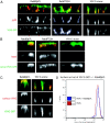
Figure 1.
Figure 1.
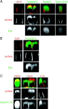
Activated Rab8 (but not dominant-negative Rab8) mislocalizes VSV-G to the apical surface. (A) Fully polarized MDCK cells were microinjected with cDNAs of ts045 VSV-G GFP and T7-tagged Rab8Q67L (dominant active) or Rab8T22N (dominant negative) at 200 ng/μl, incubated at the nonpermissive temperature of 40°C to accumulate VSV-G in the ER for 2 h, and chased for 2 h at the permissive temperature of 31°C in the presence of cycloheximide. The cells were fixed, permeabilized, and processed for IF. Indirect IF of an endogenous basolateral protein using the anti-gp58 antibody (red, second row), followed by Alexa® 568 secondary antibody. The cells were analyzed by confocal microscopy and a representative x-z section is shown. (B) Polarized MDCK cells were microinjected with the cDNAs of apical ts045 VSV-G–G3 with T7-Rab8Q67L or T7-Rab8T22N under the same pulse-chase conditions as above. Cells were processed for IF as above. Rab8 expression (red, second row) was monitored using a mouse anti-T7 tag antibody followed by anti–mouse Alexa® 568 secondary antibody. (C) ts045 VSV-G–GFP was microinjected with T7-Rab8Q67L under the same pulse-chase conditions as in A, but the chase is followed by fixation without permeabilization. The cells are immunolabeled using an antibody, TK-G, against the ectodomain of VSV-G followed by Alexa® 568 secondary antibody. (D) Confluent MDCK cells were infected overnight at 40°C with adenoviruses encoding ts045 VSV-G–GFP and T7-Rab8Q67L. After a 2-h chase at 31°C in the presence of cycloheximide, the cells were trypsinized, fixed without permeabilization, and immunolabeled using the TK-G antibody followed by an anti–mouse phytoerythrin secondary antibody. FACS® analysis shows that the efficiency of surface expression of VSV-G is unchanged in the presence of Rab8Q67L.
Figure 2.
Figure 2.
Activated Rab8 causes the missorting of VSV-G to the apical surface in the biosynthetic pathway. (A) Polarized MDCK cells were infected overnight with VSV-G–GFP and Rab8Q67L adenoviruses and pulse-labeled with [35S]Met/Cys for 15 min. Cells were then incubated at 37°C in medium containing fivefold excess methionine and cysteine for the indicated times. After the chase, cells were placed in ice-cold PBS++ and biotinylated on either the apical (A) or basolateral (B) surfaces. VSV-G was immunoprecipitated using the P5D4 antibody, and the antibody complexes were pulled down with protein G–Sepharose. After spinning down the beads, 20% of the immunoprecipitated protein was set aside as the “total,” whereas the remaining 80% was applied to neutravidin beads in order to isolate the biotinylated membranes. All samples were run on 10% SDS-PAGE gels. The gels were dried and quantitative autoradiography was performed. (B) Graphical representation of the quantitation of the signal of total VSV-G at the surface in the absence or presence of activated Rab8 from A. VSV-G at the apical (AP) surface is in blue and VSV-G at the basolateral (BL) side is in red.
Figure 3.
Figure 3.
Activated Rab8 missorts only AP-1B cargo. (A–C) Fully polarized MDCK cells were microinjected with the cDNAs encoding activated GFP-Rab8 (Rab8Q67L; A, second column), nonprenylated GFP-Rab8 (Rab8ΔC; A, third column), activated GFP-Rab11 (Rab11Q70L; A, fourth column), or dominant-negative GFP-Cdc42 (Cdc42T17N) (C) with LDLR (A and C, red) or FcR (B and C, red). After injection, the cells were incubated at 37°C for 1 h, at 20°C for 2.5 h, and finally at 37°C for 2 h in the presence of cycloheximide. Cells were fixed without permeabilization and stained for surface LDLR (C7) or FcR (24G2), followed with Alexa® 568 secondary antibodies. GTPases are visualized by GFP fluorescence. Images are representative confocal z-sections.
Figure 4.
Figure 4.
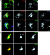
Rab8 is localized to the perinuclear region, associating with recycling endosomes. (A and B) MDCKT (stably transfected with cDNA for Tfn receptor) cells grown on coverslips were microinjected with 50 ng/μl wild-type GFP-Rab8 and were incubated at 37°C for 2 h. Cells were processed for IF and stained for the Golgi (A; GM130, red) and the TGN (B; γ-adaptin [red] arrows indicate colocalization of γ-adaptin with Rab8, and absence of colocalization with furin [blue]). (C) MDCKT cells were induced for 14 h with butyrate to express Tfn receptor, then microinjected with cDNA for 50 ng/μl wild-type GFP-Rab8 and incubated at 37°C for 2 h. After cold-binding Alexa® 594–Tfn, cells were incubated for 22 min at 37°C to accumulate Tfn in recycling endosomes. Colocalization of Tfn, Rab8, and γ-adaptin (arrow). Tfn, red; Rab8, green; γ-adaptin, blue. (D) Three-dimensional reconstruction of confocal serial sections, x-y plane of a representative cell as in C. (E) Same cell from D cut sagittally (white line in first panel of D) and rotated ∼45° to view colocalization of Tfn with Rab8.
Figure 5.
Figure 5.
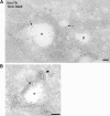
Rab8 colocalizes with Tfn in recycling endosomes. (A and B) Rab8 (10-nm gold) and Tfn (5-nm gold) are localized on endosomes (arrows) and vesicle bud (arrowhead) in MDCKT cells infected with GFP-Rab8 adenovirus. GFP-Rab8 and Alexa® 488-Tfn were visualized using anti-GFP and anti-Alexa® 488 antibodies, respectively, followed by IgG secondary antibodies and protein A–gold. e, endosome; g, Golgi. Bar, 100 nm.
Figure 6.
Figure 6.
Activated Rab8 disrupts γ-adaptin localization in MDCK cells, but does not disrupt Golgi- or furin-containing regions of the TGN. (A) Top: MDCK cells grown on coverslips were microinjected with the cDNA for T7- Rab8Q67L (200 ng/μl), incubated at 37°C for 2 h, fixed, permeabilized, and stained with anti-γ-adaptin antibody 100/3 (red, second column) or anti-T7 (green, third column). Arrows, cells expressing Rab8Q67L where γ-adaptin localization is disrupted; arrowheads, cells not expressing Rab8 with normal γ-adaptin localization. Bottom: cells were injected with cDNA for nonprenylated, activated GFP-Rab8ΔC (green, third column). Arrowheads, cells expressing Rab8ΔC have normal γ-adaptin localization (red, second column). (B) MDCK cells were microinjected with cDNA of T7-Rab8Q67L, incubated at 37°C for 2 h, and processed for IF with the TGN antibody anti-furin (A; top, second column), for the Golgi with anti-giantin (B; bottom, second column), and for T7-Rab8 (A and B, third column).
Figure 7.
Figure 7.
Activated Rab8 causes the selective disruption of AP-1B complexes. (A) LLC-PK1 cells were transfected with cDNAs for GFP-Rab8Q67L and TGN-38, and then were processed for IF with γ-adaptin 100/3 (red, second column) and TGN-38 using anti-TGN-38 antibody (blue, fourth column). Top, wild-type LLC-PK1 cells (AP-1A+/AP-1B−); bottom, LLC-PK1 stably transfected with μ1B gene (AP-1A+/AP-1B+). Arrows, colocalization of γ-adaptin with TGN-38. Notice there is no change in γ-adaptin localization in cells that express only AP-1A (top) even in the presence of Rab8Q67L expression. (B) Embryonic fibroblast cells expressing only AP-1B (AP-1A−/AP-1B+) were transfected as above with cDNA for GFP-Rab8Q67L or GFP vector alone (green). Mouse anti-γ-adaptin antibody clone 88 (red). Arrowheads, nontransfected cells with normal γ-adaptin localization.

Similar articles

Cited by

References

    1. Aroeti, B., and K.E. Mostov. 1994. Polarized sorting of the polymeric immunoglobulin receptor in the exocytotic and endocytotic pathways is controlled by the same amino acids. EMBO J. 13:2297–2304. - PMC - PubMed
    1. Carroll, K.S., J. Hanna, I. Simon, J. Krise, P. Barbero, and S.R. Pfeffer. 2001. Role of Rab9 GTPase in facilitating receptor recruitment by TIP47. Science. 292:1373–1376. - PubMed
    1. Cohen, D., A. Musch, and E. Rodriguez-Boulan. 2001. Selective control of basolateral membrane protein polarity by cdc42. Traffic. 2:556–564. - PubMed
    1. Drubin, D.G., and W.J. Nelson. 1996. Origins of cell polarity. Cell. 84:335–344. - PubMed
    1. Erickson, J.W., C. Zhang, R.A. Kahn, T. Evans, and R.A. Cerione. 1996. Mammalian Cdc42 is a brefeldin A-sensitive component of the Golgi apparatus. J. Biol. Chem. 271:26850–26854. - PubMed

Publication types

MeSH terms