. 2004 Jan;15(1):162-75.
doi: 10.1091/mbc.e03-07-0535. Epub 2003 Oct 17.

rsly1 binding to syntaxin 5 is required for endoplasmic reticulum-to-Golgi transport but does not promote SNARE motif accessibility

Affiliations

rsly1 binding to syntaxin 5 is required for endoplasmic reticulum-to-Golgi transport but does not promote SNARE motif accessibility

Antionette L Williams et al. Mol Biol Cell. 2004 Jan.

Abstract

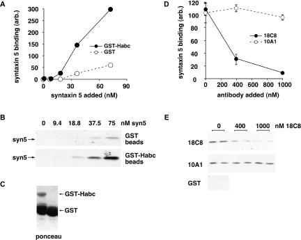
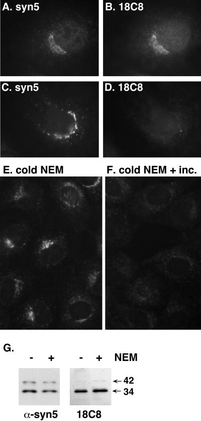
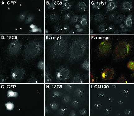
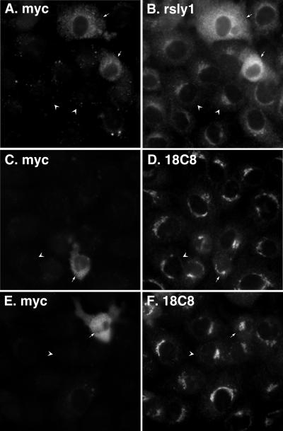
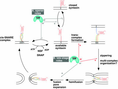
Although some of the principles of N-ethylmaleimide-sensitive factor attachment protein receptor (SNARE) function are well understood, remarkably little detail is known about sec1/munc18 (SM) protein function and its relationship to SNAREs. Popular models of SM protein function hold that these proteins promote or maintain an open and/or monomeric pool of syntaxin molecules available for SNARE complex formation. To address the functional relationship of the mammalian endoplasmic reticulum/Golgi SM protein rsly1 and its SNARE binding partner syntaxin 5, we produced a conformation-specific monoclonal antibody that binds only the available, but not the cis-SNARE-complexed nor intramolecularly closed form of syntaxin 5. Immunostaining experiments demonstrated that syntaxin 5 SNARE motif availability is nonuniformly distributed and focally regulated. In vitro endoplasmic reticulum-to-Golgi transport assays revealed that rsly1 was acutely required for transport, and that binding to syntaxin 5 was absolutely required for its function. Finally, manipulation of rsly1-syntaxin 5 interactions in vivo revealed that they had remarkably little impact on the pool of available syntaxin 5 SNARE motif. Our results argue that although rsly1 does not seem to regulate the availability of syntaxin 5, its function is intimately associated with syntaxin binding, perhaps promoting a later step in SNARE complex formation or function.


Figures

Figure 2.
Figure 2.
18C8 binds to the syntaxin 5 SNARE motif mutually exclusively with ER/Golgi SNAREs. (A) Purified bacterially expressed GST or GST-membrin was immobilized on glutathione beads and mixed with soluble syntaxin 5 SNARE motif and rbet1 cytoplasmic domain. Shown is a quantification of syntaxin 5 bound to the GST and GST-membrin beads after washing with buffer and immunoblotting. GST and GST-membrin beads were reacted either in the absence of mAb (filled bars) or the presence of equal concentrations of the indicated mAb (open bars). (B) ER/Golgi quaternary complexes were formed from syntaxin 5 SNARE motif, membrin, rbet1, and sec22b in solution and purified by gel filtration as described previously (Xu et al., 2000). Purified ER/Golgi quaternary complex (open bars), or a lesser amount of syntaxin 5 SNARE motif in isolation (filled bars), were subjected to immunoprecipitation with 18C8 and protein A-Sepharose beads at the indicated antibody concentrations, and syntaxin 5 in the immunoprecipitated pellets was quantified by immunoblotting after SDS-PAGE and transfer to nitrocellulose. (C) Autoradiogram of the blot that was quantified to produce B. Shown are the immunoprecipitation input lanes (left) and the immunoprecipitated pellets (right).
Figure 3.
Figure 3.
18C8 inhibits binding between the syntaxin 5 SNARE motif and Habc domain. (A) Purified bacterially expressed GST (open symbols) or GST-syntaxin 5 Habc domain (filled symbols) was immobilized on glutathione beads and mixed with soluble syntaxin 5 SNARE motif at the indicated concentrations. SNARE motif bound to the beads after buffer washes was quantified by immunoblotting. (B) Autoradiogram of the blot that was quantified to produce A. (C) Ponceau stain of the immunoblot lanes to which no soluble SNARE motif was added; a high proportion of the protein in the GST-Habc preparation was GST. (D) Purified bacterially expressed GST-syntaxin 5 Habc domain was immobilized on glutathione beads and mixed with soluble syntaxin 5 SNARE motif at the highest concentration on the curve in A, in the presence of increasing concentrations of purified 18C8 (filled symbols) or 10A1 (open symbols). SNARE motif bound to the beads after buffer washes was quantified by immunoblotting after SDS-PAGE and transfer to nitrocellulose. (E) Autoradiogram of the blot that was quantified to produce D.
Figure 9.
Figure 9.
Quantitation of 18C8 staining intensities reveals that dissociation of rsly1–syntaxin 5 interactions causes a modest increase in 18C8 accessibility. (A) Demonstration of ratiometric quantitation of 18C8 staining intensity relative to rsly1 staining (left) and polyclonal anti-syntaxin 5 staining (right). Digitonin-permeabilized cells were fixed and stained after incubation on ice with buffer (open bars), with buffer containing purified NSF, α-SNAP and MgATP (gray bars), or after a 37°C NEM treatment as in Figure 4 (filled bars). Plotted is the ratio of Golgi area 18C8 staining intensity to that of anti-rsly1 or anti-syntaxin 5, averaged over ∼120 cells per condition as described in MATERIALS AND METHODS. (B) Ratiometric quantitation of 18C8 staining intensity relative to rsly1 staining (left) and polyclonal anti-syntaxin 5 staining (right) in untransfected (open bars) and syntaxin 5 (1-43)-GFP-transfected cells (solid bars). Nontransfected and transfected cells were from the same coverslips. For both A and B, the means are plotted plus or minus SE.
Figure 11.
Figure 11.
rsly1 must bind stoichiometrically to a fillable membrane site to function in ER-to-Golgi transport. (A) Endoglycosidase H analysis of VSVG ts045 protein in permeabilized NRK cells after transport incubations containing the indicated concentrations of the indicated control (mouse, rabbit) or immune (α-rsly1, 18C8) Fab fragments. Endo H-resistant (HR) and -sensitive (HS) bands are indicated (arrows). Control reactions lacking any Fabs are shown above. (B) Quantitation of the experiment from A (main axis) and also a separate experiment (histogram) in which a partially inhibitory concentration of α-rsly1 intact IgG was tested in the absence (left bar) or presence (right bar) of excess purified GST-rsly1. (C) Permeabilized NRK cells were either preincubated on ice with or without anti-rsly1 antibodies (bars 5–9) or else incubated at 32°C immediately (bars 2–4) with regular transport cocktail (reg.) or cocktail supplemented with a 100-fold excess of soluble purified recombinant rsly1 (rec. rsly1) or a 10-fold excess of partially purified native liver rsly1 (liv. rsly1). The preincubated cells (bars 5–9) were subsequently washed twice and resuspended in regular transport cocktail (reg.) or cocktail supplemented with α-rsly1 antibodies (+Ab) or excess rsly1-containing cocktails (rec. rsly1 and liv. rsly1). VSVG transport was quantified after 90 min at 32°C as in B. Plotted values are means of duplicate reactions plus or minus SE. (D) Immunoblots demonstrating the quantity of rsly1 present in washed, permeabilized NRK cells used for transport, the normal rat liver cytosol used for transport, and the indicated dilutions of the purified recombinant and partially purified cytosolic rsly1 used in B.
Figure 1.
Figure 1.
A set of monoclonal antibodies directed against the syntaxin 5 SNARE motif. Antibodies from tissue culture supernatants from the indicated hybridomas (above) were purified by protein A or protein G-Sepharose and used to immunoblot identical lanes of crude rat brain membranes separated by SDS-PAGE and transferred to nitrocellulose. The migration of molecular weight marker proteins are shown (left), as are the positions of the 42-kDa (syn 5 42) and 34-kDa (syn 5 34) endogenous syntaxin 5 00oforms and a commonly seen syntaxin 5 degradation fragment at 32 kDa [syn 5 32 (deg.)].
Figure 4.
Figure 4.
18C8 stains only free, uncomplexed syntaxin 5 in fixed NRK cells. (A–D) NRK cells were either incubated in control medium (A and B) or in medium containing 50 μM NEM (C and D) for 5 min at 37°C in a CO2 incubator before fixation with paraformaldehyde and immunostaining with 18C8 and polyclonal anti-syntaxin 5 as described in MATERIALS AND METHODS. (E and F) NRK cells were incubated in medium containing 100 μM NEM for 5 min on ice and then washed several times with NEM-free medium and either fixed on ice (E) or incubated at 37°C for 5 min and then fixed on ice (F). After fixation, the cells were immunostained with purified 18C8. (G) Cells that had undergone control or NEM incubations were placed on ice, lysed, separated by SDS-PAGE, and immunoblotted using 18C8 or polyclonal anti-syntaxin 5 antisera.
Figure 5.
Figure 5.
18C8-available syntaxin 5 is nonuniformly and focally l/calized. Fixed NRK cells were double-stained with 18C8 and polyclonal anti-syntaxin 5 antibodies by using FITC- and Texas Red-labeled secondary antibodies, respectively. Images were collected for both filter sets every 0.2 μm through the cell, and the image stacks were optically deconvolved using an algorithm that removes no light from the stack and involves no arbitrary user inputs. Single optical sections of three Golgi regions, from three different cells are shown for the FITC channel (A, D, and G), the Texas Red channel (B, E, and H) and merged images (C, F, and I).
Figure 6.
Figure 6.
rsly1 is l/calized to the Golgi region independently of the oligomeric state of syntaxin 5. Fixed NRK cells were immunostained using an affinity-purified anti-rsly1 antiserum under control conditions (A and C), after 50 μM NEM treatment as in Figure 4 (D), or in the presence of an excess of purified bacterially produced GST-rsly1 (B). A and B are from a separate experiment from C and D.
Figure 7.
Figure 7.
Expression of syntaxin 5 (1-43)-GFP dissociates Golgi rsly1 staining from that of 18C8. NRK cells were transfected with syntaxin 5 (1-43)-GFP, fixed, and immunostained with the indicated primary antibodies followed by cy3- and cy5-labeled secondary antibodies. Images shown used filter sets for GFP (A and G), cy3 (B, D, and H), and cy5 (C, E, and I) or a merge of cy3 and cy5 (F). D, E, and F are magnified views of the boxed region in B and C. Arrowheads mark staining in nontransfected cells, and arrows mark staining in transfected cells.
Figure 8.
Figure 8.
Expression of syntaxin 5 (1-43)-GFP does not significantly alter 18C8 staining intensity relative to polyclonal anti-syntaxin 5 staining. NRK cells were transfected with syntaxin 5 (1-43)-GFP, fixed, and immunostained with 18C8 or polyclonal anti-syntaxin 5 antibodies followed by cy3- and cy5–labeled secondary antibodies, respectively. Images shown used filter sets for GFP (A), cy3 (B and D) and cy5 (C and E). D and E are magnified views of the boxed region in B and C. Arrowheads demonstrate staining in nontransfected cells, short arrows demonstrate staining in low-to-moderately-expressing cells, and long arrows demonstrate staining in highly expressing cells.
Figure 10.
Figure 10.
Overexpression of myc-rsly1 does not significantly change 18C8 accessibility of syntaxin 5. NRK cells were transfected with myc-rsly1, fixed, and immunostained with anti-myc (A, C, and E), anti-rsly1 (B), or 18C8 (D and F) antibodies. Overexpressing cells (arrows) displayed similar 18C8 staining to that of surrounding nontransfected cells (arrowheads).
Figure 12.
Figure 12.
Syntaxin 5 binding is essential for rsly1 function in ER-to-Golgi transport. (A) VSVG transport was monitored in the presence of the indicated concentrations of GST (filled circles) or thrombin-cleaved GST-syntaxin 5 (1-43) (open circles). (B) Transport was monitored under control conditions (open bars) or in the presence of the indicated concentrations of synthetic peptides corresponding to syntaxin 5 amino acids 1–27 (UM-1, solid bars) or the same peptide containing T7A and F10A mutations (UM-2, gray bars). Plotted are the mean transport values after 90 min of incubation at 32°C, plus or minus SE.
Figure 13.
Figure 13.
Schematic of possible mechanisms of action of SM proteins in the SNARE cycle and membrane fusion. Known or hypothetical steps in the SNARE cycle are represented with black arrows. Potential SM protein roles that are consistent with our data are indicated with red arrows and roles that are inconsistent or less consistent with our data are indicated with gray arrows. Potential SM protein roles are grouped for illustration purposes into opener roles (A) and late-stage roles (B). The opener roles are inconsistent with our staining experiments (e.g., Figures 9 and 10), because they would predict a decrease in available syntaxin 5 SNARE motif when syntaxin 5–rsly1 interactions were blocked, and an increase when rsly1 was overexpressed. Among the potential late-stage roles, a role in promotion of fusion pore expansion or other lipidic events is less consistent with our data than the other illustrated roles, because they would not necessarily require syntaxin 5–rsly1 interactions, whereas our transport experiments (Figures 11 and 12) demonstrated this requirement for rsly1 function. The role indicated by “multicomplex organization” is not explicitly illustrated because very little is known about what this may entail; one suggestion would be arrangement of multiple SNARE complexes around a central fusion site. We illustrate only potentially required, positive roles. Negative roles such as stabilization of closed syntaxin would not predict the strict requirement for rsly1 in ER-to-Golgi transport (Figure 11). Note that rsly1 could potentially perform more than one of the indicated functions.

Similar articles

Cited by

References

    1. Bracher, A., and Weissenhorn, W. (2002). Structural basis for the Golgi membrane recruitment of Sly1p by Sed5p. EMBO J. 21, 6114–6124. - PMC - PubMed
    1. Bryant, N.J., and James, D.E. (2001). Vps45p stabilizes the syntaxin homologue Tlg2p and positively regulates SNARE complex formation. EMBO J. 20, 3380–3388. - PMC - PubMed
    1. Bryant, N.J., and James, D.E. (2003). The Sec1p/Munc18 (SM) protein, Vps45p, cycles on and off membranes during vesicle transport. J. Cell Biol. 161, 691–696. - PMC - PubMed
    1. Carr, C.M., Grote, E., Munson, M., Hughson, F.M., and Novick, P.J. (1999). Sec1p binds to SNARE complexes and concentrates at sites of secretion. J. Cell Biol. 146, 333–344. - PMC - PubMed
    1. Dulubova, I., Yamaguchi, T., Gao, Y., Min, S.W., Huryeva, I., Sudhof, T.C., and Rizo, J. (2002). How Tlg2p/syntaxin 16 `snares' Vps45. EMBO J. 21, 3620–3631. - PMC - PubMed

Publication types

MeSH terms

LinkOut - more resources