

Discs-Large and Strabismus are functionally linked to plasma membrane formation
- PMID: 14562058
- PMCID: PMC3471657
- DOI: 10.1038/ncb1055
Discs-Large and Strabismus are functionally linked to plasma membrane formation
Abstract
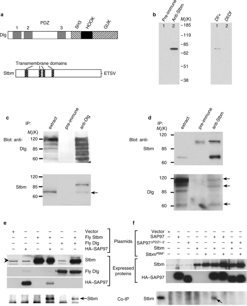
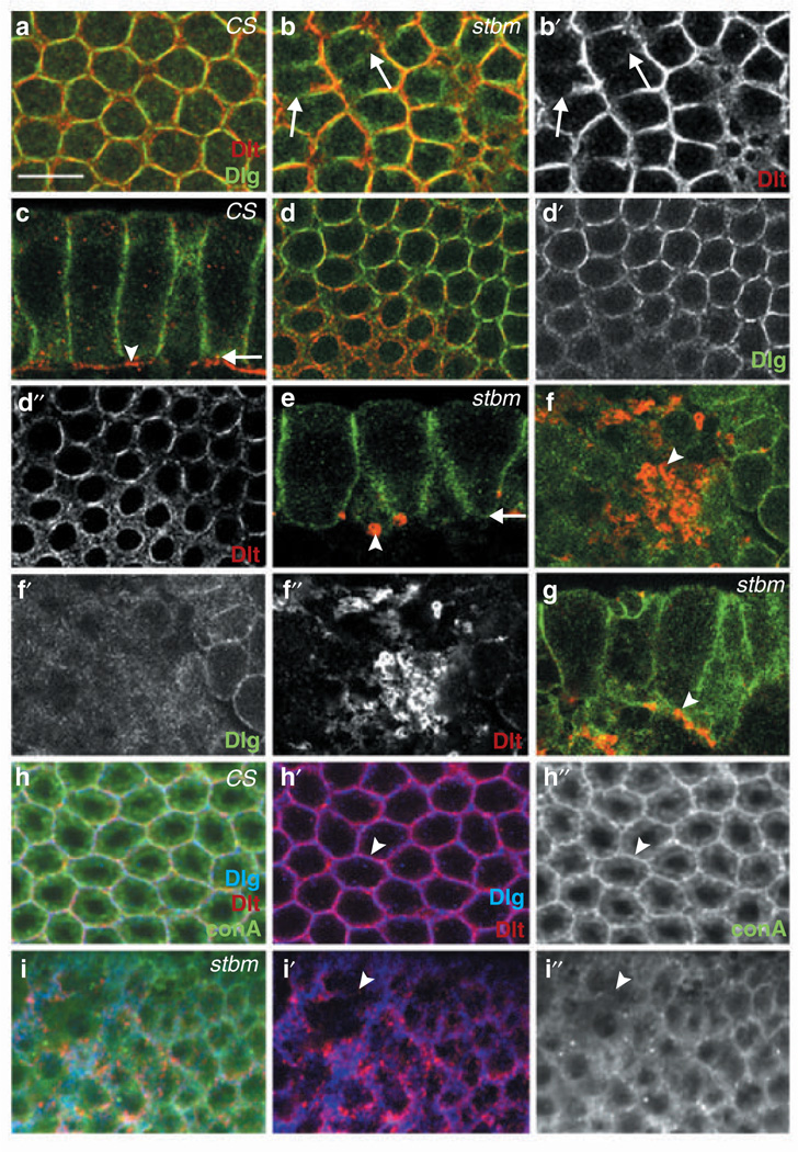
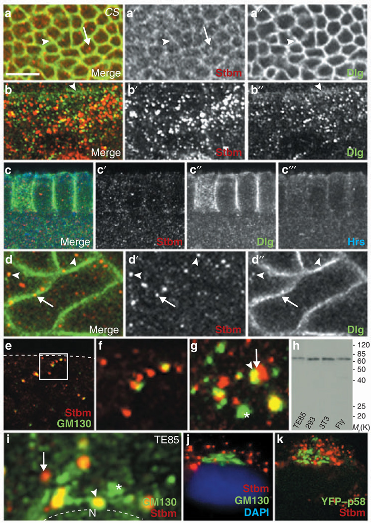
During early embryogenesis in Drosophila melanogaster, extensive vesicle transport occurs to build cell boundaries for 6,000 nuclei. Here we show that this important process depends on a functional complex formed between the tumour suppressor and adaptor protein Discs-Large (Dlg) and the integral membrane protein Strabismus (Stbm)/Van Gogh (Vang). In support of this idea, embryos with mutations in either dlg or stbm displayed severe defects in plasma membrane formation. Conversely, overexpression of Dlg and Stbm synergistically induced excessive plasma membrane formation. In addition, ectopic co-expression of Stbm (which associated with post-Golgi vesicles) and the mammalian Dlg homologue SAP97/hDlg promoted translocation of SAP97 from the cytoplasm to both post-Golgi vesicles and the plasma membrane. This effect was dependent on the interaction between Stbm and SAP97. These findings suggest that the Dlg-Stbm complex recruits membrane-associated proteins and lipids from internal membranes to sites of new plasma membrane formation.
Figures


Similar articles
-
The planar cell polarity protein Strabismus promotes Pins anterior localization during asymmetric division of sensory organ precursor cells in Drosophila.Development. 2004 Jan;131(2):469-78. doi: 10.1242/dev.00928. Development. 2004. PMID: 14701683
-
Craniofacial dysmorphogenesis including cleft palate in mice with an insertional mutation in the discs large gene.Mol Cell Biol. 2001 Mar;21(5):1475-83. doi: 10.1128/MCB.21.5.1475-1483.2001. Mol Cell Biol. 2001. PMID: 11238884 Free PMC article.
-
Prickle and Strabismus form a functional complex to generate a correct axis during planar cell polarity signaling.EMBO J. 2003 Sep 1;22(17):4409-20. doi: 10.1093/emboj/cdg424. EMBO J. 2003. PMID: 12941693 Free PMC article.
-
Dlg, Scribble and Lgl in cell polarity, cell proliferation and cancer.Bioessays. 2003 Jun;25(6):542-53. doi: 10.1002/bies.10286. Bioessays. 2003. PMID: 12766944 Review.
-
Control of tumourigenesis by the Scribble/Dlg/Lgl polarity module.Oncogene. 2008 Nov 24;27(55):6888-907. doi: 10.1038/onc.2008.341. Oncogene. 2008. PMID: 19029932 Review.
Cited by
-
Loss- or Gain-of-Function Mutations in ACOX1 Cause Axonal Loss via Different Mechanisms.Neuron. 2020 May 20;106(4):589-606.e6. doi: 10.1016/j.neuron.2020.02.021. Epub 2020 Mar 12. Neuron. 2020. PMID: 32169171 Free PMC article.
-
Nuf, a Rab11 effector, maintains cytokinetic furrow integrity by promoting local actin polymerization.J Cell Biol. 2008 Jul 28;182(2):301-13. doi: 10.1083/jcb.200712036. Epub 2008 Jul 21. J Cell Biol. 2008. PMID: 18644888 Free PMC article.
-
Planar cell polarity in Drosophila.Organogenesis. 2011 Jul-Sep;7(3):165-79. doi: 10.4161/org.7.3.18143. Epub 2011 Jul 1. Organogenesis. 2011. PMID: 21983142 Free PMC article. Review.
-
CASK deletion in intestinal epithelia causes mislocalization of LIN7C and the DLG1/Scrib polarity complex without affecting cell polarity.Mol Biol Cell. 2009 Nov;20(21):4489-99. doi: 10.1091/mbc.e09-04-0280. Epub 2009 Sep 2. Mol Biol Cell. 2009. PMID: 19726564 Free PMC article.
-
Zygotically controlled F-actin establishes cortical compartments to stabilize furrows during Drosophila cellularization.J Cell Sci. 2008 Jun 1;121(11):1815-24. doi: 10.1242/jcs.025171. Epub 2008 May 6. J Cell Sci. 2008. PMID: 18460582 Free PMC article.
References
-
- Woods DF, Bryant PJ. The discs-large tumor suppressor gene of Drosophila encodes a guanylate kinase homolog localized at septate junctions. Cell. 1991;66:451–464. - PubMed
-
- Wolff T, Rubin G. strabismus, a novel gene that regulates tissue polarity and cell fate decisions in Drosophila. Development. 1998;125:1149–1159. - PubMed
Publication types
MeSH terms
Substances
Grants and funding
LinkOut - more resources
Full Text Sources
Other Literature Sources
Molecular Biology Databases
