. 2003 Nov;23(21):7875-86.
doi: 10.1128/MCB.23.21.7875-7886.2003.

Molecular mechanism for a role of SHP2 in epidermal growth factor receptor signaling

Affiliations

Molecular mechanism for a role of SHP2 in epidermal growth factor receptor signaling

Yehenew M Agazie et al. Mol Cell Biol. 2003 Nov.

Abstract

The Src homology 2-containing phosphotyrosine phosphatase (SHP2) is primarily a positive effector of receptor tyrosine kinase signaling. However, the molecular mechanism by which SHP2 effects its biological function is unknown. In this report, we provide evidence that defines the molecular mechanism and site of action of SHP2 in the epidermal growth factor-induced mitogenic pathway. We demonstrate that SHP2 acts upstream of Ras and functions by increasing the half-life of activated Ras (GTP-Ras) in the cell by interfering with the process of Ras inactivation catalyzed by Ras GTPase-activating protein (RasGAP). It does so by inhibition of tyrosine phosphorylation-dependent translocation of RasGAP to the plasma membrane, to its substrate (GTP-Ras) microdomain. Inhibition is achieved through the dephosphorylation of RasGAP binding sites at the level of the plasma membrane. We have identified Tyr992 of the epidermal growth factor receptor (EGFR) to be one such site, since its mutation to Phe renders the EGFR refractory to the effect of dominant-negative SHP2. To our knowledge, this is the first report to outline the site and molecular mechanism of action of SHP2 in EGFR signaling, which may also serve as a model to describe its role in other receptor tyrosine kinase signaling pathways.


Figures
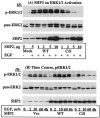
FIG. 1.
FIG. 1.
Effect of the C/S protein on ERK1/2 phosphorylation. (A) COS-1 cells were transfected with various amounts of an expression vector for the WT or C/S protein, incubated for 36 h in 10% serum-containing medium, serum starved for 12 h, and then stimulated with 100 ng of EGF/ml for 10 min. Total-cell lysates containing equivalent amounts of protein were separated on a 10% polyacrylamide gel and then analyzed by immunoblotting with the indicated antibodies. (B) Time course studies of ERK1/2 phosphorylation. A constant amount of the indicated expression plasmids was transfected into COS-1 cells. Following transfection, cells were treated as for panel A and then left unstimulated or stimulated with 100 ng of EGF/ml for the indicated time points. Total-cell lysates containing equivalent amounts of protein were analyzed by immunoblotting with the indicated antibodies. (C) Quantitation of phospho-ERK1/2 band intensities. The same set of lysates as in panel B was used to determine band intensities for ERK1/2 phosphorylation as described in Materials and Methods. IB, immunoblot.
FIG. 1.
FIG. 1.
Effect of the C/S protein on ERK1/2 phosphorylation. (A) COS-1 cells were transfected with various amounts of an expression vector for the WT or C/S protein, incubated for 36 h in 10% serum-containing medium, serum starved for 12 h, and then stimulated with 100 ng of EGF/ml for 10 min. Total-cell lysates containing equivalent amounts of protein were separated on a 10% polyacrylamide gel and then analyzed by immunoblotting with the indicated antibodies. (B) Time course studies of ERK1/2 phosphorylation. A constant amount of the indicated expression plasmids was transfected into COS-1 cells. Following transfection, cells were treated as for panel A and then left unstimulated or stimulated with 100 ng of EGF/ml for the indicated time points. Total-cell lysates containing equivalent amounts of protein were analyzed by immunoblotting with the indicated antibodies. (C) Quantitation of phospho-ERK1/2 band intensities. The same set of lysates as in panel B was used to determine band intensities for ERK1/2 phosphorylation as described in Materials and Methods. IB, immunoblot.
FIG. 2.
FIG. 2.
Effect of C/S protein on V12-H-Ras-induced ERK1/2 phosphorylation. COS-1 cells were transfected with a constant amount of an expression vector for V12-H-Ras and a varying amount for the C/S protein. Following transfection, cells were incubated for 36 h in 10% serum-containing medium and for a further 12 h in the absence of serum. Total-cell lysates containing equivalent amounts of protein were separated on a 10% polyacrylamide gel and then analyzed by immunoblotting with the indicated antibodies. IB, immunoblot.
FIG. 3.
FIG. 3.
Effect of the C/S protein on Ras activation. (A) COS-1 cells were cotransfected with two different concentrations of an expression vector for SHP2 (WT or the C/S protein) and H-Ras. (B) Time course studies of Ras activation. COS-1 cells were cotransfected with a constant amount of WT-H-Ras and the WT or C/S protein. In either case, cells were incubated for 36 h in 10% serum-containing medium, serum starved for 12 h, and then stimulated with 100 ng of EGF/ml for 10 min in panel A or the indicated time points in panel B. Total-cell lysates (TCL) prepared from these cells were subjected to affinity precipitation (AP) with GST-RBD of Raf-1, resolved on a 10% polyacrylamide gel, and then analyzed by immunoblotting (IB) with T7 tag antibody for Ras. To examine expression of SHP2 and Ras proteins, total-cell lysates were directly analyzed with the indicated antibodies.
FIG. 4.
FIG. 4.
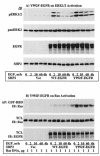
Characterization of new dominant-negative mutant of SHP2. The R/E and the C/S-R/E proteins were developed as described in Materials and Methods. An expression vector containing the WT, C/S, R/E, or C/S-R/E protein was transfected into COS-1 cells. Following transfection, cells were incubated for 36 h in 10% serum-containing medium and for a further 12 h in the absence of serum and then left unstimulated or stimulated with 100 ng of EGF/ml for 10 min. (A) Total-cell lysates were analyzed for ERK1/2 phosphorylation (top), loading control for ERK2 (second panel), level of expression of the different SHP2 proteins (third panel), or tyrosine phosphorylation pattern (bottom). (B) Time course study with PDGF stimulation (10 ng/ml). The same SHP2 constructs were transfected into NIH 3T3 cells, incubated under the same conditions as in panel A, and then stimulated with PDGF for the indicated time points. Lysates prepared from these cells were analyzed for ERK1/2 activation (top), loading control for ERK2 (middle), and level of expression of the different SHP2 proteins (bottom). (C) Time course study with FGF stimulation (100 ng/ml). All the procedures were the same as in panel B except that the ligand was FGF instead of PDGF. TCL, total-cell lysates; IB, immunoblot.
FIG. 4.
FIG. 4.
Characterization of new dominant-negative mutant of SHP2. The R/E and the C/S-R/E proteins were developed as described in Materials and Methods. An expression vector containing the WT, C/S, R/E, or C/S-R/E protein was transfected into COS-1 cells. Following transfection, cells were incubated for 36 h in 10% serum-containing medium and for a further 12 h in the absence of serum and then left unstimulated or stimulated with 100 ng of EGF/ml for 10 min. (A) Total-cell lysates were analyzed for ERK1/2 phosphorylation (top), loading control for ERK2 (second panel), level of expression of the different SHP2 proteins (third panel), or tyrosine phosphorylation pattern (bottom). (B) Time course study with PDGF stimulation (10 ng/ml). The same SHP2 constructs were transfected into NIH 3T3 cells, incubated under the same conditions as in panel A, and then stimulated with PDGF for the indicated time points. Lysates prepared from these cells were analyzed for ERK1/2 activation (top), loading control for ERK2 (middle), and level of expression of the different SHP2 proteins (bottom). (C) Time course study with FGF stimulation (100 ng/ml). All the procedures were the same as in panel B except that the ligand was FGF instead of PDGF. TCL, total-cell lysates; IB, immunoblot.
FIG. 5.
FIG. 5.
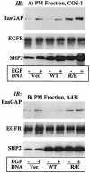
SHP2 regulates EGF-induced translocation of RasGAP to the PM. Immunoblot analysis of PMFs prepared from COS-1 cells transfected with the vector, the WT, or the R/E protein (A) or from A431 cells infected with a retrovirus expressing the same set of proteins (B). In both panels A and B, samples of PMFs containing equivalent amounts of protein were separated on a 10% polyacrylamide gel and then analyzed with anti-RasGAP antibody (top), anti-EGFR antibody as a loading control (middle), or anti-SHP2 antibody to show expression (bottom). IB, immunoblot.
FIG. 6.
FIG. 6.
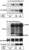
Affinity precipitation and far-Western analysis for the interaction of RasGAP with the EGFR. COS-1 cells were transfected with expression vector for the indicated SHP2 proteins. Following transfection, cells were incubated for 36 h in 10% serum-containing medium and for a further 12 h in the absence of serum and then left unstimulated or stimulated with 100 ng of EGF/ml for 10 min. PMFs were prepared as described in Materials and Methods, subjected to affinity precipitation with GST-SH2-RasGAP (A), or directly separated on a 10% polyacrylamide gel (B and C). In panels A and B, the membranes were probed first with anti-pY antibody and second with anti-EGFR after stripping. Membrane B was further probed with anti-SHP2 antibody to show expression of the different SHP2 proteins. In panel C, total PMFs were analyzed by far-Western blotting with 10 μg of GST-SH2-GAP/ml as described in Materials and Methods. Membrane C was further reprobed with anti-EGFR antibody to show the presence of approximately equal amounts of the EGFR protein in all lanes. IB, immunoblot; AP, affinity precipitation.
FIG. 6.
FIG. 6.
Affinity precipitation and far-Western analysis for the interaction of RasGAP with the EGFR. COS-1 cells were transfected with expression vector for the indicated SHP2 proteins. Following transfection, cells were incubated for 36 h in 10% serum-containing medium and for a further 12 h in the absence of serum and then left unstimulated or stimulated with 100 ng of EGF/ml for 10 min. PMFs were prepared as described in Materials and Methods, subjected to affinity precipitation with GST-SH2-RasGAP (A), or directly separated on a 10% polyacrylamide gel (B and C). In panels A and B, the membranes were probed first with anti-pY antibody and second with anti-EGFR after stripping. Membrane B was further probed with anti-SHP2 antibody to show expression of the different SHP2 proteins. In panel C, total PMFs were analyzed by far-Western blotting with 10 μg of GST-SH2-GAP/ml as described in Materials and Methods. Membrane C was further reprobed with anti-EGFR antibody to show the presence of approximately equal amounts of the EGFR protein in all lanes. IB, immunoblot; AP, affinity precipitation.
FIG. 7.
FIG. 7.
Identification of SHP2 substrate pY residue(s) on the EGFR. (A) Schematic representation of WT or the single Tyr-to-Phe mutants of EGFR used in this experiment. (B) NIH 3T3 cells were transfected with the vector or the constructs depicted in panel A and incubated for 48 h in 10% serum-containing medium and for a further 6 h in serum-free medium. Cells were then stimulated with 100 ng of EGF/ml for 10 min as indicated. Total-cell lysates (TCL) prepared from these cells were directly analyzed by immunoblotting (IB) with anti-EGFR antibody (top) or subjected to affinity precipitation (AP) with GST fusion of DM-PTP (middle) or SH2-RasGAP (bottom) and then immunoblotting with anti-EGFR antibody.
FIG. 8.
FIG. 8.
Y992F-EGFR activates ERK1/2 and Ras independently of functional SHP2. NIH 3T3 cells stably expressing the R/E protein were produced as described in Materials and Methods. (A) These cells were transfected with vector alone, WT-EGFR, or Y992F-EGFR; incubated for ∼36 h; serum starved for 12 h; and then stimulated with 100 ng of EGF/ml for the indicated time points. Lysates prepared from these cells were analyzed for ERK1/2 activation (top), for level of ERK2 as loading control (second panel), for expression of EGFR proteins (third panel), or for R/E protein expression (bottom). (B) The same cells were cotransfected with T7-tagged WT-H-Ras and WT-EGFR or Y992F-EGFR and incubated and treated under the same conditions as in panel A. Lysates prepared from these cells were assayed for GTP-Ras levels as described for Fig. 3. Shown are anti-T7 tag immunoblots for GTP-Ras level (top) and total Ras expression (middle) and anti-EGFR immunoblot for expression of EGFR proteins (bottom). IB, immunoblot; AP, affinity precipitation; TCL, total-cell lysate.
FIG. 9.
FIG. 9.
Schematic representation of the molecular mechanism of SHP2 in the EGFR signaling pathway. The five Y's represent autophosphorylation sites on the C-terminal region of the EGFR. SHP2 PTP activity dephosphorylates the most proximal pY, which corresponds to pY992, a binding site of RasGAP shown as GAP SH2.

Similar articles

Cited by

References

    1. Agazie, Y. M., and M. J. Hayman. Oncogene, in press. - PubMed
    1. Agazie, Y. M., and M. J. Hayman. 2003. Development of an efficient “substrate-trapping” mutant of Src homology phosphotyrosine phosphatase 2 and identification of the epidermal growth factor receptor, Gab1, and three other proteins as target substrates. J. Biol. Chem. 278:13952-13958. - PubMed
    1. Bardelli, A., P. Longati, D. Gramaglia, M. C. Stella, and P. M. Comoglio. 1997. Gab1 coupling to the HGF/Met receptor multifunctional docking site requires binding of Grb2 and correlates with the transforming potential. Oncogene 15:3103-3111. - PubMed
    1. Bennett, A. M., S. F. Hausdorff, A. M. O'Reilly, R. M. Freeman, and B. G. Neel. 1996. Multiple requirements for SHPTP2 in epidermal growth factor-mediated cell cycle progression. Mol. Cell. Biol. 16:1189-1202. - PMC - PubMed
    1. Carlberg, K., and L. R. Rohrschneider. 1997. Characterization of a novel tyrosine phosphorylated 100-kDa protein that binds to SHP-2 and phosphatidylinositol 3′-kinase in myeloid cells. J. Biol. Chem. 272:15943-15950. - PubMed

Publication types

MeSH terms

Substances