

Review
doi: 10.1128/jvi.77.19.10179-10185.2003.
Herpesvirus entry: an update
Affiliations
- PMID: 12970403
- PMCID: PMC228481
- DOI: 10.1128/jvi.77.19.10179-10185.2003
Item in Clipboard
Review
Herpesvirus entry: an update
J Virol.
2003 Oct.
No abstract available
Figures
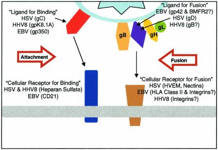
Participants in herpesvirus entry and virus-induced cell fusion. For both alphaherpesviruses and gammaherpesviruses, binding to cells can be mediated by a virion glycoprotein that is not essential for entry. The binding receptors are heparan sulfate for HSV gC and HHV-8 K8.1A and CD21 for EBV gp350 (in the case of B cells). Entry requires interaction of a viral ligand with another cell surface receptor. For HSV, virion gD is the ligand for several cell surface receptors (HVEM, nectins, 3-O-sulfated heparan sulfate), any one of which can mediate entry. For EBV entry into B cells, gp42 binds to HLA class II molecules. It should be noted that gp42 is not required for EBV infection of epithelial cells. The entry receptors in epithelial cells have not yet been identified but could include integrins. The viral ligands could be gH and/or BMRF2. For HHV-8 entry, gB can bind to one of the integrins. Any one of these interactions of a viral ligand with an entry receptor is thought to activate the fusion activity of gB and gH-gL.
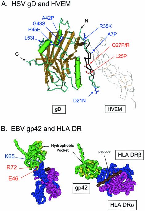
Mutational analysis of HSV and EBV entry receptors and ligands. (A) Mutations in HSV gD that influence functional interactions of gD with the various entry receptors. The features of HSV-1 gD (left; in color) and HVEM (right; in gray) shown are based on the crystal structure of gD-HVEM complexes (6). The peptide backbone of gD is shown as a tube in light blue with the beta strands and alpha-helices shown in gold and green, respectively. The peptide backbone of HVEM is shown as a wire in gray with the disulfide bonds in orange. Amino acids 7 to 15 and 24 to 32, the contact regions with HVEM, are shown in black; deletion of one or both of these regions significantly reduces the number of functional interactions of HSV-1 or HSV-2 gD with all known entry-fusion receptors except nectin-1 (48). The amino acid substitutions shown in red (Q27P/R and L25P) enhance functional interactions of HSV-1 gD with nectin-2 but do not have this effect in HSV-2 gD since the latter already has significant activity with nectin-2. The substitution Q27P/R significantly reduces functional interactions of either HSV-1 or HSV-2 gD with HVEM, whereas L25P has much less effect. However, both substitutions reduce the activity of HSV-1 gD with 3-O-sulfated heparan sulfate. The substitutions shown in dark blue represent the amino acid differences found in HSV-2 gD within the first 66 amino acids. These amino acid differences are principally responsible for the greater activity of HSV-2 gD with nectin-2 (49). Thus, the combination of these amino acid substitutions or either of two single amino acid substitutions (Q27P/R or L25P) can render HSV-1 gD more active with nectin-2. (B) Mutations in an HLA class II molecule that influence interactions of gp42 with HLA class II molecules. The structure of gp42 (green) bound to an HLA class II molecule, based on X-ray crystallography of the complex (30), is shown in side view (left) or from above (right). The HLA class II α chain (purple) and β chain (blue) are illustrated, with bound peptide (brown). The residues E46 and R72 (red labels) within HLA DRβ, which areessential for gp42 binding and EBV entry, are indicated by arrows (24). K65 (blue label) mutations can have differential effects on gp42 binding and EBV entry. When the residue is changed to alanine, there is little effect on gp42 binding and EBV entry, whereas mutation to glutamic acid completely abolishes gp42 binding and EBV entry. K65 and R72 are within the alpha helix of the β chain that forms one side of the peptide-binding groove. E46 is within a loop of the β chain that extends from the base of the peptide-binding groove. The hydrophobic pocket of gp42, consisting of I159, V184, Y185, I187, F188, Y194, F198, V201, F210, and L211, is labeled and highlighted.
Similar articles
-
Herpesviruses and heparan sulfate: an intimate relationship in aid of viral entry.J Clin Invest. 2001 Aug;108(4):503-10. doi: 10.1172/JCI13799. J Clin Invest. 2001. PMID: 11518721 Free PMC article. Review. No abstract available.
-
gH/gL supercomplexes at early stages of herpesvirus entry.Curr Opin Virol. 2016 Jun;18:1-8. doi: 10.1016/j.coviro.2016.01.010. Epub 2016 Feb 2. Curr Opin Virol. 2016. PMID: 26849495 Free PMC article. Review.
-
Structure of unliganded HSV gD reveals a mechanism for receptor-mediated activation of virus entry.EMBO J. 2005 Dec 7;24(23):4144-53. doi: 10.1038/sj.emboj.7600875. Epub 2005 Nov 17. EMBO J. 2005. PMID: 16292345 Free PMC article.
-
Herpesvirus lytic replication and the cell cycle: arresting new developments.J Virol. 2001 May;75(10):4475-81. doi: 10.1128/JVI.75.10.4475-4481.2001. J Virol. 2001. PMID: 11312317 Free PMC article. Review. No abstract available.
-
[Mechanisms of herpesvirus infection--virus entry into host cells and virus assembly].Uirusu. 2007 Dec;57(2):151-8. doi: 10.2222/jsv.57.151. Uirusu. 2007. PMID: 18357753 Review. Japanese.
Cited by
-
Role of Filopodia in HSV-1 Entry into Zebrafish 3-O-Sulfotransferase-3-Expressing Cells.Open Virol J. 2013 Apr 5;7:41-8. doi: 10.2174/1874357901307010041. Print 2013. Open Virol J. 2013. PMID: 23667409 Free PMC article.
-
KSHV (HHV8) vaccine: promises and potential pitfalls for a new anti-cancer vaccine.NPJ Vaccines. 2022 Sep 20;7(1):108. doi: 10.1038/s41541-022-00535-4. NPJ Vaccines. 2022. PMID: 36127367 Free PMC article. Review.
-
Herpes simplex virus 1 glycoprotein M and the membrane-associated protein UL11 are required for virus-induced cell fusion and efficient virus entry.J Virol. 2013 Jul;87(14):8029-37. doi: 10.1128/JVI.01181-13. Epub 2013 May 15. J Virol. 2013. PMID: 23678175 Free PMC article.
-
KEGG orthology-based annotation of the predicted proteome of Acropora digitifera: ZoophyteBase - an open access and searchable database of a coral genome.BMC Genomics. 2013 Jul 26;14:509. doi: 10.1186/1471-2164-14-509. BMC Genomics. 2013. PMID: 23889801 Free PMC article.
-
The precise function of alphaherpesvirus tegument proteins and their interactions during the viral life cycle.Front Microbiol. 2024 Jul 2;15:1431672. doi: 10.3389/fmicb.2024.1431672. eCollection 2024. Front Microbiol. 2024. PMID: 39015737 Free PMC article. Review.
References
-
- Akula, S. M., N. P. Pramod, F. Z. Wang, and B. Chandran. 2001. Human herpesvirus 8 envelope-associated glycoprotein B interacts with heparan sulfate-like moieties. Virology 284:235-249. - PubMed
-
- Akula, S. M., N. P. Pramod, F.-Z. Wang, and B. Chandran. 2002. Integrin α3β1 (CD49c/29) is a cellular receptor for Kaposi's sarcoma-associated herpesvirus (KSHV/HHV-8) entry into the target cells. Cell 108:407-419. - PubMed
-
- Borza, C. M., and L. M. Hutt-Fletcher. 2002. Alternate replication in B cells and epithelial cells switches tropism of Epstein-Barr virus. Nat. Med. 8:594-599. - PubMed
Publication types
MeSH terms
Substances
Grants and funding
- CA21776/CA/NCI NIH HHS/United States
- R01 CA093444/CA/NCI NIH HHS/United States
- R01 AI049394/AI/NIAID NIH HHS/United States
- AI36293/AI/NIAID NIH HHS/United States
- AI53774/AI/NIAID NIH HHS/United States
- R01 CA062234/CA/NCI NIH HHS/United States
- R01 DE013127/DE/NIDCR NIH HHS/United States
- R37 AI036293/AI/NIAID NIH HHS/United States
- AI49394/AI/NIAID NIH HHS/United States
- CA62234/CA/NCI NIH HHS/United States
- CA93444/CA/NCI NIH HHS/United States
- R01 CA073507/CA/NCI NIH HHS/United States
- AI31494/AI/NIAID NIH HHS/United States
- CA73507/CA/NCI NIH HHS/United States
- U19 AI031494/AI/NIAID NIH HHS/United States
- R01 CA021776/CA/NCI NIH HHS/United States
LinkOut - more resources
Full Text Sources
Other Literature Sources
