. 2003 Sep 15;22(18):4866-75.
doi: 10.1093/emboj/cdg450.

A new retroelement constituted by a natural alternatively spliced RNA of murine replication-competent retroviruses

Affiliations

A new retroelement constituted by a natural alternatively spliced RNA of murine replication-competent retroviruses

Laurent Houzet et al. EMBO J. .

Erratum in

  • EMBO J. 2003 Nov 3;22(21):5962

Abstract

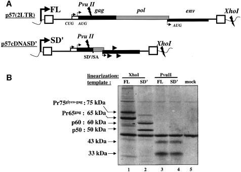
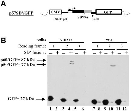
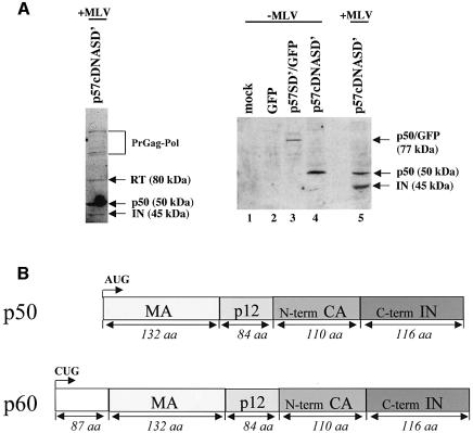
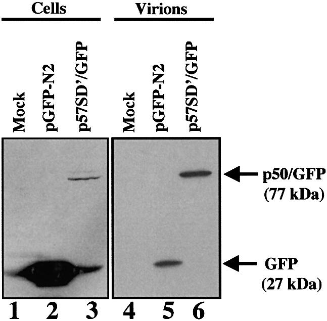
Replication of simple retroviruses depends on the recruitment of a single large primary transcript toward splicing, transport/packaging and translation regulations. In this respect, we studied the novel SD' 4.4 kb RNA of murine leukemia retroviruses (MLV) which results from alternative splicing of the primary transcript. We showed that SD' RNA was required for optimal replication since expression of a pre-spliced SD' RNA trans-complemented the impaired infectivity of a SD'-defective mutant. We monitored the fate of this novel transcript throughout early and late events of the viral life cycle. SD' RNA was specifically incorporated into virions demonstrating that the unspliced RNA was not the unique viral RNA present in virions. Furthermore, SD' RNA was reverse transcribed and its DNA copy integrated into the host genome, thus constituting a new splice donor-associated retroelement (SDARE) in infected cells. Finally, we showed that SD' mRNA encoded a 50 kDa polyprotein, and to a lower extent an additional 60 kDa polyprotein, which harbored Gag and integrase domains.


Figures

None
Fig. 1. Effect of SD′ mutation on virus replication in vector production system and trans-complementation assay. (A) Schematic maps of plasmids used in the virus vector production system and expressing the viral components independently. Location of splice donor (SD) and acceptor (SA) sites is shown. Mutations are indicated by asterisks and inactivated functions are crossed. The two versions of the genomic vector are represented: wt, wild type; mutant, inactivated SD′ site (referred to F1 mutation; Dejardin et al., 2000). (B) Structure of plasmid allowing prespliced SD′ RNA and used in trans-complementation assays. (C) Titration of virus stocks on NIH3T3 target cells. The vector pseudotypes of retroviruses shared common capsid and envelope proteins; only the nature of the genomic vector varied. In trans-complementation assays, the additional vector is noted as +SD′ for the p57cDNASD′ vector. Titers are given in AP focus-forming units per milliliter. Experiments were performed at least three times for each virus in parallel and each test was performed in quadruple. The bars indicate the standard error of the mean of each series.
None
Fig. 2. Measure of SD′ RNA content in replication-competent MLV infected M.dunni cells and virions by real-time RT-PCR. (A) Analysis of RT-PCR products by agarose gel electrophoresis. Viral RNA components were extracted from either infected cells or virions, and were analyzed by quantitative RT-PCR. From a unique RT reaction, four different PCRs were performed specific to each RNA species: genomic (FL), alternatively spliced (SD′), canonically spliced (SD) and control GAPDH mRNA. Approximate positions of the primers used to amplify the different RNA species are indicated by the numbered arrows. Mock samples correspond to similar experiments conducted with non-infected cells. Amplified samples were loaded on agarose gel and stained by ethidium bromide. Each RNA species detected is indicated on the left side of the gel, with corresponding oligonucleotide pairs identified by numbers on the right. (B) Results of quantitative RT-PCR experiments. Relative levels of FL (fixed to 100), SD′ and SD RNA in both chronically infected cells and virions were determined as described in Materials and methods. Values represent average of at least three independent RT-PCR assays with standard deviations. Absolute quantification values (parentheses) are expressed as RNA copy number per infected cell. (C) RNA encapsidation efficiency of each viral RNA species. Final encapsidation levels were calculated as the ratio of viral to cellular RNA values obtained in (B) and expressed as a percentage.
None
Fig. 3. Analysis of proviral DNA in the MLV-infected M.dunni cells by real-time PCR. Cellular DNA was extracted from 4 h infected or from chronically or mock-infected M.dunni cells. The same amount of cellular DNA (50 ng) was submitted to quantitative PCR amplifications using the same specific primers as used in previous RT-PCR experiments (see Materials and methods). Results were normalized to canonical full-length proviral DNA arbitrarily fixed to 100. Results are presented as means ± SD (error bars) of three independent measurements. Absolute quantification values (parentheses) are expressed as proviral DNA copy number per cell.
None
Fig. 4. In vitro transcription/translation of cDNASD′. (A) Schematic representation of p57(2LTR) and p57cDNASD′ plasmids used for in vitro transcription of FL and SD′ RNA, respectively. In both cases, transcription starts at the T7 promotor (bold broken arrow) located upstream at the 5′ viral LTR (rectangular boxes) and ends either at the XhoI or PvuII restriction sites as noted on the map. The positions of translation initiation codons of Glyco-Gag, Gag and Env proteins are indicated by thin broken arrows. Putative additional initiation sites of p57cDNASD′ are indicated by arrowheads. (B) Translated products from SD′ mRNA in rabbit reticulocyte lysate. Template plasmids described in (A) were used for one-step in vitro transcription/translation assays after linearization with XhoI (lanes 1 and 2) or PvuII (lanes 3 and 4) and resulting [35S]methionine-labeled proteins were electrophoresed on polyacrylamide gel. The background of the reactions was controlled by the missing DNA vector (lane 5).
None
Fig. 5. Search for SD′ mRNA ORF ex vivo. (A) Schematic representation of p57SD′/GFP construct. SpeI-SacII fragment from p57cDNASD′ was fused to the NheI-SacII cloning sites of vectors expressing GFP in three frames. Arrows indicate initiation codon positions of Glyco-Gag, Gag and Env. (B) Analysis of proteins fused to the N-terminus of GFP by immunoblotting. The three constructs were transfected into NIH3T3 (lanes 1–6) or 293T (lanes 7–12) cells, and cellular protein extracts were analyzed by western blot with an anti-GFP antibody as described in Materials and methods. Reading frame and GFP plasmids, fused (+) or not fused (–) to SD′, are noted at the top.
None
Fig. 6. Characterization of SD′ gene product. (A) Western blot analysis of cellular SD′ gene products. The p50 protein was detected with an anti-Pol antibody in infected NIH3T3 cells transfected with p57cDNASD′ (left panel). The right panel shows western blot analysis by using an anti-IN antibody. NIH3T3 cells (lane 1) were transfected with empty GFP vector (lane 2), p57SD′/GFP (lane 3) and p57cDNASD′ (lane 4). Infected NIH3T3 cells were transfected with p57cDNASD′ (lane 5). (B) Structure of p50 and p60 polyproteins encoded by SD′ mRNA.
None
Fig. 7. Localization of p50/60 protein fused to GFP in NIH3T3 infected with MLV. Infected cells cultured on coverslips were transfected with p57SD′/GFP clone grown for 2 days and analyzed for direct fluorescence localization. (A) Cells were photographed on a Leica scanning microscope using a 100× oil immersion objective with a GFP or Hoechst staining filter. Images were deconvoluted using Huygens 2 software. Arrows indicate plasma membrane (m) and perinuclear (pn) localization of fusion protein. Hoechst nuclear staining appears in blue. (B) Detailed analysis of membrane localization. Three-dimensional visualization of the cell section shown as an open rectangle in (A) was obtained by Imaris software analysis. Fusion protein (green) is localized at the plasma membrane all around the cell.
None
Fig. 8. Detection of p50/GFP protein in MLV particles. Same amount of NIH3T3 infected cells were transfected with p57SD′/GFP or pGFP-N2 vector. To allow quantitative analysis proteins were extracted from 5 × 105 cells (lanes 1–3) and from corresponding cell-free pelleted supernatant (lanes 4–6) and analyzed by western blotting using an anti-GFP antibody. Mock-transfected cells (lanes 1 and 4) and cells transfected with GFP vector (lanes 2 and 5) were used as controls.

Similar articles

Cited by

References

    1. Aldovini A. and Young,R.A. (1990) Mutations of RNA and protein sequences involved in human immunodeficiency virus type 1 packaging result in production of non infectious virus. J. Virol., 64, 1920–1926. - PMC - PubMed
    1. Alin K. and Goff,S.P. (1996a) Amino acid substitutions in the CA protein of Moloney murine leukemia virus that block early events in infection. Virology, 222, 339–351. - PubMed
    1. Alin K. and Goff,S.P. (1996b) Mutational analysis of interactions between the Gag precursor proteins of murine leukemia viruses. Virology, 216, 418–424. - PubMed
    1. Audit M., Dejardin,J., Hohl,B., Sidobre,C., Hope,T.J., Mougel,M. and Sitbon,M. (1999) Introduction of a cis-acting mutation in the capsid-coding gene of moloney murine leukemia virus extends its leukemogenic properties. J. Virol., 73, 10472–10479. - PMC - PubMed
    1. Berlioz C. and Darlix,J.L. (1995) An internal ribosomal entry mechanism promotes translation of murine leukemia virus gag polyprotein precursors. J. Virol., 69, 2214–2222. - PMC - PubMed

Publication types

MeSH terms