. 2003 Sep;15(9):2058-75.
doi: 10.1105/tpc.013896.

Involvement of the secretory pathway and the cytoskeleton in intracellular targeting and tubule assembly of Grapevine fanleaf virus movement protein in tobacco BY-2 cells

Affiliations

Involvement of the secretory pathway and the cytoskeleton in intracellular targeting and tubule assembly of Grapevine fanleaf virus movement protein in tobacco BY-2 cells

Céline Laporte et al. Plant Cell. 2003 Sep.

Abstract

Grapevine fanleaf virus (GFLV) is one of a large class of plant viruses whose cell-to-cell transport involves the passage of virions through tubules composed of virus-encoded movement protein (MP). The tubules are embedded within modified plasmodesmata, but the mechanism of targeting of MP to these sites is unknown. To study intracellular GFLV MP trafficking, a green fluorescent protein-MP fusion (GFP:MP) was expressed in transgenic tobacco BY-2 suspension cells under the control of an inducible promoter. We show that GFP:MP is targeted preferentially to calreticulin-labeled foci within the youngest cross walls, where it assembles into tubules. During cell division, GFP:MP colocalizes in the cell plate with KNOLLE, a cytokinesis-specific syntaxin, and both proteins are linked physically, as shown by coimmunoprecipitation of the two proteins from the same microsomal fraction. In addition, treatment with various drugs has revealed that a functional secretory pathway, but not the cytoskeleton, is required for tubule formation. However, correct GFP:MP targeting to calreticulin-labeled foci seems to be cytoskeleton dependent. Finally, biochemical analyses have revealed that at least a fraction of the MP behaves as an intrinsic membrane protein. These findings support a model in which GFP:MP would be transported to specific sites via Golgi-derived vesicles along two different pathways: a microtubule-dependent pathway in normal cells and a microfilament-dependent default pathway when microtubules are depolymerized.


Figures

Figure 1.
Figure 1.
Kinetics of GFP:MP Induction with Dexamethasone in 2B15 Cells. Total proteins from wild-type cells 24 h after induction (lane C), noninduced 2B15 cells (lane 1), and induced 2B15 cells (lanes 2 to 8) were examined by immunoblot analysis using affinity-purified anti-MP immunoglobulins. Induction times were 0, 2, 4, 6, 8, 10, 16, and 24 h (hpi; lanes 1 to 8, respectively). The arrowhead indicates the position of GFP:MP on the gel. Molecular mass markers are indicated at right.
Figure 2.
Figure 2.
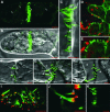
CLSM Analysis of 2B15 Cells 24 h after Induction. (A) to (C) Localization of GFP:MP in living cells. (A) Bright fluorescent thread-like structures (arrowheads) are visible in cross walls. Fluorescent punctate bodies are observed in the cytoplasm (arrows). (B) Composite image between (A) and a differential interference contrast (DIC) image. (C) Detailed view of a cross wall from which numerous oriented fluorescent thread-like structures arise (GFP:MP channel and DIC image merged). (D) and (E) Localization of GFP:MP in N. benthamiana leaf cells. Leaves were induced 3 days after agroinfiltration and observed 24 h after induction. (D) Fluorescent threads are observed in walls between two epidermal cells. Chloroplasts are shown in red. (E) Details of the boxed region shown in (D). (F) to (H) Behavior of the fluorescent thread-like structures during plasmolysis. Mannitol (0.45 M) was added and plasmolysis was followed with the microscope as a function of time before the addition of mannitol (F), at 4.5 min of plasmolysis (G), and at 10 min of plasmolysis (H). Hechtian strands that connect the retracting protoplast plasma membrane to the cell wall are visible (double arrowheads). Fluorescent threads are present within Hechtian strands and remain attached to the cell wall during plasmolysis. Excessive tension provoked the breakage of some tubules during plasmolysis (single arrowhead). The GFP:MP channel and the DIC image are merged in all images. (I) to (K) Calreticulin immunolocalization in GFP:MP-expressing cells. Induced cells containing green fluorescent threads (arrowheads) were fixed and labeled with calreticulin antibodies (red signals). The GFP:MP and calreticulin-specific channels are merged in the images. (I) The cells were oriented so that the cross wall appears in face view. (J) Side view of a cross wall at higher magnification. Asterisks indicate typical calreticulin-labeled punctate structures in the cross walls. Fluorescent threads emerge from the calreticulin-labeled foci, as judged by the presence of yellow spots in the merged images (open arrows). (K) Control induced cell in which the anti-calreticulin antibody was omitted. (D), (E), and (I) represent projections of 19, 19, and 23 optical 0.45-μm sections, respectively. All other images represent single 0.45-μm optical sections. Bars = 5 μm except in (D) and (E) (10 μm) and (J) (2 μm).
Figure 3.
Figure 3.
Transmission Electron Microscopy Images of Plasmodesmata and GFP:MP Tubules in Cross Walls of 2B15 Cells Before and 24 h after Induction with Dexamethasone. (A) Before induction. Normal plasmodesmata (arrows) are visible in cross walls from noninduced cells. The inset shows a higher magnification view of a plasmodesma in the horizontal orientation. (B) to (E) Twenty-four hours after induction. (B) Oriented tubules associated with cross walls from induced cells (white arrowheads). Note the electrolucent cell wall protrusions surrounding the basal part of the tubules. (C) and (D) Detailed view of a tubule in longitudinal section (C) and in cross-section (D). Note the electron-dense material that fills the central cavity (white arrowhead) and the plasma membrane lining the exterior of the tubules (black arrowheads). (E) Immunogold labeling (10-nm gold particles) of a tubule with GFP-specific antiserum. The white arrows indicate gold beads. Bars = 500 nm except in (A) inset, (C), and (D) (200 nm).
Figure 4.
Figure 4.
Preferential Targeting of GFP:MP Protein to Young Walls and the Cell Plate. (A) Tubules assemble preferentially in odd-numbered cross walls from living BY-2 cell chains 24 h after induction (arrowheads). (B) and (C) Tubule formation within caffeine-treated (10 mM) cells 24 h after induction. (B) Tubules are present within the youngest cross walls in a chain of four cells that have not undergone cell division during the caffeine treatment (mononucleated cells). (C) Tubules formed exclusively within the aborted plane of division in binucleated cells that have undergone mitosis during caffeine treatment. N, nucleus. (D) GFP:MP targeting to the cell plate in a BY-2 cell at late anaphase stage, 24 h after induction. (E) Three-dimensional projection of the cell shown in (D). (F) and (G) Three-dimensional projections showing GFP:MP (green), microtubules (red), and the nuclei after 4′,6-diamidino-2-phenylindole staining (blue) in immunolabeled cells at late anaphase (F) and late telophase (G). (H) to (J) GFP:MP and KNOLLE colocalize in the plane of division. (H) and (I) GFP:MP distribution (H) and KNOLLE immunolabeling (I) in a cell at anaphase stage. (J) Merged image showing the partial colocalization (yellow signal) of GFP:MP and KNOLLE within the cell plate. (K) Immunolabeling of the MP in GFLV-infected BY-2 cells at late anaphase stage. The MP (green) accumulates almost exclusively within the cell plate formed between the separated chromosomes (blue). Single optical sections are shown in (A) to (D) and (H) to (K). (E), (F), and (G) show projections of 18, 22, and 44 sections, respectively. All optical sections were 0.45 μm thick. In (A) to (D), the GFP:MP channel and differential interference contrast images have been merged. Bars = 5 μm except in (A) (50 μm) and (B), (D), and (E) (10 μm).
Figure 5.
Figure 5.
Involvement of the Endomembrane System and the Cytoskeleton in GFP:MP Targeting. (A) Effect of BFA on GFP:MP distribution. BFA (10 μg/mL) was added 8 h after induction to block the secretory pathway, and cells were observed 16 h later. GFP:MP tubule formation is inhibited, and fluorescence is cytoplasmic. (B) Effect of oryzalin on tubule formation. A total of 10 μM oryzalin was added 8 h after induction to depolymerize microtubules. Cells were observed 40 h later. Tubules formed over the entire cell surface instead of being localized specifically within the cross wall (arrows). (C) to (F) Effect of plasmolysis on tubules in oryzalin-treated cells. Cells were observed at least 1 h after the addition of mannitol, when the lateral plasma membrane also had retracted from the side walls. (C) Oryzalin-treated cells after plasmolysis. Protoplasts have retracted from the cell wall, giving rise to erect tubules that are not attached to the cross wall (cf. with [B], in which the tubules are appressed to the cell wall). The position of the cross wall is indicated by arrows. (D) Tubules have retracted from the walls together with the protoplast during plasmolysis. The tubules are erect and embedded within Hechtian strands connected to cross walls or side walls. (E) Differential interference contrast (DIC) image showing Hechtian strands (double arrowheads) connected to a side wall (asterisk). (F) Tubules are present within the Hechtian strands attached to side walls. Note that the tubules are not attached to the walls (asterisk). (G) An oryzalin-treated induced cell fixed with glutaraldehyde and immunolabeled with tubulin antibodies (red). Microtubules are completely disassembled (red), whereas GFP:MP tubules (green) are visible at the cell surface. (H) Distribution of GFP:MP tubules (green) and calreticulin-labeled structures (red) in an oryzalin-treated cell. Tubules formed over the entire cell surface, and most emerged from calreticulin-labeled spots (open arrows). (I) Orthogonal view of the calreticulin-labeled structures in two neighboring wild-type BY-2 cells. Numerous calreticulin-labeled spots are present over the entire cell surface. (J) to (L) Effects of the simultaneous disruption of microtubules and microfilaments on tubule formation. (J) A total of 10 μM oryzalin and 2 μM latrunculin B were added 8 h after induction to depolymerize both microtubules and actin filaments. Cells were observed 40 h later. Under these conditions, tubules accumulated preferentially in the nucleus periphery. (K) Cells containing aster-like tubules. (L) Detailed view of the boxed region in (K). (M) Calreticulin-labeled structures in a cell treated with oryzalin and latrunculin. (N) to (P) Aster-like tubules (N) and cytoplasmic calreticulin aggregates (O) in a cell treated with oryzalin and latrunculin. (P) shows the corresponding merged image. Single optical sections are shown in (A), (D) to (G), (J), and (K). (B), (C), (H), (L), and (N) to (P) show projections of 27, 40, 7, 16, 19, and 45 sections, respectively. All optical sections were 0.45 μm thick except for those in (N) to (P) (0.1 μm). In (D) to (F), (J), and (K), the GFP:MP channel and DIC images have been merged. Bars = 5 μm except in (A) to (C), (G), (J), (K), and (M) (10 μm).
Figure 6.
Figure 6.
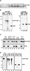
Biochemical Analysis of the GFP:MP Protein. (A) Subcellular distribution of GFP:MP and KNOLLE (KN) in synchronized 2B15 cells as determined by dot blot analysis of fractions obtained after sedimentation in a 20 to 60% linear sucrose density gradient. Ten-microliter aliquots of each gradient fraction were probed with MP antibodies or with KNOLLE immunoglobulins. Fraction numbers are indicated at top, and the sucrose concentration in each fraction is indicated at bottom. (B) Cell fractionation analysis of GFP:MP in extracts of noninduced and induced 2B15 cells and of MP in extracts from agroinfiltrated N. benthamiana leaves. Fractions correspond to the supernatant (S) and pellet (P) after centrifugation of cleared S10 extracts at 100,000g. Equal amounts (20 μg) of total protein from the S and P fractions were examined by immunoblot analysis using anti-MP (lanes 1 to 6) or anti-KNOLLE (lanes 7 to 10) antibodies. Lanes 1 and 2, noninduced 2B15 cells (−Dex); lanes 3 and 4, induced 2B15 cells 24 h after induction (+Dex); lanes 5 and 6, induced leaves 24 h after induction. Arrowheads indicate the positions of the 68-kD GFP:MP, the 38-kD MP, and KNOLLE proteins. Molecular mass markers (kD) are indicated at left. (C) Membrane association of GFP:MP. Aliquots (250 μg) of the 100,000g P fraction were supplemented with 1 M NaCl (lanes 1 and 2), Na2CO3, pH 11.5 (lanes 3 and 4), 1% Triton X-100 (lanes 5 and 6), or buffer (lanes 7 and 8), incubated on ice for 60 min, and centrifuged again at 100,000g to give the Sw and Pw washed fractions. In lanes 9 and 10, 1 mg of protein from the P fraction was supplemented with 1% Triton X-114 in Tris buffer, incubated on ice, and centrifuged. The supernatant was incubated further at 37°C and centrifuged again to separate the detergent phase (DP) and the aqueous phase (AP). All fractions were examined by immunoblot analysis using anti-MP (top gel) or anti-KNOLLE (bottom gel) antibodies. Positions of the GFP:MP and KNOLLE proteins and molecular mass markers (kD) are indicated at right and left, respectively. (D) Protease susceptibility of GFP:MP. Aliquots (100 μg) of the P and S fractions of induced 2B15 cells were incubated with proteinase K. As a control, soluble GFP:MP was incubated without (lane 1) or with (lane 2) added proteinase K (12 μg) in the absence of Triton X-100. In lanes 3 to 10, P fractions from induced cells were incubated without (lanes 3, 5, 7, and 9) or with (lanes 4, 6, 8, and 10) proteinase K (16 μg) and in the absence (−T 1%; lanes 3, 4, 7, and 8) or the presence (+T 1%; lanes 5, 6, 9, and 10) of 1% Triton X-100. Aliquots (20 μg) were examined by immunoblot analysis using anti-MP (lanes 1 to 6) or anti-KNOLLE (lanes 7 to 10) antibodies.
Figure 7.
Figure 7.
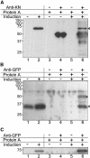
Coimmunoprecipitation of in Vivo–Expressed GFP:MP and KNOLLE. The 100,000g P fractions from noninduced and induced 2B15 cells were examined by immunoblot analysis with antibodies directed against GFP (A), KNOLLE (KN; [B]), or MP (C) either directly (lanes 1 and 2) or after immunoprecipitation with antibodies directed against KNOLLE ([A], lanes 3 to 6) or GFP ([B] and [C], lanes 3 to 6). The immunoprecipitation treatments performed on the different samples (with or without antibodies) are indicated in the tables above the gels. Arrowheads at right mark the positions of GFP:MP in (A) and KNOLLE in (B). In (A) lane 4, the band corresponds to the ∼50-kD anti-KNOLLE heavy chain immunoglobulins, which were recognized in lanes 4 and 6 by the goat anti-mouse immunoglobulins coupled to horseradish peroxidase used for immunodetection. The asterisk in (B) indicates an uncharacterized protein immunoprecipitated by the GFP antibodies and recognized by the KNOLLE antibodies. The positions of molecular mass markers (in kD) are indicated at left.

Similar articles

Cited by

References

    1. Akashi, T., Izumi, K., Nagano, E., Enomoto, M., Mizuno, K., and Shibaoka, H. (1988). Effects of propyzamide on tobacco cell microtubules in vivo and in vitro. Plant Cell Physiol. 29, 1053–1062.
    1. Aoyama, T., and Chua, N. (1997). A glucocorticoid-mediated transcriptional induction system in transgenic plants. Plant J. 11, 605–612. - PubMed
    1. Baluška, F., Samaj, J., Napier, R., and Volkmann, D. (1999). Maize calreticulin localizes preferentially to plasmodesmata in root apex. Plant J. 19, 481–488. - PubMed
    1. Bauly, J., Sealy, I., Macdonald, H., Brearley, J., Droge, S., Hillmer, S., Robinson, D., Venis, M., Blatt, M., Lazarus, C., and Napier, R. (2000). Overexpression of auxin-binding protein enhances the sensitivity of guard cells to auxin. Plant Physiol. 124, 1229–1238. - PMC - PubMed
    1. Bertens, P., Wellink, J., Goldbach, R., and van Kammen, A. (2000). Mutational analysis of the cowpea mosaic virus movement protein. Virology 267, 199–208. - PubMed

Publication types

MeSH terms