. 2003 Sep 1;162(5):765-72.
doi: 10.1083/jcb.200305077.

Dynamic phosphoregulation of the cortical actin cytoskeleton and endocytic machinery revealed by real-time chemical genetic analysis

Affiliations

Dynamic phosphoregulation of the cortical actin cytoskeleton and endocytic machinery revealed by real-time chemical genetic analysis

Mariko Sekiya-Kawasaki et al. J Cell Biol. .

Abstract

We used chemical genetics to control the activity of budding yeast Prk1p, which is a protein kinase that is related to mammalian GAK and AAK1, and which targets several actin regulatory proteins implicated in endocytosis. In vivo Prk1p inhibition blocked pheromone receptor endocytosis, and caused cortical actin patches to rapidly aggregate into large clumps that contained Abp1p, Sla2p, Pan1p, Sla1p, and Ent1p. Clump formation depended on Arp2p, suggesting that this phenotype might result from unregulated Arp2/3-stimulated actin assembly. Electron microscopy/immunoelectron microscopy analysis and tracking of the endocytic membrane marker FM4-64 revealed vesicles of likely endocytic origin within the actin clumps. Upon inhibitor washout, the actin clumps rapidly disassembled, and properly polarized actin patches reappeared. Our results suggest that actin clumps result from blockage at a normally transient step during which actin assembly is stimulated by endocytic proteins. Thus, we revealed tight phosphoregulation of an intrinsically dynamic, actin patch-related process, and propose that Prk1p negatively regulates the actin assembly-stimulating activity of endocytic proteins.


Figures
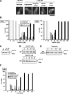
Figure 1.
Figure 1.
Initial characterization of ark1 Δ prk1- as mutants. (A) To observe the actin morphology, cells were stained with rhodamine-phalloidin. The range of actin morphologies of ark1Δ prk1-as3 cells treated with 1NA-PP1 for 2 min is shown. A mock-treated cell is also shown. (B) Actin morphology of small-budded ark1Δ prk1-as1 (AS1) and ark1Δ prk1-as3 (AS3) cells was scored as a function of 1NA-PP1 concentration in a blind study (n ≥ 200 for each sample). The cells were treated with the inhibitor for 1 h before fixation. (C and D) ark1Δ PRK1 (WT), ark1Δ prk1-as1, and ark1Δ prk1-as3 cells were cultured without 1NA-PP1 (C), or with the indicated concentration of 1NA-PP1 for 30 min (D), and then processed for anti-Ent1p Western blotting. (E) Ent1 phosphorylation as a function of time in ark1Δ prk1-as1 cells treated with 80 μM 1NA-PP1. (C–E) Phosphorylated (Ent1-P) and unphosphorylated (Ent1) forms of Ent1p are indicated. (F) Actin morphology of small-budded ark1Δ prk1-as3 cells in the presence of 40 μM 1NA-PP1 at indicated time points (n ≥100). (G) Actin morphology of ark1Δ prk1-as3 and ark1Δ prk1-as3 arp2–1. The cells were mock treated or treated with 1NA-PP1 at 25°C for 15 min before fixation. Strains: ark1Δ PRK1, DDY2547; ark1Δ prk1-as1, DDY2595; ark1Δ prk1-as3, DDY2597; ark1Δ prk1-as3 arp2–1, DDY2610. Bars, 5 μm.
Figure 1.
Figure 1.
Initial characterization of ark1 Δ prk1- as mutants. (A) To observe the actin morphology, cells were stained with rhodamine-phalloidin. The range of actin morphologies of ark1Δ prk1-as3 cells treated with 1NA-PP1 for 2 min is shown. A mock-treated cell is also shown. (B) Actin morphology of small-budded ark1Δ prk1-as1 (AS1) and ark1Δ prk1-as3 (AS3) cells was scored as a function of 1NA-PP1 concentration in a blind study (n ≥ 200 for each sample). The cells were treated with the inhibitor for 1 h before fixation. (C and D) ark1Δ PRK1 (WT), ark1Δ prk1-as1, and ark1Δ prk1-as3 cells were cultured without 1NA-PP1 (C), or with the indicated concentration of 1NA-PP1 for 30 min (D), and then processed for anti-Ent1p Western blotting. (E) Ent1 phosphorylation as a function of time in ark1Δ prk1-as1 cells treated with 80 μM 1NA-PP1. (C–E) Phosphorylated (Ent1-P) and unphosphorylated (Ent1) forms of Ent1p are indicated. (F) Actin morphology of small-budded ark1Δ prk1-as3 cells in the presence of 40 μM 1NA-PP1 at indicated time points (n ≥100). (G) Actin morphology of ark1Δ prk1-as3 and ark1Δ prk1-as3 arp2–1. The cells were mock treated or treated with 1NA-PP1 at 25°C for 15 min before fixation. Strains: ark1Δ PRK1, DDY2547; ark1Δ prk1-as1, DDY2595; ark1Δ prk1-as3, DDY2597; ark1Δ prk1-as3 arp2–1, DDY2610. Bars, 5 μm.
Figure 2.
Figure 2.
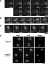
Reversible actin clump formation from cortical actin patches upon inhibition of Prk1p activity. A and C are the selected frames from Video 1 and Video 2, respectively. (A–C) ark1Δ prk1-as1 cells expressing Abp1-GFP were imaged. (A and B) Cells before or after treatment with 1NA-PP1 for the indicated times. (A) The two Abp1 patches (top, arrowheads) or already formed clumps (bottom, arrowheads) fuse to become one entity (asterisks) upon addition of 1NA-PP1. (B) An Abp1 clump (arrowhead) moves from the daughter to the mother cell. (C) Cells were pretreated with 1NA-PP1 for 30 min, followed by washout of the inhibitor. An Abp1 clump at the neck (arrowhead) disappears during the time course. (D) ark1Δ prk1-as1 cells expressing Abp1-CFP (red) and Sla2-YFP (green) were imaged after a 10-min treatment with 1NA-PP1. Mock-treated cells are also shown. The actin clumps are marked with arrowheads. Exposure times for each panel: 1 s for A and C, 66 ms for B, and 300–700 ms for D. Strains: ark1Δ prk1-as1 ABP1-GFP, DDY2600 (A and C) and DDY2601 (B); ark1Δ prk1-as1 ABP1-CFP SLA2-YFP, DDY2603 (D). Bars, 5 μm.
Figure 3.
Figure 3.
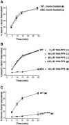
Defective receptor-mediated endocytosis upon inhibition of Prk1p activity. Internalization of 35S-labeled α-factor was measured. In A and B, the time course of internalization was started after 30 min incubation with 1NA-PP1 or mock treatment. (A) ark1Δ PRK1 (WT) and ark1Δ prk1-as3 (AS3) cells without 1NA-PP1. (B) ark1Δ PRK1 (WT) and ark1Δ prk1-as3 (AS3) cells treated with 1NA-PP1. (C) ark1Δ PRK1(WT) and ark1Δ prk1 D159A cells. Error bars represent the SD from at least three experiments. The experiment with ark1Δ PRK1 in B was performed twice. Strains: ark1Δ PRK1, DDY2607; ark1Δ prk1-as3, DDY2608; ark1Δ prk1 D159A, DDY2609.
Figure 4.
Figure 4.
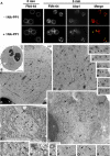
Actin-associated membrane accumulation upon inhibition of Prk1p activity. (A) FM4-64 labeling of ark1Δ prk1-as3 cells expressing Abp1-GFP. Cells were treated with media containing FM4-64 and 1NA-PP1 in a flow chamber. FM4-64 (red) and Abp1-GFP (green) were visualized at 0 and 5 min. Mock-treated cells are also shown. (B–J) Conventional EM of cells post-treated with 1% tannic acid. (B) A wild-type cell shows normal, unremarkable morphology. (C–H) ark1Δ prk1Δ cells reveal electron-dense areas (encircled by dashed line in C) containing a heterogeneous population of ∼100-nm vesicles (shown at higher magnification in D and indicated by arrows in C and D), as well as numerous microfilament profiles (marked by arrowheads in D–H). (I and J) ark1Δ prk1-as3 cells treated with 1NA-PP1 for 10 min also reveal ∼100-nm vesicles and microfilament profiles (marked by arrowheads). For more examples, also see Fig. S1. (K–N) EM of cells fixed by high pressure freezing and freeze substitution. ark1Δ prk1Δ cells reveal ribosome exclusion areas (encircled by dashed line in K) containing ∼100-nm vesicles (some of which are indicated by arrows in K–N). Ribosomes appear as small, uniform, electron-dense spots distributed throughout most of the cytoplasm. (O–Q) Indirect immunogold labeling of ultrathin cryosections of ark1Δ prk1Δ cells demonstrates the localization of actin within a slightly electron-dense/vesicle-enriched region (encircled by dashed line in O). In many instances, vesicles/membranous elements are detected within actin-labeled region (arrows in O and Q; boxed areas P and Q). (R) Double-indirect immunogold labeling with ark1Δ prk1Δ SLA1-GFP cells reveals occasional Sla1p-containing vesicles (5-nm gold, arrowheads) amid a clustered/electron-dense actin region (10-nm gold). Strains: ark1Δ prk1-as3 ABP1-GFP, DDY2606 (A); Wild-type, DDY904 (B); ark1Δ prk1Δ, DDY2541 (C–H and O–Q) and DDY1564 (K–N); ark1Δ prk1-as3, DDY2597 (I and J); ark1Δ prk1Δ SLA1-GFP, DDY2611 (R). n, nucleus; V, vacuole; pm, plasma membrane. Bars: (A) 5 μm; (B) 1 μm; (C) 0.2 μm; (D) 0.5 μm; (E–J) 0.1 μm; (K) 0.5 μm; (L–R) 0.1 μm.
Figure 5.
Figure 5.
Model for Ark-kinase function in endocytosis. (A) In wild-type cells, Prk1p promotes proper disassembly of the actin-associated endocytic complex. Phosphatase(s) might be responsible for reformation and/or activation of the endocytic complex. Ark1p, a closely related homologue of Prk1p, is likely to perform an overlapping function. (B) When Ark1p and Prk1p are inhibited, vesicles associated with actin filaments and endocytic proteins accumulate. See text for further discussion.

Similar articles

Cited by

References

    1. Ayscough, K.R., J. Stryker, N. Pokala, M. Sanders, P. Crews, and D.G. Drubin. 1997. High rates of actin filament turnover in budding yeast and roles for actin in establishment and maintenance of cell polarity revealed using the actin inhibitor latrunculin-A. J. Cell Biol. 137:399–416. - PMC - PubMed
    1. Bishop, A.C., K. Shah, Y. Liu, L. Witucki, C. Kung, and K.M. Shokat. 1998. Design of allele-specific inhibitors to probe protein kinase signaling. Curr. Biol. 8:257–266. - PubMed
    1. Bishop, A.C., O. Buzko, and K.M. Shokat. 2001. Magic bullets for protein kinases. 2001. Trends Cell Biol. 11:167–172. - PubMed
    1. Conner, S.D., and S.L. Schmid. 2002. Identification of an adaptor-associated kinase, AAK1, as a regulator of clathrin-mediated endocytosis. J. Cell Biol. 156:921–929. - PMC - PubMed
    1. Cope, M.J., S. Yang, C. Shang, and D.G. Drubin. 1999. Novel protein kinases Ark1p and Prk1p associate with and regulate the cortical actin cytoskeleton in budding yeast. J. Cell Biol. 144:1203–1218. - PMC - PubMed

Publication types

MeSH terms