Coordinate cytokine gene expression in vivo following induction of tuberculous pleurisy in guinea pigs
- PMID: 12874302
- PMCID: PMC166002
- DOI: 10.1128/IAI.71.8.4271-4277.2003
Coordinate cytokine gene expression in vivo following induction of tuberculous pleurisy in guinea pigs
Abstract
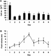
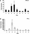
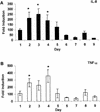
Tuberculous pleurisy is a severe inflammatory response induced by Mycobacterium tuberculosis organisms that have escaped from lung granulomata into the pleural space during pulmonary infection. We have used the guinea pig model of tuberculous pleurisy to examine several aspects of the immune response to this antigen-specific inflammatory event. Pleurisy was induced by injection of heat-killed M. tuberculosis H37Rv directly into the pleural space of guinea pigs previously vaccinated with M. bovis BCG. Four animals were euthanized each day over a period of 9 days. Fluid in the pleural cavity was analyzed for transforming growth factor beta 1 (TGF-beta 1) and total interferon (IFN) protein levels. In addition, RNA was obtained from pleural cells and examined for TGF-beta 1, tumor necrosis factor alpha (TNF-alpha), IFN-gamma, and interleukin-8 (IL-8) expression by real-time PCR. Finally, pleural cells were examined for the ability to proliferate in response to concanavalin A and purified protein derivative (PPD) in vitro. In the pleural fluid, TGF-beta 1 protein concentrations increased over the course of the inflammatory response while IFN protein levels were not significantly altered. Expression of TGF-beta 1 mRNA peaked on days 3 and 4, and IFN-gamma mRNA expression peaked on day 3 and then returned to background levels. TNF-alpha mRNA expression was highest on days 2 to 4, and IL-8 mRNA levels remained elevated between days 2 and 5, peaking on day 3 before returning to background levels. PPD-induced proliferative responses were evident by day 3 and remained present throughout the study. Analysis of cytokine expression during tuberculous pleurisy may lead to a better understanding of the self-healing nature of this manifestation of tuberculosis.
Figures


Similar articles
-
Effect of neutralizing transforming growth factor beta1 on the immune response against Mycobacterium tuberculosis in guinea pigs.Infect Immun. 2004 Mar;72(3):1358-63. doi: 10.1128/IAI.72.3.1358-1363.2004. Infect Immun. 2004. PMID: 14977939 Free PMC article.
-
Expression of interferon-gamma and tumour necrosis factor-alpha messenger RNA does not correlate with protection in guinea pigs challenged with virulent Mycobacterium tuberculosis by the respiratory route.Immunology. 2009 Sep;128(1 Suppl):e296-305. doi: 10.1111/j.1365-2567.2008.02962.x. Epub 2008 Nov 7. Immunology. 2009. PMID: 19016908 Free PMC article.
-
Altered inflammatory responses following transforming growth factor-beta neutralization in experimental guinea pig tuberculous pleurisy.Tuberculosis (Edinb). 2008 Sep;88(5):430-6. doi: 10.1016/j.tube.2008.05.001. Epub 2008 Jun 13. Tuberculosis (Edinb). 2008. PMID: 18555747
-
[Commemorative lecture of receiving Imamura Memorial Prize. Analysis of cellular immunity against tuberculosis in man with special reference to tuberculous pleurisy and cytokines].Kekkaku. 1996 Oct;71(10):591-6. Kekkaku. 1996. PMID: 8936994 Review. Japanese.
-
[Some problems concerning local cellular immunity in tuberculosis].Kekkaku. 1995 Oct;70(10):595-600. Kekkaku. 1995. PMID: 8523853 Review. Japanese.
Cited by
-
The PseEF efflux system is a virulence factor of Pseudomonas syringae pv. syringae.J Microbiol. 2012 Feb;50(1):79-90. doi: 10.1007/s12275-012-1353-9. Epub 2012 Feb 27. J Microbiol. 2012. PMID: 22367941
-
Guinea pig neutrophils infected with Mycobacterium tuberculosis produce cytokines which activate alveolar macrophages in noncontact cultures.Infect Immun. 2007 Apr;75(4):1870-7. doi: 10.1128/IAI.00858-06. Epub 2007 Feb 5. Infect Immun. 2007. PMID: 17283104 Free PMC article.
-
Mechanisms of T-lymphocyte accumulation during experimental pleural infection induced by Mycobacterium bovis BCG.Infect Immun. 2008 Dec;76(12):5686-93. doi: 10.1128/IAI.00133-08. Epub 2008 Sep 22. Infect Immun. 2008. PMID: 18809659 Free PMC article.
-
Strain-dependent CNS dissemination in guinea pigs after Mycobacterium tuberculosis aerosol challenge.Tuberculosis (Edinb). 2011 Sep;91(5):386-9. doi: 10.1016/j.tube.2011.07.003. Epub 2011 Aug 9. Tuberculosis (Edinb). 2011. PMID: 21831713 Free PMC article.
-
Lung macrophages from bacille Calmette-Guérin-vaccinated guinea pigs suppress T cell proliferation but restrict intracellular growth of M. tuberculosis after recombinant guinea pig interferon-gamma activation.Clin Exp Immunol. 2007 Aug;149(2):387-98. doi: 10.1111/j.1365-2249.2007.03425.x. Epub 2007 Jun 12. Clin Exp Immunol. 2007. PMID: 17565610 Free PMC article.
References
-
- Allen, J. C., and M. A. Apicella. 1968. Experimental pleural effusion as a manifestation of delayed hypersensitivity to tuberculin PPD. J. Immunol. 101:481-487. - PubMed
-
- Antony, V. B., J. W. Hott, S. L. Kunkel, S. W. Godbey, M. D. Burdick, and R. M. Strieter. 1995. Pleural mesothelial cell expression of C-C (monocyte chemotactic peptide) and C-X-C (interleukin 8) chemokines. Am. J. Respir. Cell Mol. Biol. 12:581-588. - PubMed
Publication types
MeSH terms
Substances
Grants and funding
LinkOut - more resources
Full Text Sources

