. 2003 Jul;112(2):170-80.
doi: 10.1172/JCI16603.

The antitumor effects of IFN-alpha are abrogated in a STAT1-deficient mouse

Affiliations

The antitumor effects of IFN-alpha are abrogated in a STAT1-deficient mouse

Gregory B Lesinski et al. J Clin Invest. 2003 Jul.

Abstract

IFN-alpha activates the signal transducer and activator of transcription (STAT) family of proteins; however, it is unknown whether IFN-alpha exerts its antitumor actions primarily through a direct effect on malignant cells or by stimulating the immune system. To investigate the contribution of STAT1 signaling within the tumor, we generated a STAT1-deficient melanoma cell line, AGS-1. We reconstituted STAT1 into AGS-1 cells by retroviral gene transfer. The resulting cell line (AGS-1STAT1) showed normal regulation of IFN-alpha-stimulated genes (e.g., H2k, ISG-54) as compared with AGS-1 cells infected with the empty vector (AGS-1MSCV). However, mice challenged with the AGS-1, AGS-1STAT1, and AGS-1MSCV cell lines exhibited nearly identical survival in response to IFN-alpha treatment, indicating that restored STAT1 signaling within the tumor did not augment the antitumor activity of IFN-alpha. In contrast, STAT1-/- mice could not utilize exogenous IFN-alpha to inhibit the growth of STAT1+/+ melanoma cells in either an intraperitoneal tumor model or in the adjuvant setting. The survival of tumor-bearing STAT1-/- mice was identical regardless of treatment (IFN-alpha or PBS). Additional cell depletion studies demonstrated that NK cells mediated the antitumor effects of IFN-alpha. Thus, STAT1-mediated gene regulation within immune effectors was necessary for mediating the antitumor effects of IFN-alpha in this experimental system.


Figures

Figure 1
Figure 1
Characterization of the AGS-1 cell line. AGS-1 has the appearance of a malignant melanoma by (a) hematoxylin and eosin staining and immunohistochemistry for (b) S-100 and (c) HMB-45. (d) Immunoblot analysis confirmed that the AGS-1 cell line was STAT1 deficient (lane 3). Cell lysates from the A431 cell line (lane 1) and B16F1 cell line (lane 2) were used as positive controls. The protein content of the lysates was quantitated, and equal amounts were loaded onto the gel. (e) EMSA analysis revealed no activation of STAT1 in IFN-α–treated AGS-1 cells. Lysates from IFN-α–stimulated B16F1 cells were used as positive controls in this assay. SIS, SIS-inducible element. (f) Flow cytometric analysis of H2k expression. Treatment of B16F1 cells for 48 hours with 104 U/ml muIFN-α resulted in increased H2k expression. Loss of STAT1 significantly reduced the induction of H2k in the AGS-1 cell line after IFN-α treatment. The minor induction of H2k expression by IFN-α in the AGS-1 cell line can be attributed to STAT1-independent signaling through NF-κB (28, 29). (g) Northern blot analysis indicated a loss of ISG-54 gene regulation in AGS-1 cells after stimulation with 104 U/ml muIFN-α for 16 hours. Probes specific for GAPDH (housekeeping gene) were used as controls and did not vary across samples. (h) Northern blot findings were confirmed by PCR using primers specific for muISG-54 and showed increased ISG-54 expression only in B16F1 cells (positive control) after IFN-α treatment. Primers specific for 28s rRNA (housekeeping gene) were used as a controls in this assay. Neg, negative control.
Figure 2
Figure 2
Antiproliferative effects of IFN-α in murine melanoma cell lines. Proliferation of the B16F1 and AGS-1 cell lines was measured by the MTT assay. IFN-α inhibits proliferation of B16F1 melanoma cells in a dose-dependent manner. In contrast, the antiproliferative effects of IFN-α were absent in the AGS-1 cell line at all concentrations tested. Results represent the mean of triplicate wells and are expressed as optical density at 570 nm ± SE.
Figure 3
Figure 3
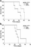
Survival of mice bearing the STAT1-deficient AGS-1 murine melanoma cell line. IFN-α treatment (open triangles) prolonged the survival of C57BL/6 mice (n = 6 mice per group) challenged with the STAT1-deficient AGS-1 melanoma cell line as compared with PBS-treated mice (filled circles) (P = 0.008). These results are representative of two separate determinations.
Figure 4
Figure 4
Reconstitution of functional STAT1 in the AGS-1 cell line. (a) Immunoblot analysis of lysates from AGS-1STAT1 cells using an anti-STAT1 antibody indicated that expression of STAT1 protein was restored. Ctrl, control. (b) The ability of STAT1 protein from AGS-1STAT1 cells to undergo phosphorylation at Tyr-701 was confirmed by immunoblotting with an anti-P-STAT1 monoclonal antibody. (c) Overnight stimulation with IFN-α (103 U/ml) resulted in enhanced H2k expression on AGS-1STAT1 but not AGS-1MSCV melanoma cells. Shaded histograms represent fluorescence intensity of H2k-PE staining in IFN-α–stimulated EGFP-positive cells. White histograms represent H2k-PE staining in PBS-stimulated EGFP-positive cells. Determination of H2k-positive cells was based on isotype control antibodies (M1), which fell within the first log of fluorescence. (d) IFN-α inhibits proliferation of the AGS-1STAT1 cell line in a dose-dependent manner. In contrast, the antiproliferative effects of IFN-α were not evident in AGS-1MSCV cells. Results represent the mean of triplicate wells and are expressed as optical density at 570 nm ± SE. (e) The ability of the AGS-1STAT1 cell line to regulate ISGF3 gene transcription in response to IFN-α was confirmed by PCR. Total cellular RNA was isolated from cell lines (B16F1, AGS-1, AGS-1STAT1, and AGS-1MSCV) after overnight treatment with 104 U/ml of murine IFN-α and converted to cDNA using standard methods. PCR primers specific for muISG-54 detected increased ISG-54 expression in only the B16F1 (positive control) and AGS-1STAT1 cell lines after IFN-α treatment. Primers specific for 28s rRNA (housekeeping gene) were used as controls.
Figure 5
Figure 5
The antitumor effects of IFN-α are not enhanced after reconstitution of STAT1 signaling within the melanoma cell. (a) IFN-α treatment (open triangles) significantly (P < 0.01) prolonged the survival of C57BL/6 mice (n = 6 mice per group) bearing AGS-1 cells infected with empty vector (AGS-1MSCV) as compared with PBS-treated mice (filled circles). (b) IFN-α treatment (open triangles) also significantly (P < 0.01) prolonged the survival of C57BL/6 mice (n = 6 mice per group) challenged with the STAT1-reconstituted AGS-1 melanoma cell line (AGS-1STAT1) as compared with PBS-treated mice (filled circles). These results are representative of two separate determinations.
Figure 6
Figure 6
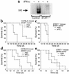
Absence of STAT1 in the host results in decreased signaling in response to IFN-α and decreased survival after tumor challenge. (a) A DNA probe specific for activated murine STAT1 reacted with whole-cell lysates from IFN-α–treated splenocytes of C57BL/6 but not STAT1-deficient mice. Similar results were obtained in other tissues, including liver and kidney (data not shown). STAT1–/– and C57BL/6 mice (n = 10 mice per group) were injected intraperitoneally with 106 B16F1 melanoma cells. As compared with PBS-treated controls (filled circles), IFN-α treatment (2 × 10⁴ U per day) (open triangles) prolonged the survival of C57BL/6 mice but not STAT1–/– mice challenged with B16F1 melanoma cells (b and c) or JB/MS melanoma cells (n = 6 mice per group) (d and e).
Figure 7
Figure 7
Histochemical analysis. B16F1 tumors from C57BL/6 mice treated with (a) PBS or (b) IFN-α and STAT1-deficient mice treated with (c) PBS or (d) IFN-α were stained with hematoxylin and eosin and analyzed at ×40. A striking degree of tumor cell necrosis was observed in STAT1-deficient mice treated with either PBS or IFN-α. A similar degree of necrosis was observed in PBS-treated C57BL/6 mice. Notably, the least degree of tumor cell necrosis was observed in C57BL/6 mice treated with IFN-α, which was the only group exhibiting prolonged survival.
Figure 8
Figure 8
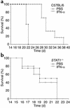
Effects of IFN-α on the survival of C57BL/6 and STAT1-deficient mice bearing locoregional B16F10 metastases. IFN-α treatment (open triangles) significantly prolonged the survival of (a) C57BL/6 mice (P = 0.002) but not (b) STAT1-deficient mice (P = 0.73) bearing B16F10 locoregional metastases as compared with PBS-treated (filled circles) controls. There were 10 mice in each group.
Figure 9
Figure 9
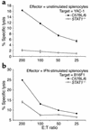
Deficient splenocyte cytotoxic activity in STAT1–/– mice. (a) Unstimulated splenocytes from STAT1–/– mice (open circles) and wild-type C57BL/6 mice (filled circles) were tested for cytotoxic activity against the NK-sensitive YAC-1 cell line in a standard 4-hour 51Cr release assay. (b) Splenocytes from STAT1–/– mice (open circles) and wild-type C57BL/6 mice (filled circles) were tested for cytotoxic activity against the B16F1 murine melanoma cell line in a standard 4-hour 51Cr release assay after stimulation with 104 U/ml IFN-α for 18 hours.
Figure 10
Figure 10
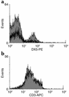
IFN-α treatment leads to increased NK cells within the PECs of mice bearing B16F1 melanoma tumors. (a) Flow cytometric analysis of PECs from C57BL/6 mice bearing B16F1 tumors revealed the presence of a significant NK cell infiltrate in IFN-α–treated mice (shaded histogram) but not in PBS-treated mice (white histogram). (b) In contrast, CD3-positive cells were present in PECs from both PBS-treated and IFN-α–treated mice. Each analysis was performed on at least 10,000 cells gated in the region of the lymphocyte population on the basis of light scatter properties (forward scatter versus side scatter). Fluorescence data were shown on four-decade log scales, and instrument settings were adjusted in reference to PECs stained with appropriate isotype control antibodies. Each histogram is representative of PECs from five mice.
Figure 11
Figure 11
The antitumor effects of IFN-α are abrogated in NK-cell depleted SCID mice. (a) IFN-α treatment (open triangles) significantly prolonged the survival of SCID mice bearing B16F1 tumors in the intraperitoneal position when compared with PBS-treated (filled circles) SCID mice (P < 0.01). IFN-α treatment (open triangles) did not prolong the survival of NK cell–depleted SCID mice bearing B16F1 tumors in the intraperitoneal position (b) as compared with PBS-pretreated (filled circles) SCID mice (c). All studies using SCID mice used six mice per treatment arm.

Similar articles

Cited by

References

    1. Kirkwood JM, Ibrahim JG, Sondak VK, Ernstoff MS, Ross M. Interferon alfa-2a for melanoma metastases. Lancet. 2002;359:978–979. - PubMed
    1. Belardelli F, Ferrantini M, Proietti E, Kirkwood JM. Interferon-alpha in tumor immunity and immunotherapy. Cytokine Growth Factor Rev. 2002;13:119–134. - PubMed
    1. Kirkwood JM, et al. High- and low-dose interferon alfa-2b in high-risk melanoma: first analysis of intergroup trial E1690/S9111/C9190. J. Clin. Oncol. 2000;18:2444–2458. - PubMed
    1. Agarwala SS, Kirkwood JM. Potential uses of interferon alpha 2 as adjuvant therapy in cancer. Ann. Surg. Oncol. 1995;2:365–371. - PubMed
    1. Biron CA. Interferons alpha and beta as immune regulators — a new look. Immunity. 2001;14:661–664. - PubMed

Publication types

MeSH terms