. 2003 Jul;14(7):2972-83.
doi: 10.1091/mbc.e03-01-0858.

Inducible systemic RNA silencing in Caenorhabditis elegans

Affiliations

Inducible systemic RNA silencing in Caenorhabditis elegans

Lisa Timmons et al. Mol Biol Cell. 2003 Jul.

Abstract

Introduction of double-stranded RNA (dsRNA) can elicit a gene-specific RNA interference response in a variety of organisms and cell types. In many cases, this response has a systemic character in that silencing of gene expression is observed in cells distal from the site of dsRNA delivery. The molecular mechanisms underlying the mobile nature of RNA silencing are unknown. For example, although cellular entry of dsRNA is possible, cellular exit of dsRNA from normal animal cells has not been directly observed. We provide evidence that transgenic strains of Caenorhabditis elegans transcribing dsRNA from a tissue-specific promoter do not exhibit comprehensive systemic RNA interference phenotypes. In these same animals, modifications of environmental conditions can result in more robust systemic RNA silencing. Additionally, we find that genetic mutations can influence the systemic character of RNA silencing in C. elegans and can separate mechanisms underlying systemic RNA silencing into tissue-specific components. These data suggest that trafficking of RNA silencing signals in C. elegans is regulated by specific physiological and genetic factors.


Figures
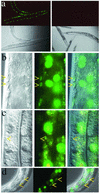
Figure 1.
Figure 1.
Transgenes expressing gfp hairpins are effective triggers for RNAi. (a) GFP fluorescence (top two panels) and Nomarski (bottom two panels) images of worms harboring an integrated myo-3::GFP transgene expressing GFP in muscle. (The GFP reporters used in all these experiments were tagged with a nuclear localization signal.) The animals depicted on the right harbor an additional myo-3::gfp hairpin transgene array capable of expressing dsgfp RNA in muscle. The images on the left depict the normal accumulation pattern of GFP expressed from the reporter transgene. The images on the right demonstrate that the myo-3::gfp hairpin construct can deliver enough dsgfp RNA to mediate effective silencing of GFP in muscle cells. (b) Nomarski (left), GFP fluorescence (middle), and composite (right) images of an animal with a rpL28::GFP transgene expressing GFP in all cells. Arrows point to muscle nuclei with GFP fluorescence. Larger fluorescent nuclei are polyploid gut nuclei. (c) Nomarski (left), GFP fluorescence (middle), and composite (right) images of an animal with two transgenes: the myo-3::gfp hairpin transgene as in (a, right) and the rpL28::GFP transgene reporter used in b. Arrow points to muscle nucleus that fails to express GFP. Nuclei nearby are not affected. (d) Nomarski (left), GFP fluorescence (middle), and composite (right) images of an animal with a let-858::GFP transgene expressing GFP in all cells and a vit-2::gfp hairpin transgene expressing dsgfp RNA in the gut. Arrows point to nuclei that show reduced GFP accumulation in gut. Two explanations are possible for the lack of interference in other gut cells: the extrachromosomal vit-2::gfp hairpin transgene array may have been mitotically segregated out of the unaffected cells; and this promoter has previously been demonstrated to drive expression in a dynamic pattern (MacMorris et al., 1994; our unpublished observations).
Figure 2.
Figure 2.
Assessments of dsRNA cell autonomy. The models depict the experimental design to address the autonomous nature of RNA silencing signals and predicts the RNAi phenotypes of transgenic animals if in vivo-transcribed RNA silencing signals remain cell autonomous (left) or are capable of cellular exit (right). A transgene expressing a gfp hairpin in a tissue-specific manner was introduced into animals expressing a GFP reporter in all cells by standard genetic crosses. Model A predicts that animals would not exhibit systemic silencing for GFP if dsRNA cannot exit the tissue of origin. Cell autonomous RNAi (white) is exhibited as tissue-specific loss of GFP. Model B predicts that animals would exhibit systemic silencing for GFP if RNA silencing signals can exit the cells of origin. (a) The GFP expression pattern in an animal harboring a chromosomally integrated let-858::GFP transgene. (b) A transgene with myo-3::gfp hairpin sequences that delivers dsgfp RNA in muscle (Figure 1, a and c) was crossed into the strain represented in a. (c) A transgene with vit-2::gfp hairpin sequence that delivers dsgfp RNA in gut (Figure 1d) was crossed into animals harboring let-858::GFP. (d) A transgene with unc-119::gfp hairpin sequences that delivers dsgfp RNA in neurons (see MATERIALS AND METHODS) was crossed into animals harboring let-858::GFP. Images were captured using a 40× objective so the overall GFP fluorescence of animals could be compared. RNA silencing was not generally observed in non-hairpin–expressing cells as evidenced by uniform GFP fluorescence in somatic cells.
Figure 3.
Figure 3.
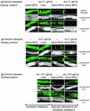
Systemic RNAi is inducible. Exposing cells to unrelated dsRNAs might facilitate intracellular release of an RNA silencing signal derived from an in vivo transcribed gfp hairpin. Pairs of images are representative of each experiment; GFP fluorescence images are above, Nomarski below. Two different integrated transgenes expressing GFP in all cells were used in these experiments: a let-858::GFP transgene (a, b, c, d, i, j, k, l, q, r, and s) or a rpL28::GFP transgene (e, f, g, h, m, n, o, p, t, u, and v); GFP is the RNAi target in these experiments. Similar results were observed for each reporter. A second transgene driving tissue-specific expression of a gfp hairpin dsRNA was present in some of the strains used. Animals were soaked in high concentrations of dstetA RNA or in water, and GFP fluorescence was monitored in the soaked animals (a, b, e, f, i, j, m, n, q, and t) or in the resulting progeny (c, d, g, h, k, l, o, p, r, s, u, and v). (The soaked animals were allowed a recovery period of 12–18 h before imaging. The accumulation of GFP in these control animals was not severely affected under these harsh conditions.) Animals in b, d, f, and h harbored a second transgene expressing a gfp hairpin dsRNA from a myo-3 promoter in addition to the GFP-expressing transgene. Animals in j, l, n, and p have a vit-2::gfp hairpin transgene. Animals in q, s, t, and v have an unc-119::gfp hairpin transgene. Soaking GFP-expressing animals in dstetA RNA does not affect GFP fluorescence (a, e, i, and m). Soaking doubly transgenic animals in control solutions does not elicit reduced GFP fluorescence (b, f, j, n, q, and t). Two progeny classes arise from doubly transgenic animals: both classes are homozygous for the GFP reporter transgene; one class inherits the gfp hairpin transgene (d, h, l, p, s, and v); the second class does not (c, g, k, o, r, and u) (Stinchcomb et al., 1985). (Animals in c, g, k, o, r, and u were recovered after photography and allowed to produce progeny. The gfp hairpin transgene was not observed in the progeny of these animals.) When doubly transgenic animals are soaked in dstetA RNA, both classes of progeny exhibit some loss of GFP fluorescence. The effects in singly transgenic progeny of soaked animals (c, g, k, o, r, and u; Table 1) may result from induced maternal deposition of gfp hairpin into these animals while resident as a germline/embryo in the treated hermaphrodite, implying exit of gfp hairpin RNAs from the maternal somatic cells. Loss of GFP expression is not observed in tissues that have been previously reported to be intractable for RNAi (neurons and some pharyngeal cells).
Figure 4.
Figure 4.
fed-1(ne309) mutant animals defective in responding to ingested dsRNAs, but are not defective in systemic RNA silencing. (A) Animals were homozygous for a chromosomally integrated rpL28::GFP transgene expressing GFP in all cells: Both wild-type animals (a, b, e, and f) and fed-1 animals (c, d, g, and h) harbor this transgene. Animals were injected with (b and d) or soaked in (f and h) dsgfp RNA. RNAi (loss of fluorescence) was observed in wild-type using both methods of delivery (b and f). All cells of fed-1 animals exhibit RNAi when the animal is injected (d), but not when the animals are soaked (h) in dsgfp RNA. (The animals depicted were progeny of animals that had been injected into the gut; similar results are obtained from germline injections.) (B) fed-1 is not defective in dissemination of RNA silencing signals. An additional transgene (myo-3::gfp hairpin) was crossed into the animals in Figure 3A. Left, the gfp hairpin expressed in muscle does not elicit robust RNA silencing in cells other than muscle in a wild-type animal. Right, the gfp hairpin expressed in muscle exhibits systemic RNA silencing in a fed-1(ne309) background. Diagram, the phenotypic analyses suggest that fed-1 mutant is defective in uptake of dsRNA from gut sources (red arrows) has an intact RNA silencing mechanism in most cells and is not inhibited (possibly hyperactive) in dissemination of cell-intrinsic RNA silencing signals from somatic cells (blue arrows).
Figure 5.
Figure 5.
Processing into siRNAs is not a requirement for import into C. elegans germ cells. Wild-type (center) and rde-4(ne299) animals (left and right) were injected with dsunc-22 RNA. The injected animals were placed individually on culture plates. Where indicated (center and right), wild-type males were also added to plates. The males harbored a PD8160 transgene (expressing GFP), thus allowing identification of cross-progeny, and only plates with cross-progeny were scored in these instances. The progeny were scored for the presence of an unc-22 phenotype (twitching). In plates where twitching was observed, 10–80% of progeny animals exhibited a strong twitching phenotype. The variation in numbers of affected progeny may reflect differences in the volume of dsRNA injected into each parent animal. (The n represents the number of plates scored and also represents the numbers of animals surviving injection or the numbers of surviving animals that produced cross-progeny expressing GFP.)

Similar articles

Cited by

References

    1. Alexopoulou, L., Holt, A.C., Medzhitov, R., and Flavell, R.A. (2001). Recognition of double-stranded RNA and activation of NF-kappaB by Toll-like receptor 3. Nature 413, 732-738. - PubMed
    1. Barber, G.N. (2001). Host defense, viruses and apoptosis. Cell Death Differ. 8, 113-126. - PubMed
    1. Baulcombe, D. (2002). RNA silencing. Curr. Biol. 12, R82-R84. - PubMed
    1. Bernstein, E., Caudy, A.A., Hammond, S.M., and Hannon, G.J. (2001). Role for a bidentate ribonuclease in the initiation step of RNA interference. Nature 409, 363-366. - PubMed
    1. Boerjan, W., Bauw, G., Van Montagu, M., and Inze, D. (1994). Distinct phenotypes generated by overexpression and suppression of S-adenosyl-l-methionine synthetase reveal developmental patterns of gene silencing in tobacco. Plant Cell 6, 1401-1414. - PMC - PubMed

Publication types

Substances