. 2003 Jul 7;162(1):125-37.
doi: 10.1083/jcb.200302083.

Hrs regulates early endosome fusion by inhibiting formation of an endosomal SNARE complex

Affiliations

Hrs regulates early endosome fusion by inhibiting formation of an endosomal SNARE complex

Wei Sun et al. J Cell Biol. .

Abstract

Movement through the endocytic pathway occurs principally via a series of membrane fusion and fission reactions that allow sorting of molecules to be recycled from those to be degraded. Endosome fusion is dependent on SNARE proteins, although the nature of the proteins involved and their regulation has not been fully elucidated. We found that the endosome-associated hepatocyte responsive serum phosphoprotein (Hrs) inhibited the homotypic fusion of early endosomes. A region of Hrs predicted to form a coiled coil required for binding the Q-SNARE, SNAP-25, mimicked the inhibition of endosome fusion produced by full-length Hrs, and was sufficient for endosome binding. SNAP-25, syntaxin 13, and VAMP2 were bound from rat brain membranes to the Hrs coiled-coil domain. Syntaxin 13 inhibited early endosomal fusion and botulinum toxin/E inhibition of early endosomal fusion was reversed by addition of SNAP-25(150-206), confirming a role for syntaxin 13, and establishing a role for SNAP-25 in endosomal fusion. Hrs inhibited formation of the syntaxin 13-SNAP-25-VAMP2 complex by displacing VAMP2 from the complex. These data suggest that SNAP-25 is a receptor for Hrs on early endosomal membranes and that the binding of Hrs to SNAP-25 on endosomal membranes inhibits formation of a SNARE complex required for homotypic endosome fusion.


Figures
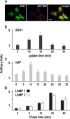
Figure 1.
Figure 1.
EGF is transported through endocytic compartments after internalization. (A) Colocalization of early endosomes with EGF-TMR in HeLa cells. The distribution of EEA1 (A, green), EGF-TMR (B, red), and the merged images (C) shows colocalization of EEA1-labeled early endosomes and EGF-TMR. (B) Percentage of EEA1-labeled early endosomes colocalized with EGF-TMR–containing vesicles after various incubation times. (C) Percentage of rab 7–labeled late endosomes colocalized with EGF-TMR–containing vesicles at various chase times after a 15-min pulse of EGF-TMR. (D) Percentage of LAMP 1/2–labeled lysosomes colocalized with EGF-TMR–containing vesicles at various chase times after a 15-min pulse of EGF-TMR. The error bars in B–D show SEM. Bar, 8 μm.
Figure 2.
Figure 2.
Characterization of a FRET-based assay measuring homotypic compartment fusion. (A) Fusion reactions were performed without the indicated component or on ice (0°C) to assess requirements for the reaction. There are significant differences between the complete homotypic fusion reactions and all other conditions in which a component has been omitted or altered (*, P ≤ 0.001) by ANOVA and Fisher test. Raw data after background subtraction are as follows. Early endosome fusion: no donor, 14.1 ± 3.5; no acceptor, 15.4 ± 5.0; no ATP, 37.3 ± 16.0; no cytosol, 28.3 ± 3.6; ice, 10.7 ± 4.3; and complete homotypic reaction, 302.5 ± 7.7. Late endosome fusion: no donor, 16.3 ± 3.4; no acceptor, 13.1 ± 12.8; no ATP, 15.6 ± 10.8; no cytosol, 26.6 ± 7.8; ice, 12.8 ± 4.8; and complete homotypic reaction, 312.1 ± 6.3. Lysosome fusion: no donor, 28.4 ± 1.4; no acceptor, 21.0 ± 6.2; no ATP, 23.9 ± 8.2; no cytosol, 24.3 ± 7.0; ice, 13.3 ± 1.2; and complete homotypic reaction, 325.3 ± 16.0. (B) Energy transfer is dependent on EGF-labeled donor and acceptor membranes. When both fluorescent tags are present on EGF and endosomes are labeled and fused a FRET signal occurs. However, labeling the acceptor population of endosomes with transferrin-TMR reduces the FRET signal to background levels (*, P ≤ 0.01). The error bars show SEM. (C) The optimal incubation time for fusion reactions was determined by examining the extent of fusion after incubating the reactions for various amounts of time at 37°C. The amount of fusion increased until 60 min of incubation, after which no further significant increase in fusion is observed. (D) The fusion reaction volume was increased to dilute the concentration of donor/acceptor membranes while the concentrations of ATP and cytosol were maintained at a constant level. A decrease in FRET signal was observed concomitant with an incremental increase in the reaction volume. (E) Syntaxin 13 and 7 affect homotypic endosome fusion. Syntaxin 13 inhibited early endosome homotypic fusion. Syntaxin 7 inhibited homotypic lysosome fusion. These data are consistent with previously published reports (see Introduction and Results). *, P ≤ 0.01.
Figure 3.
Figure 3.
Morphology of endosomal membranes before and after fusion reactions. Membranes were obtained as described for homotypic reactions of early endosomes (see Materials and methods) and fixed in 3% glutaraldehyde (A, donor) or after incubation with acceptor membranes, cytosol, and ATP regenerating system (B, after fusion). The diameter of all membrane bound profiles was measured on images from donor and fused samples (C). The mean diameter of donor compartments was 58.3 ± 1.7 nm (n = 123) and postfusion compartment diameter was 188.5 ± 7.3 nm (n = 96; *, P ≤ 0.0001). The error bars show SEM. Bars: (A and B) 100 nm.
Figure 3.
Figure 3.
Morphology of endosomal membranes before and after fusion reactions. Membranes were obtained as described for homotypic reactions of early endosomes (see Materials and methods) and fixed in 3% glutaraldehyde (A, donor) or after incubation with acceptor membranes, cytosol, and ATP regenerating system (B, after fusion). The diameter of all membrane bound profiles was measured on images from donor and fused samples (C). The mean diameter of donor compartments was 58.3 ± 1.7 nm (n = 123) and postfusion compartment diameter was 188.5 ± 7.3 nm (n = 96; *, P ≤ 0.0001). The error bars show SEM. Bars: (A and B) 100 nm.
Figure 4.
Figure 4.
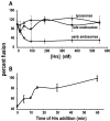
Hrs inhibits early endosome fusion at an early stage in the fusion reaction. (A) Hrs inhibits early endosome fusion in a dose-dependent and saturable manner (diamond), without having a significant effect on late endosome (square) or lysosome (triangle). (B) Hrs-induced inhibition of early endosome fusion occurs most prominently when Hrs is added to the reactions within the first 10 min of incubation. If Hrs is added after the first 10 min of the fusion reaction, its ability to inhibit the reaction reduced.
Figure 5.
Figure 5.
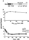
Determination of the domain of Hrs required for the inhibition of early endosome fusion. (A) The linear structure of Hrs highlighting some protein motifs. (B) The NH2-terminal half of Hrs, including the VHS and FYVE domains, had no effect on early endosome fusion. (C) Although both helical domains of Hrs inhibit early endosome fusion with similar efficacy to the full-length protein, the minimal fragment of Hrs that is both necessary and sufficient for the recapitulation of the inhibition of early endosome fusion observed with full-length Hrs is the Q-SNARE domain Hrs(515–562).
Figure 6.
Figure 6.
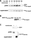
Hrs binds saturably to early endosomal membranes. Hrs and Hrs(449–562) bind to early endosomal membranes. Early endosomes were purified on a discontinuous sucrose gradient and contain the marker protein EEA-1. Full-length Hrs binds saturably to early endosomal membranes (A). SNAP-25(150–206) inhibited the binding of Hrs to endosomal membranes (B). Hrs(449–562) also binds to endosomal membranes (C), suggesting that the coiled-coil domain of Hrs is sufficient for endosomal membrane binding. Hrs or Hrs(449–562) is not found in the pellet in the absence of membranes.
Figure 7.
Figure 7.
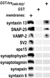
Affinity chromatography of brain membranes using immobilized Hrs (449–562). A SNARE complex consisting of syntaxin 13, SNAP-25, and VAMP2 has been previously reported to be present on early endosomal membranes (Prekeris et al., 1998). Hrs has been previously reported to bind to SNAP-25 (Bean et al., 1997) requiring aa 515–562 (Tsujimoto and Bean, 2000). Candidate proteins were examined by Western analysis on blots obtained from the affinity column eluate. SNAP-25, syntaxin 13, and VAMP2 bound to the column, whereas SV2, eps15, synaptophysin, synaptotagmin, synapsin, syntaxin 6, and rab 5, rab 15, and EEA-1 (not depicted) did not bind to Hrs (449–562).
Figure 8.
Figure 8.
BoNT/E inhibits early endosomal fusion in a SNAP-25 (150–206 )–dependent manner. (A) BoNT/E dose dependently and saturably inhibited early endosome fusion. (B) The inhibition of early endosome fusion by BoNT/E is reversed by addition of SNAP-25(150–206) supporting a role for SNAP-25 in endosome fusion.
Figure 9.
Figure 9.
Hrs decreases the efficiency of early endosomal syntaxin13–SNAP-25–VAMP2 complex formation. Quantitation of the amount of Hrs (A, expressed in optical density units) or VAMP (B, expressed as a percentage of VAMP incorporated in the absence of Hrs) incorporated into the 7 S complex in the presence of increasing amounts of Hrs is shown. (C) Glutathione-immobilized GST or GST–syntaxin 13, SNAP-25, and VAMP2 were incubated in the absence (lane 1) and presence of increasing amounts of Hrs. Samples were processed, and immunoblots were visualized with 125I-secondary antibodies and phosphorimaging.
Figure 9.
Figure 9.
Hrs decreases the efficiency of early endosomal syntaxin13–SNAP-25–VAMP2 complex formation. Quantitation of the amount of Hrs (A, expressed in optical density units) or VAMP (B, expressed as a percentage of VAMP incorporated in the absence of Hrs) incorporated into the 7 S complex in the presence of increasing amounts of Hrs is shown. (C) Glutathione-immobilized GST or GST–syntaxin 13, SNAP-25, and VAMP2 were incubated in the absence (lane 1) and presence of increasing amounts of Hrs. Samples were processed, and immunoblots were visualized with 125I-secondary antibodies and phosphorimaging.
Figure 10.
Figure 10.
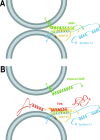
Model for the mechanism by which Hrs inhibits early endosome fusion. (A) Pairing of SNAREs (VAMP2, SNAP-25, and syntaxin 13) from opposing membranes results in helical bundle formation required for membrane fusion. (B) Hrs inhibits early endosome fusion by preventing SNARE complex formation. Hrs binds to SNAP-25 using its Q-SNARE–containing second coiled coil and inhibits VAMP2 from binding to the SNAP-25–syntaxin13 complex. N, NH2; C, COOH.

Similar articles

Cited by

References

    1. Advani, R.J., H.R. Bae, J.B. Bock, D.S. Chao, Y.C. Doung, R. Prekeris, J.S. Yoo, and R.H. Scheller. 1998. Seven novel mammalian SNARE proteins localize to distinct membrane compartments. J. Biol. Chem. 273:10317–10324. - PubMed
    1. Antonin, W., C. Holroyd, R. Tikkanen, S. Honing, and R. Jahn. 2000. The R-SNARE endobrevin/VAMP-8 mediates homotypic fusion of early endosomes and late endosomes. Mol. Biol. Cell. 11:3289–3298. - PMC - PubMed
    1. Asao, H., Y. Sasaki, T. Arita, N. Tanaka, K. Endo, H. Kasai, T. Takeshita, Y. Endo, T. Fujita, and K. Sugamura. 1997. Hrs is associated with STAM, a signal-transducing adaptor molecule. Its suppressive effect on cytokine-induced cell growth. J. Biol. Chem. 272:32785–32791. - PubMed
    1. Banerjee, A., J.A. Kowalchyk, B.R. DasGupta, and T.F. Martin. 1996. SNAP-25 is required for a late postdocking step in Ca2+-dependent exocytosis. J. Biol. Chem. 271:20227–20230. - PubMed
    1. Barbieri, M.A., R.L. Roberts, A. Gumusboga, H. Highfield, C. Alvarez-Dominguez, A. Wells, and P.D. Stahl. 2000. Epidermal growth factor and membrane trafficking. EGF receptor activation of endocytosis requires Rab5a. J. Cell Biol. 151:539–550. - PMC - PubMed

Publication types

MeSH terms

Substances