. 2003 Aug 15;551(Pt 1):139-53.
doi: 10.1113/jphysiol.2003.044784. Epub 2003 Jun 17.

Pyramidal cell communication within local networks in layer 2/3 of rat neocortex

Affiliations

Pyramidal cell communication within local networks in layer 2/3 of rat neocortex

Carl Holmgren et al. J Physiol. .

Abstract

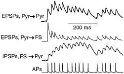
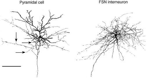
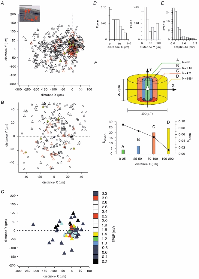
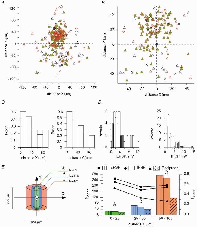
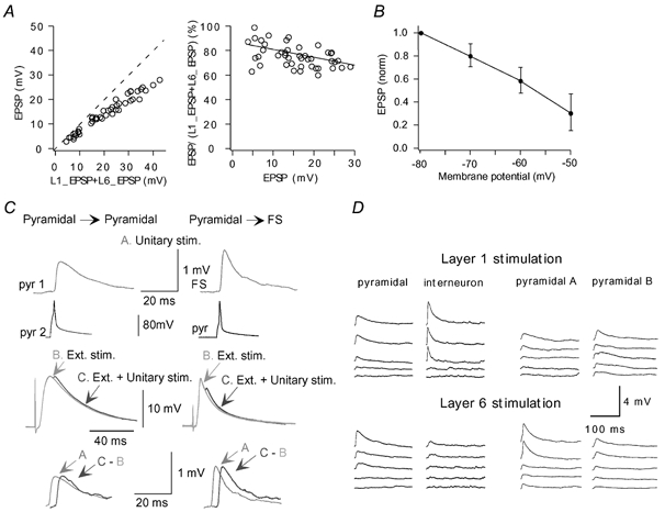
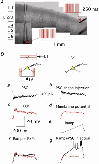
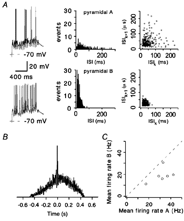
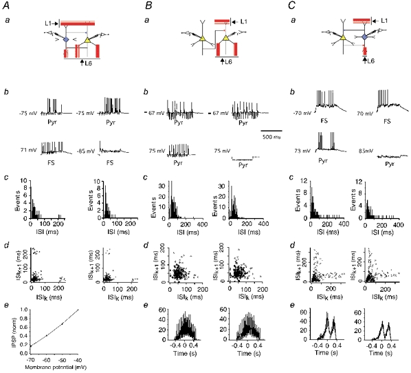
The extent to which neocortical pyramidal cells function as a local network is determined by the strength and probability of their connections. By mapping connections between pyramidal cells we show here that in a local network of about 600 pyramidal cells located within a cylindrical volume of 200 microm x 200 microm of neocortical layer 2/3, an individual pyramidal cell receives synaptic inputs from about 30 other pyramidal neurons, with the majority of EPSP amplitudes in the 0.2-1.0 mV range. The probability of connection decreased from 0.09 to 0.01 with intercell distance (over the range 25-200 microm). Within the same volume, local interneuron (fast-spiking non-accommodating interneuron, FS)-pyramidal cell connections were about 10 times more numerous, with the majority of connections being reciprocal. The probability of excitatory and inhibitory connections between pyramidal cells and FS interneurons decreased only slightly with distance, being in the range 0.5-0.75. Pyramidal cells in the local network received strong synaptic input during stimulation of afferent fibres in layers 1 and 6. Minimal-like stimulation of layer 1 or layer 6 inputs simultaneously induced postsynaptic potentials in connected pyramidal cells as well as in pyramidal-FS cell pairs. These inputs readily induced firing of pyramidal cells, although synaptically connected cells displayed different firing patterns. Unitary EPSPs in pyramidal-pyramidal cell pairs did not detectably alter cell firing. FS interneurons fire simultaneously with pyramidal cells. In pyramidal-FS cell pairs, both unitary EPSPs and IPSPs efficiently modulated cell firing patterns. We suggest that computation in the local network may proceed not only by direct pyramidal-pyramidal cell communication but also via local interneurons. With such a high degree of connectivity with surrounding pyramidal cells, local interneurons are ideally poised to both coordinate and expand the local pyramidal cell network via pyramidal-interneuron-pyramidal communication.


Figures

Figure 4
Figure 4. Summation of unitary EPSPs and IPSPs in pyramidal-pyramidal and pyramidal-FS cell pairs
Postsynaptic potential profiles in three types of connection induced by a random pattern of APs in presynaptic cells. Traces represent the average of 50-100 sweeps normalized to the maximum amplitude during the train.
Figure 1
Figure 1. A representative pyramidal cell and interneuron after reconstruction using confocal laser-scanning microscopy
Images were taken over a 460.6 μm × 460.6 μm surface area. Arrows point to cuts in the pyramidal cell axon. Scale bar = 200 μm.
Figure 2
Figure 2. Mapping of pyramidal cell connections in layer 2/3 of neocortex
A, the inset demonstrates the orientation of X- (tangential) and Y- (radial) axes in a slice. The blue triangle with (0, 0) coordinates corresponds to a pyramidal cell patched with the immobile pipette or, in the case of a synaptic connection, to the postsynaptic cell. From 542 cell pairs, synaptic connections were found in 61 cell pairs (red triangles), with 7 pairs connected reciprocally (green filled triangles). All other cells, depicted as black triangles, were not connected. B, the area of highest cell density, from Fig. 1A, on an expanded scale. C, mapping of EPSP amplitudes of connected pyramidal cells. Numbers on the scale bar on the right show the maximal EPSP amplitude represented by each coloured square. D, connection probability (Pconn), with distance, of pyramidal cells in the tangential (X) and radial (Y) directions. E, distribution of EPSP amplitudes in pyramidal-pyramidal cell pairs. F, upper panel, numbers in the diagram (N) show the total pyramidal cell number that would be in each volume (values calculated taking a pyramidal cell density of 100 000 pyramidal cells mm−3). Lower panel, connection probability (Pconn), from the mapping of pyramidal connectivity (A), depending on the distance between cells (black rectangles) and the calculated number of pyramidal cells synaptically connected (Nconn) to a single postsynaptic pyramidal cell (bars) in volumes shown in the upper panel.
Figure 3
Figure 3. Mapping of pyramidal-interneuron (FS) connections in layer 2/3 of neocortex
A, the interneuron (blue trapezium) is set in the centre (0, 0) with pyramidal cells depicted as triangles. In most cases, pyramidal cells innervated FS neurons (red triangles). Moreover, the majority of connections were reciprocal (green filled triangles). Blue triangles correspond to the unidirectional inhibitory connections. B, central part of the map with dimensions similar to those in Fig. 2B for pyramidal cells. Contrary to pyramidal-pyramidal cell connectivity, the majority of pyramidal cells are interconnected with the interneuron. C, connection probability (Pconn), with distance, of reciprocal pyramidal-FS connections in the tangential (X) and radial (Y) directions. D, distributions of EPSP and IPSP amplitudes measured at resting potential. E, left, numbers (N) in the diagram show the total pyramidal cell number that would be in each volume, calculated by taking a pyramidal cell density of 100 000 pyramidal cells mm−3 (FS cell central). Right, probabilities (Pconn), from the mapping of pyramidal-FS connectivity (A), of excitatory (rectangles), inhibitory (circles) and reciprocal (triangles) connections. Bars indicate the calculated number of corresponding connections (Nconn) formed by the interneuron with pyramidal cells in the volumes shown on the left.
Figure 5
Figure 5. Neuronal responses to afferent stimulation in L1 and L6
A, EPSPs induced in a pyramidal cell by L1 or L6 stimulation summate sub-linearly. Left, EPSPs recorded during simultaneous L1, L6 stimulation are shown versus summed EPSPs recorded during separate stimulation of L1 or L6. Right, the degree of non-linearity did not show strong dependence on the EPSP amplitudes, changing from 85 % to 68 % over the amplitude range 3.5-29 mV. B, dependence of EPSP amplitudes, evoked by extracellular stimulation (Ext. stim.), on membrane potential. EPSPs were normalized to EPSP amplitude at -80 mV. C, transient membrane depolarization initiates mechanisms supporting unitary EPSPs. Unitary EPSPs (A) were recorded in pyramidal-pyramidal and pyramidal-FS cell connections. Large EPSPs (B) were induced by L1, L6 stimulation. Then unitary EPSPs were recorded on the background of large EPSPs induced by L1, L6 stimulation (C). Lower traces compare unitary EPSPs (A) with those extracted from C. D, incrementing (30 μA step) stimulation of L1 or L6 afferent fibres induced simultaneous responses in synaptically connected cells in pyramidal-FS and pyramidal-pyramidal pairs. Each trace shows the average of 10-30 sweeps.
Figure 6
Figure 6. Dendritic integration in slices during intense synaptic activity
A, example of the location of patch pipettes and extracellular electrodes in a slice. Random stimulation patterns applied to the extracellular electrodes are depicted in red. B, dendritic conductance is considerably increased during synaptic activity induced by afferent stimulation. a, postsynaptic currents recorded in a pyramidal cell during random extracellular stimulation; b, corresponding PSC-shape current injected in the same pyramidal cell soma in the absence of afferent stimulation; c, postsynaptic potential recorded into a pyramidal cell during random extracellular stimulation; d, membrane potential in response to a PSP-shape current injection; e, membrane potential in response to a current ramp (0.2 nA s−1) injection; f, membrane potential (black) recorded during simultaneous afferent stimulation and current ramp injection (membrane potential during only afferent stimulation is shown in red); g, membrane potential (black) recorded during simultaneous injection of PSP-shape current and current ramp (membrane potential during only PSP-shape current injection is shown in red). All traces are the average of 10-20 sweeps.
Figure 7
Figure 7. Firing of connected pyramidal cells in response to stimulation of afferent fibres in L1 and L6
A, synaptically connected pyramidal cells reveal distinct firing patterns (left panel) during random L1, L6 stimulation. Note that both the RP and initial strength of postsynaptic responses are similar. Corresponding ISI distributions and ISI return maps are shown in the middle and right panels, respectively. B, cross-correlation of AP firing in the pyramidal cells in A. C, mean firing rates in six pairs of synaptically connected pyramidal cells during L1, L6 stimulation.
Figure 8
Figure 8. Modulation of cell firing by unitary synaptic signalling
a, schematic diagrams of FS-pyramidal cell (A), pyramidal-pyramidal cell (B) and pyramidal cell-FS (C) microcircuits. b, investigation of the effect of unitary PSPs induced by firing of the presynaptic pyramidal cell or interneuron on the firing patterns of the postsynaptic neuron. Firing was initiated either by random L1, L6 stimulation in the postsynaptic neuron (upper panels) and in the presynaptic cell (Ab, lower panel) or by somatic current injection (Bb and Cb, lower panels). Firing of the presynaptic interneurons was prevented by somatic hyperpolarization (lower right panels). ISI distributions (c) and ISI return maps (d) in the postsynaptic cell were measured with (left) and without (right) firing of the presynaptic neuron. Ae, effect of membrane potential on unitary IPSP amplitude. IPSPs were normalized to IPSP amplitude at -40 mV. Be and Ce, cross-correlograms of neuronal firing with (left panel) or without (right panel) firing of the presynaptic neuron.

Similar articles

Cited by

References

    1. Azouz R, Gray CM. Cellular mechanisms contributing to response variability of cortical neurons in vivo. J Neurosci. 1999;19:2209–2223. - PMC - PubMed
    1. Bernander Ö, Duglas RJ, Martin KAC, Koch C. Synaptic background activity influences spatiotemporal integration in single pyramidal cells. Proc Natl Acad Sci U S A. 1991;88:11569–11573. - PMC - PubMed
    1. Borg-Graham LJ, Monier C, Fregnac Y. Visual input evokes transient and strong shunting inhibition in visual cortical neurons. Nature. 1998;393:369–373. - PubMed
    1. Buhl EH, Tamas G, Szilagyi T, Stricker C, Paulsen O, Somogyi P. Effect, number and location of synapses made by single pyramidal cells onto aspiny interneurons of cat visual cortex. J Physiol. 1997;500:689–713. - PMC - PubMed
    1. Cauli B, Audinat E, Lambolez B, Angulo M, Ropert N, Tsuzuki K, Hestrin S, Rossier J. Molecular and physiological diversity of cortical nonpyramidal cells. J Neurosci. 1997;17:3894–3906. - PMC - PubMed

Publication types

LinkOut - more resources