. 2003 Jun 16;197(12):1731-43.
doi: 10.1084/jem.20020801.

Tumors acquire inhibitor of apoptosis protein (IAP)-mediated apoptosis resistance through altered specificity of cytosolic proteolysis

Affiliations

Tumors acquire inhibitor of apoptosis protein (IAP)-mediated apoptosis resistance through altered specificity of cytosolic proteolysis

Xu Hong et al. J Exp Med. .

Abstract

Many tumors overexpress members of the inhibitor of apoptosis protein (IAP) family. IAPs contribute to tumor cell apoptosis resistance by the inhibition of caspases, and are degraded by the proteasome to allow further progression of apoptosis. Here we show that tumor cells can alter the specificity of cytosolic proteolysis in order to acquire apoptosis resistance, which promotes formation of rapidly growing tumors. Survival of tumor cells with low proteasomal activity can occur in the presence of high expression of Tri-peptidyl-peptidase II (TPP II), a large subtilisin-like peptidase that complements proteasomal activity. We find that this state leaves tumor cells unable of effectively degrading IAPs, and that cells in this state form rapidly growing tumors in vivo. We also find, in studies of apoptosis resistant cells derived from large in vivo tumors, that these have acquired an altered peptidase activity, with up-regulation of TPP II activity and decreased proteasomal activity. Importantly, we find that growth of subcutaneous tumors is limited by maintenance of the apoptosis resistant phenotype. The apoptosis resistant phenotype was reversed by increased expression of Smac/DIABLO, an antagonist of IAP molecules. Our data suggest a reversible mechanism in regulation of apoptosis resistance that drives tumor progression in vivo. These data are relevant in relation to the multitude of therapy-resistant clinical tumors that have increased levels of IAP molecules.


Figures
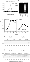
Figure 1.
Figure 1.
Resistance to serum starvation-induced apoptosis in cells adapted to reduced proteasomal activity. (a) Chymotryptic proteasomal activity in EL-4 and EL-4ad measured by cleavage of succinyl-LLVY-AMC in fractions of high molecular weight cytosolic protein eluted from a Superose 6 column. (b) DNA fragmentation measured during growth of EL-4 control and EL-4ad cells in normal cell culture medium (5% FCS) and in serum starvation medium (0.1% FCS). (c and d) Proliferation of EL-4 and EL-4ad cells in normal cell culture medium (c) versus serum starvation medium (d). (e) Western blot analysis for cytochrome c using 5 μg of cytosols from EL-4 and EL-4ad cells growing in normal (5% FCS) or serum starvation medium (0.1% FCS). (f) Western blot analysis for PARP in EL-4 and EL-4ad cells treated with etoposide.
Figure 2.
Figure 2.
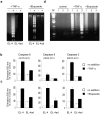
Reversible apoptosis resistance induced by adaptation to reduced proteasomal activity. (a) DNA fragmentation in EL-4 control and EL-4ad cells exposed to 10 nM TNF-α (left panel) or 25 μM Etoposide (right panel). (b and c) The activities of caspases 8, 9, and 3 in response to treatment with 10 nM TNF-α (b) or 25 μM Etoposide (c) were tested in parallel. The data represent the mean out of three independent experiments, where the background in EL-4 control cells was normalized to 100%. Standard deviation is indicated. (d) Loss of apoptosis resistance after culture in the absence of proteasomal inhibitor, as measured by DNA fragmentation in response to TNF-α or Etoposide. EL-4 control (lanes labeled 1), EL-4ad (2), and EL-4ad, i.e., EL-4ad cultured without NLVS (3) were compared in indicated lanes.
Figure 3.
Figure 3.
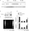
Inhibited proteasomal degradation of IAP molecules contributes to apoptosis resistance in EL-4ad cells. (a) XIAP expression in EL-4, EL-4ad, and EL-4ad cells after treatment with 25 μM Etoposide. EL-4ad denotes EL-4ad cells that were cultured in the absence of NLVS. XIAP molecules were detected by Western blot analysis of 5 μg of cellular lysates from the indicated cell lines. As loading controls, β-actin was probed. (b) Smac/DIABLO-expression in EL-4ad cells transfected with either pEF control vector or pEF-DIABLO. (c and d) Reversal of EL-4ad apoptosis resistance by Smac/DIABLO as measured by DNA fragmentation (c) and Caspase activation (d).
Figure 4.
Figure 4.
Reversal of apoptosis resistance in an expanded set of Smac/DIABLO-transfectant EL-4ad lines. (a) Proliferation of EL-4ad-pEF and EL-4ad-pEF/DIABLO cells was measured during normal (5% FCS) or conditions of serum starvation (0.1% FCS). The data represent the mean of proliferation of four independently transfected lines. (b and c). Reversal of EL-4ad apoptosis resistance in three independently transfected Smac/DIABLO-transfected lines, as measured by DNA fragmentation (b) and Caspase 3 activation following exposure to Etoposide (c).
Figure 5.
Figure 5.
Rapid tumor growth by EL-4 cells adapted to low proteasomal activity. (a-d) EL-4 and EL-4ad cells were grafted at 106 or 105 cells, as indicated in figure, in syngeneic C57Bl/6 mice and tumor size was monitored. (a and b) Tumor growth of EL-4 and EL-4ad cells in syngeneic C57Bl/6 mice. EL-4/tumor in panel a denotes cells removed from killed mice to be analyzed further in Figs. 8 and 9, (c) Tumor growth of EL-4ad cells transfected with either pEF control vector or with pEF-Smac/DIABLO. (d) Tumor growth of EL-4 and EL-4ad cells in immunodeficient PKOB/RAG1−/− mice, deficient for T and B cells as well as NK cell cytotoxic activity. The data represent the mean of at least eight mice per group in a-c, whereas four mice per group were used in d.
Figure 6.
Figure 6.
TPP II transfection confers resistance to proteasomal inhibition and reduces IAP degradation. (a and b) EL-4 cells transfected with pcDNA3 control vector or pcDNA3-TPP II, were treated either with NLVS (a) or with AdaAhx3Leu3VS (b). Whereas NLVS (reference 24) inhibits predominantly the chymotryptic activity of the proteasome, AdaAhx3Leu3VS (reference 37) inhibits all proteasomal β-subunits with comparable efficiency. Cellular proliferation was measured daily by counting live cells by trypan blue exclusion, and the data represent the mean from two independent experiments. (c) EL-4 cells transfected with either pcDNA3 or pcDNA3-TPP II were treated with Etoposide and DNA fragmentation was followed for up to 36 h. (d) Tumor growth of 106 EL-4pcDNA3 and EL-4.TPP II cells in vivo in syngeneic C57Bl/6 mice. Growth was monitored weekly, and the data represent the mean out of two independent experiments with a total of six mice per group.
Figure 7.
Figure 7.
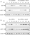
Increased expression of TPP II allows accumulation of IAP molecules. (a) EL-4.pcDNA3 and (b) EL-4.TPP II were either treated with 50 μM NLVS or not. Then, these two groups were exposed to Etoposide and degradation of XIAP and c-IAP-1 was followed by Western blot analysis of 5 μM of cellular lysate. β-actin was Western blotted as loading controls.
Figure 8.
Figure 8.
Altered activity of high molecular weight peptidases during tumor growth. (a–c) Measurements of high molecular weight peptidase activity in cell lines derived from in vitro cultures and in vivo tumors derived from (a) EL-4, (b) ALC lymphoma, and (c) B16 melanoma. Inhibitors specific for either the proteasome (NLVS; reference 24) or TPP II (Butabindide; reference 40), were used to identify the activities cleaving succinyl-LLVY-AMC and AAF-AMC. (d) Activity of high molecular weight peptidases measured in EL-4.pcDNA3 and EL-4.TPP II, experiments performed as in a–c. (e) In vitro growth rate of EL-4, EL-4ad versus EL-4/tumor cells in the presence or absence of 5 μM NLVS, as measured by counting live cells by trypan blue exclusion. (f) Western blotting of high molecular weight proteins for HC9, a proteasomal α-subunit (α3). 2, 5, or 10 μg of protein was loaded in each lane, as indicated in figure.
Figure 9.
Figure 9.
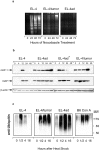
Selection of apoptosis resistance and delayed degradation of XIAP and c-IAP-1 during tumor growth (a) EL-4 control, EL-4ad, and EL-4/tumor cells were treated with 1 μM Nocodazole and induction of apoptosis was monitored by the detection of DNA fragmentation. DNA was purified from the cells and separated by a 1.8% agarose gel. (b) Western blotting of XIAP and c-IAP-1 in cells treated with etoposide for the indicated length of time. 5 μg of protein was loaded in each lane. Western blotting of β-actin was used as loading controls. (c), EL-4 control, EL-4ad, EL-4/tumor (derived from in vivo tumors), or C57Bl/6 ConA blasts were incubated for 30 min at 42°C and degradation of ubiquitin-conjugates was followed for up to 16 h with Western blot with anti-ubiquitin.
Figure 10.
Figure 10.
Induction of TPP II activity and rapid tumor growth by adaptation to cellular starvation. (a) EL-4 cells incubated in cell culture medium diluted with PBS were lysed, and high molecular weight cytosolic proteins were analysed by Western blot for TPP II expression. (b) EL-4 cells and EL-4 cells growing in starvation medium were exposed to etoposide and degradation of c-IAP-1 was followed by Western blotting analysis. (c) The high molecular weight cytosolic fractions in (a) were analyzed for cleavage of AAF-AMC. The inhibitors NLVS and AAF-CMK were included as controls. (d-e) EL-4 cells and EL-4 cells adapted to growth in starvation medium were grafted to irradiated C57Bl/6 mice at 106 (d) or 104 (e) cells per mouse.

Similar articles

Cited by

References

    1. Rock, K.L., and A.L. Goldberg. 1999. Degradation of cell proteins and the generation of MHC class I-presented peptides. Annu. Rev. Immunol. 17:739–779. - PubMed
    1. Schwartz, A.L., and A. Ciechanover. 1999. The ubiquitin-proteasome pathway and pathogenesis of human diseases. Annu. Rev. Med. 50:57–74. - PubMed
    1. Voges, D., P. Zwickl, and W. Baumeister. 1999. The 26S proteasome: a molecular machine designed for controlled proteolysis. Annu. Rev. Biochem. 68:1015–1068. - PubMed
    1. Horwich, A.L., E.U. Weber-Ban, and D. Finley. 1999. Chaperone rings in protein folding and degradation. Proc. Natl. Acad. Sci. USA. 96:11033–11040. - PMC - PubMed
    1. Dantuma, N.P., K. Lindsten, R. Glas, M. Jellne, and M.G. Masucci. 2000. Short-lived green fluorescent proteins for quantifying ubiquitin/proteasome-dependent proteolysis in living cells. Nat. Biotechnol. 18:538–543. - PubMed

Publication types

MeSH terms