. 2003 Jun;14(6):2250-61.
doi: 10.1091/mbc.e02-11-0730. Epub 2003 Apr 4.

ADP-ribosylation factor/COPI-dependent events at the endoplasmic reticulum-Golgi interface are regulated by the guanine nucleotide exchange factor GBF1

Affiliations

ADP-ribosylation factor/COPI-dependent events at the endoplasmic reticulum-Golgi interface are regulated by the guanine nucleotide exchange factor GBF1

Rafael García-Mata et al. Mol Biol Cell. 2003 Jun.

Abstract

ADP-ribosylation factor (ARF) mediated recruitment of COPI to membranes plays a central role in transport between the endoplasmic reticulum (ER) and the Golgi. The activation of ARFs is mediated by guanine nucleotide exchange factors (GEFs). Although several ARF-GEFs have been identified, the transport steps in which they function are still poorly understood. Here we report that GBF1, a member of the Sec7-domain family of GEFs, is responsible for the regulation of COPI-mediated events at the ER-Golgi interface. We show that GBF1 is essential for the formation, differentiation, and translocation of pre-Golgi intermediates and for the maintenance of Golgi integrity. We also show that the formation of transport-competent ER-to-Golgi intermediates proceeds in two stages: first, a COPI-independent event leads to the formation of an unstable compartment, which is rapidly reabsorbed in the absence of GBF1 activity. Second, the association of GBF1 with this compartment allows COPI recruitment and leads to its maturation into transport intermediates. The recruitment of GBF1 to this compartment is specifically inhibited by brefeldin A. Our findings imply that the continuous recruitment of GBF1 to spatially differentiated membrane domains is required for sustained membrane remodeling that underlies membrane traffic and Golgi biogenesis.


Figures
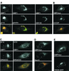
Figure 1.
Figure 1.
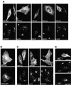
GBF1 localizes to the cis-Golgi, to ERES, and to COPI-coated cargo-containing pre-Golgi intermediates. (A) HeLa cells were stained with antibodies to GBF1 and p115, GM130, or giantin. Insets show higher magnification of boxed areas. GBF1 colocalizes significantly with p115 but only partially with giantin and GM130 (B) GBFmyc-transfected cells were stained with anti-COPI (β-COP) and anti-myc. Throughout this study we used either endogenous GBF1 or exogenously expressed myc-tagged GBF1, which behaves like wild type at low levels of expression; see supplemental material). (C) Cells were stained with anti-GBF1 and anti-ERGIC53 antibodies in control cells or in cells incubated at 15°C for 2 h. Arrowheads indicate peripheral structures that contain both GBF1 and ERGIC53. (D) GBFmyc-transfected cells were stained with anti-myc antibodies and antibodies to COPII (anti-Sec31). GBFmyc colocalizes significantly with Sec31 (arrowheads). (E) Cells were cotransfected with GBFmyc and VSV-G-GFP, incubated for 16 h at 42°C, and shifted to 32°C for 30 min. Cells were stained with anti-myc antibodies. GBFmyc is detected in pre-Golgi intermediates that contain VSV-G-GFP (arrowheads). The red signal in the merged panels correspond to GBF1 (in all figures). Bars, 10 μm.
Figure 2.
Figure 2.
Overexpression of GBF1 arrests COPI-coated transport intermediates at a late pre-Golgi step. (A) HeLa cells were transfected with GBFmyc and stained with antibodies to giantin, MannII, GM130, and β-COP. In the GalT-GFP panel, cells were cotransfected with GBFmyc and GalT-GFP and stained with anti-myc antibodies. In cells expressing high levels of GBFmyc (asterisks), the Golgi redistributes to enlarged peripheral clusters. These enlarged elements are coated with COPI (β-COP panel). In the GalT-GFP panel, the arrowheads indicate a cell expressing medium levels of GBFmyc in which the Golgi is not significantly affected. The arrow shows significant Golgi disruption in a cell expressing higher levels of GBFmyc. (B) Cells were cotransfected with GBFmyc and VSV-G-GFP, incubated for 16 h at 42°C, and then shifted to 32°C for 60 min. Cells were fixed and stained with anti-myc antibodies. VSVG-GFP accumulates at enlarged peripheral and perinuclear elements. (C) GBFmyc-transfected cells were stained with antibodies to myc and antibodies to ERES (anti-Sec31) and to VTCs (anti-p115 and anti-ERGIC53). In transfetecd cells (asterisk), the distribution of ERES is not significantly affected, whereas VTC markers redistribute to peripheral enlarged elements (arrows). Arrowheads indicate the presence of small peripheral elements similar to those found in nontransfected cells. (D) Cells were cotransfected with GBFmyc and GalT-GFP and stained with anti-myc and anti-p115 antibodies. GalT-GFP localization is less affected than that of p115. Bars, 10 μm.
Figure 3.
Figure 3.
Overexpression of an inactive mutant of GBF1 induces disassembly of the Golgi and arrests transport at an early step. (A) Sequence alignment of the F-G loop form seven Sec7 domain GEFs. Identical residues are indicated in blue, and homologous residues are in green. The essential glutamic acid residue is highlighted in yellow and the inactivating substitution is indicated by an asterisk. (B) HeLa lysates from control cells and from cells transfected with E794K were separated by SDS-PAGE, transferred to nitrocellulose membranes, and blotted with anti-GBF1 (endogenous GBF1) or anti-myc antibodies (E794K). (C) E794K-transfected cells were stained with antibodies to MannII, giantin, p115, GM130, and β-COP. (D) Cells were treated with 5 μg/ml BFA for 30 min, and processed as described in C. Both in 794K-transfected cells and in BFA-treated cells, MannII and giantin redistributed to the ER, and p115 and giantin to peripheral punctuate structures, and β-COP to the cytosol. Bars, 10 μm.
Figure 4.
Figure 4.
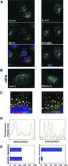
E794K localizes to a post-ER exit sites compartment. (A) HeLa cells were transfected with E794K and stained with anti-Sec31 or anti-ERGIC53 antibodies. (B) Cells were treated with 5 μg/ml BFA and stained as described in A. (C) Insets show a higher magnification of the boxed areas in A. The arrow marks a Sec31-positive structure adjacent to an E794K-positive structure. Arrowheads show two Sec31-positive structures associated to a single E794K positive structure. (D) Fluorescence intensity of the boxed area in C was plotted against pixel position (E) Double-labeled images were acquired, analyzed for signal overlap, and plotted. Bars, 10 μm.
Figure 5.
Figure 5.
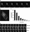
Dynamics of E794K-GFP in live cells. (A) Cells expressing E794K-GFP were imaged at 15-s intervals for 45 min. Images shown cover a period of 12:30 min. Bar, 10 μm. (B) Overlaid in the first frame of the accompanying movie are the tracings of six randomly chosen structures over a period of 45 min. (C) Population analysis of post-ER exit site structures' half-life. The half-life of individual particles (n = 78) was measured, grouped into 5-min intervals, and plotted. More than 50% of the initial structures disappeared during the first 10 min. (D) Analysis of post-ER exit sites' compartment behavior. (Blink out) The series show two particles, one disappearing in ∼1 min (arrowheads), whereas the other remains stable throughout. (buildup) The series shows a particle occurring in the vicinity of a blink-out event after the sequential disappearing of two particles. The series show two particles that come into proximity and fuse with each other (fusion). Bar, 1 μm.
Figure 6.
Figure 6.
BFA directly influences E794K localization. (A) Control cells and E794K-transfected cells were incubated with 5 μg/ml BFA for 30 min and stained with anti-GBF1 and anti-myc antibodies. Sorting of E794K into the post-ERES compartment is inhibited by BFA. (B) Cells expressing a constitutively inactive mutant of ARF1 (T31N) (HA-tagged) were stained with anti-HA and anti β-COP or anti GBF1 antibodies. Although COPI recruitment to membranes is completely abolished in T31N-transfected cells, GBF1 is still recruited to post-ERES structures. (C) Dynamics of E794K-GFP in BFA-treated cells. Cells expressing E794K-GFP were treated with 5 μg/ml BFA and imaged at 20-s intervals for 45 min. Images begin 2 min after BFA addition and extent to 14 min. BFA causes redistribution of E794K to the ER and prevents reappearance. (D) Population analysis of post-ERES structures' half-life in BFA-treated cells (n = 62). The half-life of individual particles was measured, grouped into 5-min intervals, and plotted. Bar, 10 μm.
Figure 7.
Figure 7.
GBF1 regulates COPI coating at the ER-Golgi interface and defines a novel functional stage in the progressive maturation of VTCs. Expression of an inactive GBF1 mutant arrests transport, blocks COPI recruitment to membranes, and induces the collapse of the Golgi into the ER. The effect of expressing an inactive GBF1 is analogous to that of BFA. GBF1 inactivation allows the formation of an abortive post-ER exit sites compartment (pre-COPI VTCs capable of selectively sorting some transport components. In the absence of COPI recruitment, pre-COPI VTCs continuously form and disappear, presumably by fusing with the ER (red arrow). COPII-mediated differentiation of pre-COPI VTCs represents the first step in VTC formation. GBF1 regulates the subsequent association of COPI with pre-COPI VTCs, which is required for stabilization of these structures and sorting of others proteins. The COPI-mediated differentiation of pre-COPI VTCs into VTCs is essential for subsequent transport to the Golgi. Increased expression of GBF1 causes Golgi disassembly and leads to the accumulation of enlarged COPI-coated elements in a peri-Golgi region. High levels of GBF1 promote COPI recruitment and result in the stabilization of the peri-Golgi compartment, preventing its maturation into Golgi.

Similar articles

Cited by

References

    1. Achstetter, T., Franzusoff, A., Field, C., and Schekman, R. (1988). SEC7 encodes an unusual, high molecular weight protein required for membrane traffic from the yeast Golgi apparatus. J. Biol. Chem. 263, 11711-11717. - PubMed
    1. Aridor, M., Bannykh, S.I., Rowe, T., and Balch, W.E. (1995). Sequential coupling between COPII and COPI vesicle coats in endoplasmic reticulum to Golgi transport. J. Cell Biol. 131, 875-893. - PMC - PubMed
    1. Bannykh, S.I., and Balch, W.E. (1997). Membrane dynamics at the endoplasmic reticulum-Golgi interface. J. Cell Biol. 138, 1-4. - PMC - PubMed
    1. Bannykh, S.I., Rowe, T., and Balch, W.E. (1996). The organization of endoplasmic reticulum export complexes. J. Cell Biol. 135, 19-35. - PMC - PubMed
    1. Beraud-Dufour, S., Robineau, S., Chardin, P., Paris, S., Chabre, M., Cherfils, J., and Antonny, B. (1998). A glutamic finger in the guanine nucleotide exchange factor ARNO displaces Mg2+ and the β-phosphate to destabilize GDP on ARF1. EMBO J. 17, 3651-3659. - PMC - PubMed

MeSH terms

Substances