Activation of the signal transducer and activator of transcription 1 signaling pathway in thymocytes from HIV-1-infected human thymus
- PMID: 12799548
- PMCID: PMC4415361
- DOI: 10.1097/00002030-200306130-00001
Activation of the signal transducer and activator of transcription 1 signaling pathway in thymocytes from HIV-1-infected human thymus
Abstract
Objective: To identify HIV-induced host factors in the severe combined immunodeficient (SCID)-hu Thy/Liv mouse that may contribute to HIV pathogenesis in the thymus.
Design: To identify genes specifically altered by HIV-1 infection using the cDNA microarray assay, SCID-hu Thy/Liv organs derived from the same donors were used. Therefore, no genetic variations existed between HIV and mock-infected samples. In addition, the 12-14 day post-infection timepoint was chosen because no significant thymocyte depletion was detected in HIV-infected Thy/Liv organs, so mRNA from the same cell types could be compared.
Methods: Using SCID-hu Thy/Liv mice constructed from the same donor tissues, we analysed the expression of 9183 host genes in response to HIV infection with cDNA microarrays. Expression of selected genes with more than threefold induction was confirmed by measuring RNA (reverse transcriptase-polymerase chain reaction; RT-PCR) and proteins.
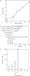
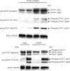
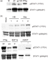
Results: HIV-1 (JD or NL4-3) infection of the SCID-hu Thy/Liv mouse led to more than threefold induction of 19 genes, 12 of which were IFN-inducible and six were unknown EST clones. We confirmed induction by RT-PCR and protein blots. Both signal transducer and activator of transcription (STAT)1 and STAT2 proteins were induced, and STAT1 was also activated by phosphorylation at the Tyr701 and Ser727 sites in human thymus infected with HIV-JD or NL4-3. Treatment of human fetal thymus organ culture or human thymocytes with recombinant HIV-1 gp120 proteins also led to induction or activation of STAT1.
Conclusion: HIV-1 infection of the thymus led to activation of the STAT1 signaling pathway in thymocytes, which may contribute to HIV-1 pathogenesis in the thymus.
Figures


Similar articles
-
HIV-1 replication and pathogenesis in the human thymus.Curr HIV Res. 2003 Jul;1(3):275-85. doi: 10.2174/1570162033485258. Curr HIV Res. 2003. PMID: 15046252 Free PMC article. Review.
-
Induction of MHC class I expression on immature thymocytes in HIV-1-infected SCID-hu Thy/Liv mice: evidence of indirect mechanisms.J Immunol. 1999 Jun 15;162(12):7555-62. J Immunol. 1999. PMID: 10358212 Free PMC article.
-
Disseminated human immunodeficiency virus 1 (HIV-1) infection in SCID-hu mice after peripheral inoculation with HIV-1.J Exp Med. 1994 Feb 1;179(2):513-22. doi: 10.1084/jem.179.2.513. J Exp Med. 1994. PMID: 8294863 Free PMC article.
-
Diphtheria toxin A gene-mediated HIV-1 protection of cord blood-derived T cells in the SCID-hu mouse model.J Hematother. 1998 Aug;7(4):319-31. doi: 10.1089/scd.1.1998.7.319. J Hematother. 1998. PMID: 9735863
-
SCID-hu mice: a model for studying disseminated HIV infection.Semin Immunol. 1996 Aug;8(4):223-31. doi: 10.1006/smim.1996.0028. Semin Immunol. 1996. PMID: 8883145 Review.
Cited by
-
A critical role for Rictor in T lymphopoiesis.J Immunol. 2012 Aug 15;189(4):1850-7. doi: 10.4049/jimmunol.1201057. Epub 2012 Jul 18. J Immunol. 2012. PMID: 22815285 Free PMC article.
-
HIV-1 replication and pathogenesis in the human thymus.Curr HIV Res. 2003 Jul;1(3):275-85. doi: 10.2174/1570162033485258. Curr HIV Res. 2003. PMID: 15046252 Free PMC article. Review.
References
-
- Courgnaud V, Laure F, Brossard A, Bignozzi C, Goudeau A, Barin F, Brechot C, et al. Frequent and early in utero HIV-1 infection. AIDS Res Hum Retroviruses. 1991;7:337–341. - PubMed
-
- Seemayer TA, Laroche AC, Russo P, Malebranche R, Arnoux E, Guerin J-M, et al. Precocious thymic involution manifested by epithelial injury in the acquired immune deficiency syndrome. Hum Pathol. 1984;15:469–474. - PubMed
-
- Kourtis AP, Ibegbu C, Nahmias AJ, Lee FK, Clark WS, Sawyer MK, Nesheim S. Early progression of disease in HIV-infected infants with thymus dysfunction. N Engl J Med. 1996;335:1431–1436. - PubMed
-
- McCune JM, Namikawa R, Kaneshima H, Shultz LD, Lieberman M, Weissman IL. The SCID-hu mouse: a model for the analysis of human hematolymphoid differentiation and function. Science. 1988;241:1632–1639. - PubMed
-
- Bonyhadi ML, Rabin L, Salimi S, Brown DA, Kosek J, McCune JM, Kaneshima H. HIV induces thymus depletion in vivo. Nature. 1993;363:728–732. - PubMed
Publication types
MeSH terms
Substances
Grants and funding
LinkOut - more resources
Full Text Sources
Medical
Molecular Biology Databases
Research Materials
Miscellaneous

