

Modeling tissue-specific signaling and organ function in three dimensions
- PMID: 12766184
- PMCID: PMC2933213
- DOI: 10.1242/jcs.00503
Modeling tissue-specific signaling and organ function in three dimensions
Abstract
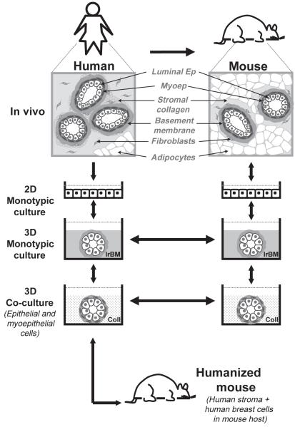
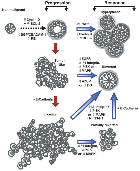
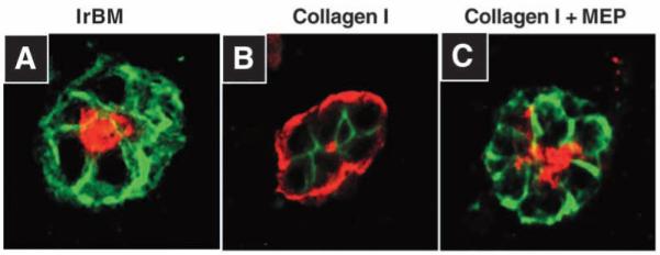
In order to translate the findings from basic cellular research into clinical applications, cell-based models need to recapitulate both the 3D organization and multicellular complexity of an organ but at the same time accommodate systematic experimental intervention. Here we describe a hierarchy of tractable 3D models that range in complexity from organotypic 3D cultures (both monotypic and multicellular) to animal-based recombinations in vivo. Implementation of these physiologically relevant models, illustrated here in the context of human epithelial tissues, has enabled the study of intrinsic cell regulation pathways and also has provided compelling evidence for the role of the stromal compartment in directing epithelial cell function and dysfunction. Furthermore the experimental accessibility afforded by these tissue-specific 3D models has implications for the design and development of cancer therapies.
Figures
Similar articles
-
3D Organotypic Culture Model to Study Components of ERK Signaling.Methods Mol Biol. 2017;1487:255-267. doi: 10.1007/978-1-4939-6424-6_19. Methods Mol Biol. 2017. PMID: 27924573
-
Organotypic culture of untransformed and tumorigenic primary mammary epithelial cells.Cold Spring Harb Protoc. 2015 May 1;2015(5):457-61. doi: 10.1101/pdb.prot078295. Cold Spring Harb Protoc. 2015. PMID: 25934933
-
The potential of bioartificial tissues in oncology research and treatment.Onkologie. 2007 Jul;30(7):388-94. doi: 10.1159/000102544. Epub 2007 Jun 27. Onkologie. 2007. PMID: 17596750 Review.
-
A novel organotypic culture model for normal human endometrium: regulation of epithelial cell proliferation by estradiol and medroxyprogesterone acetate.Hum Reprod. 2005 Apr;20(4):864-71. doi: 10.1093/humrep/deh722. Epub 2005 Jan 21. Hum Reprod. 2005. PMID: 15665014
-
Novel three-dimensional organotypic liver bioreactor to directly visualize early events in metastatic progression.Adv Cancer Res. 2007;97:225-46. doi: 10.1016/S0065-230X(06)97010-9. Adv Cancer Res. 2007. PMID: 17419948 Review.
Cited by
-
XanoMatrix surfaces as scaffolds for mesenchymal stem cell culture and growth.Int J Nanomedicine. 2016 Jun 7;11:2655-61. doi: 10.2147/IJN.S101838. eCollection 2016. Int J Nanomedicine. 2016. PMID: 27354795 Free PMC article.
-
A microengineered pathophysiological model of early-stage breast cancer.Lab Chip. 2015 Aug 21;15(16):3350-7. doi: 10.1039/c5lc00514k. Lab Chip. 2015. PMID: 26158500 Free PMC article.
-
Assessing the gene silencing potential of AuNP-based approaches on conventional 2D cell culture versus 3D tumor spheroid.Front Bioeng Biotechnol. 2024 Feb 12;12:1320729. doi: 10.3389/fbioe.2024.1320729. eCollection 2024. Front Bioeng Biotechnol. 2024. PMID: 38410164 Free PMC article.
-
Gene expression signature in organized and growth-arrested mammary acini predicts good outcome in breast cancer.Cancer Res. 2006 Jul 15;66(14):7095-102. doi: 10.1158/0008-5472.CAN-06-0515. Cancer Res. 2006. PMID: 16849555 Free PMC article.
-
In Vitro Models for Studying Invasive Transitions of Ductal Carcinoma In Situ.J Mammary Gland Biol Neoplasia. 2019 Mar;24(1):1-15. doi: 10.1007/s10911-018-9405-3. Epub 2018 Jul 28. J Mammary Gland Biol Neoplasia. 2019. PMID: 30056557 Free PMC article. Review.
References
-
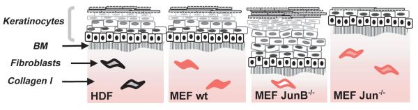
- Angel P, Szabowski A. Function of AP-1 target genes in mesenchymal-epithelial cross-talk in skin. Biochem. Pharmacol. 2002;64:949–956. - PubMed
-
- Asselineau D, Prunieras M. Reconstruction of ‘simplified’ skin: control of fabrication. Br. J. Dermatol. 1984;111(Suppl. 27):219–222. - PubMed
-
- Asselineau D, Bernhard B, Bailly C, Darmon M. Epidermal morphogenesis and induction of the 67 kD keratin polypeptide by culture of human keratinocytes at the liquid-air interface. Exp. Cell Res. 1985;159:536–539. - PubMed
-
- Bader A, Knop E, Kern A, Boker K, Fruhauf N, Crome O, Esselmann H, Pape C, Kempka G, Sewing KF. 3-D coculture of hepatic sinusoidal cells with primary hepatocytes-design of an organotypical model. Exp. Cell Res. 1996;226:223–233. - PubMed
-
- Bagutti C, Hutter C, Chiquet-Ehrismann R, Fassler R, Watt FM. Dermal fibroblast-derived growth factors restore the ability of beta(1) integrin-deficient embryonal stem cells to differentiate into keratinocytes. Dev. Biol. 2001;231:321–333. - PubMed
Publication types
MeSH terms
Grants and funding
LinkOut - more resources
Full Text Sources
Other Literature Sources
