Intracellular targeting of Gag proteins of the Drosophila telomeric retrotransposons
- PMID: 12743295
- PMCID: PMC155015
- DOI: 10.1128/jvi.77.11.6376-6384.2003
Intracellular targeting of Gag proteins of the Drosophila telomeric retrotransposons
Abstract
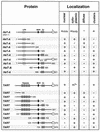
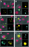
Drosophila has two non-long-terminal-repeat (non-LTR) retrotransposons that are unique because they have a defined role in chromosome maintenance. These elements, HeT-A and TART, extend chromosome ends by successive transpositions, producing long arrays of head-to-tail repeat sequences. These arrays appear to be analogous to the arrays produced by telomerase on chromosomes of other organisms. While other non-LTR retrotransposons transpose to many chromosomal sites, HeT-A and TART transpose only to chromosome ends. Although HeT-A and TART belong to different subfamilies of non-LTR retrotransposons, they encode very similar Gag proteins, which suggests that Gag proteins are involved in their unique transposition targeting. We have recently shown that both Gags localize efficiently to nuclei where HeT-A Gag forms structures associated with telomeres. TART Gag does not associate with telomeres unless HeT-A Gag is present, suggesting a symbiotic relationship in which HeT-A Gag provides telomeric targeting. We now report studies to identify amino acid regions responsible for different aspects of the intracellular targeting of these proteins. Green fluorescent protein-tagged deletion derivatives were expressed in cultured Drosophila cells. The intracellular localization of these proteins shows the following. (i) Several regions that direct subcellular localizations or cluster formation are found in both Gags and are located in equivalent regions of the two proteins. (ii) Regions important for telomere association are present only in HeT-A Gag. These are present at several places in the protein, are not redundant, and cannot be complemented in trans. (iii) Regions containing zinc knuckle and major homology region motifs, characteristic of retroviral Gags, are involved in protein-protein interactions of the telomeric Gags, as they are in retroviral Gags.
Figures


Similar articles
-
Gag proteins of the two Drosophila telomeric retrotransposons are targeted to chromosome ends.J Cell Biol. 2002 Nov 11;159(3):397-402. doi: 10.1083/jcb.200205039. Epub 2002 Nov 4. J Cell Biol. 2002. PMID: 12417578 Free PMC article.
-
Gag proteins of Drosophila telomeric retrotransposons: collaborative targeting to chromosome ends.Genetics. 2010 Mar;184(3):629-36. doi: 10.1534/genetics.109.109744. Epub 2009 Dec 21. Genetics. 2010. PMID: 20026680 Free PMC article.
-
Two retrotransposons maintain telomeres in Drosophila.Chromosome Res. 2005;13(5):443-53. doi: 10.1007/s10577-005-0993-6. Chromosome Res. 2005. PMID: 16132810 Free PMC article. Review.
-
The gag coding region of the Drosophila telomeric retrotransposon, HeT-A, has an internal frame shift and a length polymorphic region.J Mol Evol. 1996 Dec;43(6):572-83. doi: 10.1007/BF02202105. J Mol Evol. 1996. PMID: 8995054
-
Drosophila: Retrotransposons Making up Telomeres.Viruses. 2017 Jul 19;9(7):192. doi: 10.3390/v9070192. Viruses. 2017. PMID: 28753967 Free PMC article. Review.
Cited by
-
Drosophila telomeres: the non-telomerase alternative.Chromosome Res. 2005;13(5):431-41. doi: 10.1007/s10577-005-0992-7. Chromosome Res. 2005. PMID: 16132809 Review.
-
If the cap fits, wear it: an overview of telomeric structures over evolution.Cell Mol Life Sci. 2014 Mar;71(5):847-65. doi: 10.1007/s00018-013-1469-z. Epub 2013 Sep 17. Cell Mol Life Sci. 2014. PMID: 24042202 Free PMC article. Review.
-
Telomere-associated endonuclease-deficient Penelope-like retroelements in diverse eukaryotes.Proc Natl Acad Sci U S A. 2007 May 29;104(22):9352-7. doi: 10.1073/pnas.0702741104. Epub 2007 May 4. Proc Natl Acad Sci U S A. 2007. PMID: 17483479 Free PMC article.
-
Detailed mapping of the nuclear export signal in the Rous sarcoma virus Gag protein.J Virol. 2005 Jul;79(14):8732-41. doi: 10.1128/JVI.79.14.8732-8741.2005. J Virol. 2005. PMID: 15994767 Free PMC article.
-
Telomere-specific non-LTR retrotransposons and telomere maintenance in the silkworm, Bombyx mori.Chromosome Res. 2005;13(5):455-67. doi: 10.1007/s10577-005-0990-9. Chromosome Res. 2005. PMID: 16132811 Review.
References
-
- Alin, K., and S. P. Goff. 1996. Mutational analysis of interactions between the Gag precursor proteins of murine leukemia viruses. Virology 216:418-424. - PubMed
-
- Burniston, M. T., A. Cimarelli, J. Colgan, S. P. Curtis, and J. Luban. 1999. Human immunodeficiency virus type 1 Gag polyprotein multimerization requires the nucleocapsid domain and RNA and is promoted by the capsid-dimer interface and the basic region of matrix protein. J. Virol. 73:8527-8540. - PMC - PubMed
-
- Coffin, J. M., S. H. Hughes, and H. E. Varmus (ed.). 1997. Retroviruses. Cold Spring Harbor Laboratory Press, Plainview, N.Y. - PubMed
Publication types
MeSH terms
Substances
Grants and funding
LinkOut - more resources
Full Text Sources
Molecular Biology Databases

