Comparative Study
. 2003 Feb 5:1:9.
doi: 10.1186/1477-7827-1-9.

The spatiotemporal expression pattern of the bone morphogenetic protein family in rat ovary cell types during the estrous cycle

Affiliations
Comparative Study

The spatiotemporal expression pattern of the bone morphogenetic protein family in rat ovary cell types during the estrous cycle

Gregory F Erickson et al. Reprod Biol Endocrinol. .

Abstract

In the mammalian ovary, great interest in the expression and function of the bone morphogenetic protein (BMP) family has been recently generated from evidence of their critical role in determining folliculogenesis and female fertility. Despite extensive work, there is a need to understand the cellular sites of expression of these important regulatory molecules, and how their gene expression changes within the basic ovary cell types through the cycle. Here we have performed a detailed in situ hybridization analysis of the spatial and temporal expression patterns of the BMP ligands (BMP-2, -3, -3b, -4, -6, -7, -15), receptors (BMPR-IA, -IB, -II), and BMP antagonist, follistatin, in rat ovaries over the normal estrous cycle. We have found that: i) all of the mRNAs are expressed in a cell-specific manner in the major classes of ovary cell types (oocyte, granulosa, theca interstitial, theca externa, corpora lutea, secondary interstitial, vascular and ovary surface epithelium); and ii) most undergo dynamic changes during follicular and corpora luteal morphogenesis and histogenesis. The general principle to emerge from these studies is that the developmental programs of folliculogenesis (recruitment, selection, atresia), ovulation, and luteogenesis (luteinization, luteolysis) are accompanied by rather dramatic spatial and temporal changes in the expression patterns of these BMP genes. These results lead us to hypothesize previously unanticipated roles for the BMP family in determining fundamental developmental events that ensure the proper timing and developmental events required for the generation of the estrous cycle.


Figures
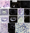
Figure 1
Figure 1
In situ hybridization of BMP-2 mRNA in the ovaries of adult cycling rats. Brightfield (A, D, F, H, J, L, N) and Darkfield (B, C, E, G, I, K, M). A, B: Sections from a DII 1100 h ovary (4X); new corpus luteum (CL-I); atretic follicle (AF); secondary interstitial cells (SIC); healthy Graafian follicle (GF); C: Negative control hybridized with sense BMP-2 cRNA probe; D, E: Secondary follicle with 3–5 layers of GCs at DII 1100 h (20X). Note positive membrana GC at periphery. F, G: Dominant follicle at DII 1100 h (10X); Positive membrana GC (MGC); Negative oocyte (O), cumulus GC (CC), periantral GC (PA) and theca (T); H, I: Atretic follicle at P 1000 h showing strongly positive GC (10X); J, K: Negative newly formed CL-I at E 0200 h (4X); Positive GF and preantral follicle (arrow); L, M: New CL-I after initiation of luteolysis at DII 1100 h showing clusters of positive cells (20X); N: Higher magnification (40X) of CL-I in L showing positive endothelial and granulosa lutein cells (GLC).
Figure 2
Figure 2
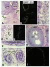
In situ hybridization of BMP-3b mRNA in ovaries of adult cycling rats. All photographs are from ovaries at DI 1100 h. Brightfield (A, C, D, F, G, H) and Darkfield (B, E, I). A, B: Low power photomicrograph showing positive theca (T), ovary surface epithelium (OSE), sex cords (SC), and CL-I (4X); C: Higher power (40X) of theca in B showing strongly positive theca interna (TI) and theca externa (TE); D, E: New CL-I showing positive signal in some theca lutein (TLC) and TE cells (20X); F: Positive OSE (40X); G: Positive SC (40X); H, I: Positive TI in healthy GF. Note negative TI of atretic AF follicle (10X).
Figure 3
Figure 3
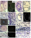
In situ hybridization of BMP-4 mRNA in ovaries of adult cycling rats. Brighfield (A, C, D, E, G, H, J, K, M, O) and Darkfield (B, F, I, L, N). A, B: Sections from P 1000 h ovaries (4X). Positive OSE, and T; weakly positive CL; negative preantral follicles (arrow heads); C: Positive SC () at P 1000 h (40X); D: Positive endothelial cells of large sinusoid (40X); E, F: Secondary follicle at P 1000 h (20X) showing positive cells (arrow heads) at periphery of T. G: High power (40X) of E showing positive theca interstitial cells (TIC) (arrows) in outer half of TI and positive cells (arrows) in the TE. H, I: Dominant follicle at P 2000 h showing positive theca (10X); J: Higher magnification (40X) of H showing positive TE and adjacent TIC in the dominant follicle. K, L: Older CL-I (10X) at E 1000 h of the next cycle showing positive TE (arrow heads); M, N: CL-I at P 1000 h (20X); showing positive TE (arrow head) and OSE only over the new CL; O: Higher magnification of CL-I showing positive OSE and underlying TE (40X).
Figure 4
Figure 4
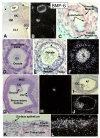
In situ hybridization of BMP-6 mRNA in ovaries of adult cycling rats. Brightfield (A, C, D, E, G, I, K) and Darkfield (B, F, H, J, L). A, B: Sections from DI 1100 h ovary (4X); positive GC in GF; preantral follicles with positive oocytes and GC (arrow heads); positive CL-I and positive OSE. C: Artery in mesovarium at P 1000 h (40X); positive endothelium. D: Early secondary follicle at E 0200 h (40X) with weakly positive oocyte; germinal vesicle (GV). E, F: Early secondary follicle at E 1000 h (20X); positive oocyte; positive peripheral membrana GC (arrow heads); negative corona radiata; G, H: Ovulating follicle at E 1000 h (4X) showing positive oocyte and negative MGC, CC, and PA cells. I, J: Atretic follicle (AF) at E 1000 h (10X) with positive oocyte (O) and GC. K, L: New CL-I at DI 1100 h (20X) showing positive TE and OSE.
Figure 5
Figure 5
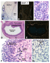
In situ hybridization of BMP-7 mRNA in ovaries of adult cycling rats. Brightfield (A, C, D, F, G) and Darkfield (B, E). A, B: Sections of DI 1100 h ovaries (4X); positive Graafian follicle (GF); positive new CL-I; negative old CL-I and SIC; C: Positive sex cords at E 0200 h (40X); D, E: Dominant follicle at P 2000 h (4X) showing positive theca interna. F: Higher magnification (40X) of D showing positive fibroblast-like cells beneath the basal lamina and adjacent positive TIC (). G: CL-I at DI 1100 h showing positive theca lutein cells (40X).
Figure 6
Figure 6
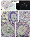
In situ hybridization of BMP-15 mRNA in ovaries of adult cycling rats. Brightfield (A, C, D, E, F, G) and Darkfield (B). A, B: Sections from a DII 1100 h ovary (4X); Positive oocytes in preantral follicles (arrow heads); C: Early primary follicle at DI 1100 h showing one cuboidal GC and a weakly positive oocyte: note most of the signal is located within the GV or nucleus (40X). D: Preantral follicle at P 1000 h (40X) in the process of forming a second layer of GC. Note a weakly positive oocyte; E: Mid secondary follicle at DII 1100 h showing a strongly positive full-grown oocyte surrounded by three to four layers of GC (20X); F: Mid cross section of a strongly positive oocyte of a dominant follicle at P 1000 h (40X). Hybridization signals are seen predominantly in the ooplasm, with some signal associated with the outer region of the nucleolus (arrow head); G: An atretic follicle at E 0200 h (40X) with a moderately positive oocyte that has resumed meiosis
Figure 7
Figure 7
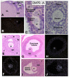
In situ hybridization of BMPR-IA mRNA in ovaries of adult cycling rats. Brighfield (A, C, D, E, G, I) and Darkfield (B, F, H, J). A, B: Sections from DII 1100 h ovaries (4X). Positive oocyte; C: Positive oocytes (O) and GC in primordial and primary follicles at P 1000 h (40X); D: Positive secondary follicle with three layers of GC at DII 1100 h (20X); E, F: Positive oocyte of an early tertiary follicle at P 1000 h. Note expression in TI (arrowheads) (10X); G, H: Preovulatory follicle at E 1000 h showing positive oocyte (O); Note weak signal in GC and TI (4X); I, J: Atretic follicle at E 1000 h showing positive O and GC (10X).
Figure 8
Figure 8
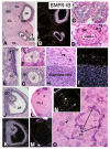
In situ hybridization of BMPR-IB mRNA in ovaries of adult cycling rats. Brightfield (A, C, D, E, F, G, H, J, L, O) and Darkfield (B, I, K, M, N). A, B: Sections from a P 2000 h ovary (4X); positive GC in dominant (DF), AF and preantral follicle (arrow head); Note positive oocyte. C: Positive sex cords at P 1000 h (40X);D: Artery at P 2000 h (40X) showing positive tunica adventita (TA) next to smooth muscle (SM); E: Positive oocytes (O) in primordial and early primary follicles at DI 1100 h (40X); F: Positive O in late primary follicle with single layer of weakly positive GC at P 2000 h (40X); G: Strongly positive O and GC in secondary follicle at E 1000 h (20X); H, I: Higher magnification (20X) of wall of DF at P 1000 h showing positive GC, TE, and a number of TIC in the outer zone of TI; J, K: Positive AF at P 1000 h (10X); L, M: CL-I at DII 1100 h showing positive TE (arrow heads); N: CL-I at P 2000 h showing positive TE at periphery (arrow head) and clusters of positive cells scattered within the CL (); O: Higher magnification (40X) of N showing positive endothelial (EC) and a few adjacent weakly positive granulosa lutein cells (arrow head).
Figure 9
Figure 9
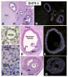
In situ hybridization of BMPR-II mRNA in ovaries of adult cycling rats. Brightfield (A, C, D, F, H) and Darkfield (B, E, G). A, B: Sections from DI 1100 h ovaries (4X). Positive new CL-I; positive GC in GF, AF, and preantral follicles (arrow heads); negative secondary interstitial cells (SIC); C: Early secondary follicle showing positive GC surrounding weakly positive oocyte (40X); D, E: Dominant preovulatory follicle at E 0200 h (4X); positive MGC and negative oocyte (O) surrounded by weakly positive CC. F, G: Atretic follicle at P 2000 h (10X) showing positive GC. H: Higher magnification of new CL-I in panel A (40X); moderately positive lutein cells; negative endothelial cells (EC) and theca externa (TE).
Figure 10
Figure 10
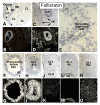
In situ hybridization of FS mRNA in ovaries of adult cycling rats. Brightfield (A, C, E, F, H, J, L, N) and Darkfield (B, D, G, I, K, M, O). A, B: Sections from a DI 1100 h ovary (4X) showing strongly positive GC in GF and weakly positive GC in preantral follicles (arrow heads); C, D: Positive SIC at P 2000 h (4X); E: Higher magnification of panel C showing high density of silver grains (black dots) over the SIC (40X); F to O: FS expression in CL-I during luteogenesis (4X) i.e. luteinization (E 0200 h, E 1000 h, DI 1100 h) and luteolysis (DII 1100 h, P 2000 h).

Similar articles

Cited by

References

    1. Shimasaki S, Moore RK, Erickson GF, Otsuka F. The role of bone morphogenetic proteins in ovarian function. Reproduction. - PubMed
    1. Attisano L, Wrana JL. Smads as transcriptional co-modulators. Curr Opin Cell Biol. 2000;12:235–243. doi: 10.1016/S0955-0674(99)00081-2. - DOI - PubMed
    1. Takao M, Hino J, Takeshita N, Konno Y, Nishizawa T, Matsuo H, Kangawa K. Identification of rat bone morphogenetic protein-3b (BMP-3b), a new member of BMP-3. Biochem Biophys Res Commun. 1996;219:656–662. doi: 10.1006/bbrc.1996.0289. - DOI - PubMed
    1. Shimasaki S, Zachow RJ, Li D, Kim H, Iemura S-I, Ueno N, Sampath K, Chang RJ, Erickson GF. A functional bone morphogenetic protein system in the ovary. Proc Natl Acad Sci U S A. 1999;96:7282–7287. doi: 10.1073/pnas.96.13.7282. - DOI - PMC - PubMed
    1. Lyons KM, Pelton RW, Hogan BL. Patterns of expression of murine Vgr-1 and BMP-2a RNA suggest that transforming growth factor-β-like genes coordinately regulate aspects of embryonic development. Genes Dev. 1989;3:1657–1668. - PubMed

Publication types

MeSH terms

Substances