. 2003 Apr 28;161(2):359-69.
doi: 10.1083/jcb.200211097.

Stu2p, the budding yeast member of the conserved Dis1/XMAP215 family of microtubule-associated proteins is a plus end-binding microtubule destabilizer

Affiliations

Stu2p, the budding yeast member of the conserved Dis1/XMAP215 family of microtubule-associated proteins is a plus end-binding microtubule destabilizer

Mark van Breugel et al. J Cell Biol. .

Abstract

The Dis1/XMAP215 family of microtubule-associated proteins conserved from yeast to mammals is essential for cell division. XMAP215, the Xenopus member of this family, has been shown to stabilize microtubules in vitro, but other members of this family have not been biochemically characterized. Here we investigate the properties of the Saccharomyces cerevisiae homologue Stu2p in vitro. Surprisingly, Stu2p is a microtubule destabilizer that binds preferentially to microtubule plus ends. Quantitative analysis of microtubule dynamics suggests that Stu2p induces microtubule catastrophes by sterically interfering with tubulin addition to microtubule ends. These results reveal both a new biochemical activity for a Dis1/XMAP215 family member and a novel mechanism for microtubule destabilization.


Figures

Figure 1.
Figure 1.
Recombinant, baculovirus-derived Stu2p and endogenous Stu2p show similar hydrodynamic properties. (A) Coomassie blue–stained SDS-polyacrylamide gel showing ∼7.5 μg of purified, baculovirus-derived Stu2p. (B) Determination of the Stokes radius of baculovirus-derived and endogenous Stu2p in yeast extracts using gel filtration on a Superose 6 column. The fractions with recombinant Stu2p (top) or yeast extract (bottom) were separated by SDS-PAGE, and Stu2p was detected by Western blotting using a polyclonal Stu2p antibody. The corresponding positions of selected markers of known Stokes radius are indicated by arrows. (C) Determination of the S value of recombinant and endogenous Stu2p in yeast extracts. Recombinant Stu2p and endogenous Stu2p in yeast extracts were spun through a linear 5–40% sucrose gradient. Fractions were analyzed by SDS-PAGE and Western blotting using a polyclonal Stu2p antibody. (Top) Western blot for recombinant Stu2p; (bottom) Western blot for endogenous Stu2p. Corresponding positions of markers with known S value are indicated by arrows.
Figure 2.
Figure 2.
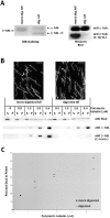
Stu2p does not strongly require the COOH terminus of β-tubulin for binding to microtubules. (A) Mock- or subtilisin- digested microtubules analyzed by SDS-PAGE and Coomassie blue staining (left) or by Western blotting (right) using a monoclonal α-tubulin antibody (top) or a monoclonal β-tubulin antibody whose epitope lies within the COOH terminus of β-tubulin (bottom). (B, top) Mock- and subtilisin-digested microtubules visualized by immunofluorescence. Bar, 5 μm. (B, bottom) Increasing amounts of mock- or subtilisin-digested microtubules were incubated with 18 nM Stu2p and bound separated from unbound Stu2p by centrifugation. Equivalent amounts of the supernatants (S) and the pellets (P) were analyzed by SDS-PAGE and Western blotting using a polyclonal Stu2p antibody (top), a monoclonal α-tubulin antibody (middle), or a monoclonal β-tubulin antibody whose epitope lies within the COOH terminus of β-tubulin (bottom). (C) Plot showing the percentage of Stu2p bound to mock- or subtilisin-digested microtubules at different microtubule concentrations.
Figure 3.
Figure 3.
Stu2p decreases microtubule length in vitro. (A) Microtubule asters at increasing Stu2p concentrations visualized by incorporation of rhodamine tubulin. Shown are representative examples of asters at 0, 0.1, 0.33, and 1 μM Stu2p. Bar, 4 μm. (B) Plot showing the distribution of aster radii at each Stu2p concentration. (C) Stu2p does not cause spontaneous tubulin nucleation in solution. Increasing amounts of Stu2p were incubated with 22 μM tubulin for 10 min at 29°C. Reactions were spun through a 40% glycerol cushion, and equivalent amounts of supernatants (S) and pellets (P) were analyzed by SDS-PAGE and Coomassie blue staining.
Figure 4.
Figure 4.
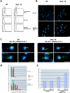
Stu2p can destabilize cytoplasmic microtubules in vivo. Wild-type or stu2–10 cells were HU arrested at the permissive temperature, shifted to the restrictive temperature, and the cytoplasmic microtubule length was determined by indirect immunofluorescence and three dimensional tracking. (A) FACS® profiles of cycling wild-type or stu2–10 cells at 25°C (top) that were arrested at 25°C with HU before (middle) or after shifting the cells to 37°C (bottom). (B) Projected indirect antitubulin immunofluorescence of wild-type or stu2–10 cells arrested at 25°C with HU before (top) or after shifting the cells to 37°C (bottom). Green, microtubules; blue, DNA. Bar, 5 μm. (C) Projected indirect antitubulin immunofluorescence of wild-type (left) or stu2–10 cells (right) arrested at 25°C with HU after shifting the cells to 37°C. Close-ups of four representative cells. Green, microtubules; blue, DNA. Bar, 5 μm. (D) Length distribution (in 1-μm intervals) of cytoplasmic microtubules of wild-type and stu2–10 cells arrested at 25°C with HU before (top) or after shifting the cells to 37°C (bottom). Green, wild-type microtubules; red, stu2–10 microtubules. (E) Average length of cytoplasmic microtubules of wild-type or stu2–10 cells arrested at 25°C with HU before and after shifting the cells to 37°C. Error bars represent SEM (P < 0.05).
Figure 5.
Figure 5.
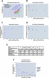
Stu2p induces catastrophes by decreasing microtubule growth rate. Real-time VE-DIC analysis of microtubules in the presence of increasing amounts of Stu2p. (A) Three examples of traces of individual microtubules at 0, 62.5, and 500 nM Stu2p. (B) Stu2p inhibits microtubule growth rate. A plot showing the averaged growth rates of microtubules at different concentrations of Stu2p. Error bars represent the SEM (P ≤ 0.05). (C) Stu2p increases microtubule catastrophe frequency. A plot showing the microtubule catastrophe frequency at different concentrations of Stu2p. (D) Stu2p decreases microtubule shrinkage rate. A plot showing the averaged shrinkage rates of microtubules at different concentrations of Stu2p. Error bars represent the SEM (P ≤ 0.05). (E) The derived dynamic parameters of microtubule polymerization in the presence of increasing amounts of Stu2p. The rescue frequency could not be determined. (F) Stu2p does not directly stimulate microtubule catastrophes. A plot showing the microtubule growth rate catastrophe frequency relation in pure tubulin (varying tubulin). The growth rates and corresponding catastrophe frequencies with varying amounts of Stu2p (as summarized in E) were added to this plot. Growth rate and corresponding catastrophe frequency at increasing concentrations of pure tubulin were obtained by VE-DIC, individual microtubule traces were binned in 0.06 μm/min growth rate intervals, and the catastrophe frequency in each interval was calculated subsequently.
Figure 6.
Figure 6.
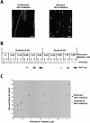
Stu2p binds preferentially to microtubule ends. (A) Antitubulin immunofluorescence of taxol stabilized microtubules before and after shearing with a tip sonicator. Bar, 5 μm. (B) Stu2p binds to a higher extent to sheared microtubules. Increasing amounts of unsheared or sheared microtubules were incubated with 19 nM Stu2p, and bound Stu2p separated from unbound Stu2p by centrifugation. Equivalent amounts of supernatants (S) and pellets (P) were analyzed by SDS-PAGE and Western blotting using a polyclonal Stu2p antibody (top) or a monoclonal tubulin antibody (bottom). (C) Plot showing the percentage of Stu2p bound to microtubules at different microtubule concentrations.
Figure 7.
Figure 7.
Stu2p binds preferentially to microtubule plus ends. Visualization of Stu2p binding to microtubules using directly Cy3-labeled Stu2p and polarity-marked microtubules. Stu2p intensity was scanned in the rhodamine channel from the minus end along the length of microtubules, and signals higher than 10% of the median intensity were scored with their location. (A) Representative examples of polarity-marked microtubules in the absence or in the presence of 16 nM Stu2p. Polarity marked microtubules are in green, and Stu2p is in red. The minus end of microtubules is brighter than the plus end. Bar, 3 μm. (B) Histogram showing the number of Stu2p spots per microtubule. (C) Histogram showing the percentage of Stu2p signals found in 10% intervals from the minus ends of the microtubules to their plus ends.

Similar articles

Cited by

References

    1. Akhmanova, A., C.C. Hoogenraad, K. Drabek, T. Stepanova, B. Dortland, T. Verkerk, W. Vermeulen, B.M. Burgering, C.I. De Zeeuw, F. Grosveld, and N. Galjart. 2001. Clasps are CLIP-115 and -170 associating proteins involved in the regional regulation of microtubule dynamics in motile fibroblasts. Cell. 104:923–935. - PubMed
    1. Arnal, I., E. Karsenti, and A. Hyman. 2000. Structural transitions at microtubule ends correlate with their dynamic properties in Xenopus egg extracts. J. Cell Biol. 149:767–774. - PMC - PubMed
    1. Ashford, A., S.S.L. Andersen, and A. Hyman. 1998. Preparation of tubulin from bovine brain. Cell Biology: A Laboratory Handbook. 2nd edition. Vol. 2. Julio E. Cells, editor. Academic Press, Inc., Orlando, FL. 205–212.
    1. Belmont, L., and T. Mitchison. 1996. Identification of a protein that interacts with tubulin dimers and increases the catastrophe rates of microtubules. Cell. 84:623–631. - PubMed
    1. Berlin, V., C.A. Styles, and G.R. Fink. 1990. BIK1, a protein required for microtubule function during mating and mitosis in Saccharomyces cerevisiae, colocalizes with tubulin. J. Cell Biol. 111:2573–2586. - PMC - PubMed

MeSH terms