

Intra-articular hyaluronan (hyaluronic acid) and hylans for the treatment of osteoarthritis: mechanisms of action
- PMID: 12718745
- PMCID: PMC165033
- DOI: 10.1186/ar623
Intra-articular hyaluronan (hyaluronic acid) and hylans for the treatment of osteoarthritis: mechanisms of action
Abstract
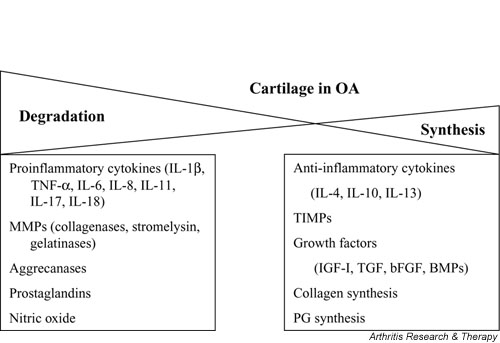
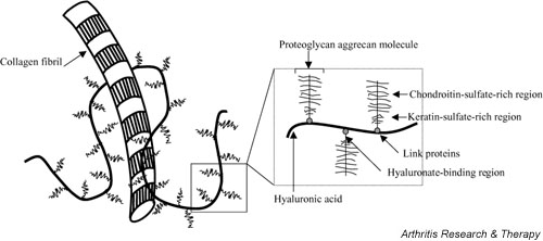
Although the predominant mechanism of intra-articular hyaluronan (hyaluronic acid) (HA) and hylans for the treatment of pain associated with knee osteoarthritis (OA) is unknown, in vivo, in vitro, and clinical studies demonstrate various physiological effects of exogenous HA. HA can reduce nerve impulses and nerve sensitivity associated with the pain of OA. In experimental OA, this glycosaminoglycan has protective effects on cartilage, which may be mediated by its molecular and cellular effects observed in vitro. Exogenous HA enhances chondrocyte HA and proteoglycan synthesis, reduces the production and activity of proinflammatory mediators and matrix metalloproteinases, and alters the behavior of immune cells. Many of the physiological effects of exogenous HA may be a function of its molecular weight. Several physiological effects probably contribute to the mechanisms by which HA and hylans exert their clinical effects in knee OA.
Figures
Similar articles
-
Potential mechanism of action of intra-articular hyaluronan therapy in osteoarthritis: are the effects molecular weight dependent?Semin Arthritis Rheum. 2002 Aug;32(1):10-37. doi: 10.1053/sarh.2002.33720. Semin Arthritis Rheum. 2002. PMID: 12219318 Review.
-
The role of intra-articular hyaluronan (Sinovial) in the treatment of osteoarthritis.Rheumatol Int. 2011 Apr;31(4):427-44. doi: 10.1007/s00296-010-1660-6. Epub 2010 Nov 28. Rheumatol Int. 2011. PMID: 21113807 Review.
-
Platelet Rich Plasma and Hyaluronic Acid Blend for the Treatment of Osteoarthritis: Rheological and Biological Evaluation.PLoS One. 2016 Jun 16;11(6):e0157048. doi: 10.1371/journal.pone.0157048. eCollection 2016. PLoS One. 2016. PMID: 27310019 Free PMC article.
-
Hyaluronic Acid Versus Platelet-Rich Plasma: A Prospective, Double-Blind Randomized Controlled Trial Comparing Clinical Outcomes and Effects on Intra-articular Biology for the Treatment of Knee Osteoarthritis.Am J Sports Med. 2017 Feb;45(2):339-346. doi: 10.1177/0363546516665809. Epub 2016 Oct 21. Am J Sports Med. 2017. PMID: 28146403 Clinical Trial.
-
The role of hyaluronic acid (hyaluronan) in health and disease: interactions with cells, cartilage and components of synovial fluid.Clin Exp Rheumatol. 1994 Jan-Feb;12(1):75-82. Clin Exp Rheumatol. 1994. PMID: 8162648 Review.
Cited by
-
Pressure sensing of lysosomes enables control of TFEB responses in macrophages.Nat Cell Biol. 2024 Aug;26(8):1247-1260. doi: 10.1038/s41556-024-01459-y. Epub 2024 Jul 12. Nat Cell Biol. 2024. PMID: 38997458
-
Notochord Cells in Intervertebral Disc Development and Degeneration.J Dev Biol. 2016 Mar;4(1):3. doi: 10.3390/jdb4010003. Epub 2016 Jan 21. J Dev Biol. 2016. PMID: 27252900 Free PMC article.
-
Diacerein-Loaded Hyaluosomes as a Dual-Function Platform for Osteoarthritis Management via Intra-Articular Injection: In Vitro Characterization and In Vivo Assessment in a Rat Model.Pharmaceutics. 2021 May 21;13(6):765. doi: 10.3390/pharmaceutics13060765. Pharmaceutics. 2021. PMID: 34063749 Free PMC article.
-
Ultrasound-guided viscosupplementation of subacromial space in elderly patients with cuff tear arthropathy using a high weight hyaluronic acid: prospective open-label non-randomized trial.Eur Radiol. 2011 Jan;21(1):182-7. doi: 10.1007/s00330-010-1894-4. Epub 2010 Jul 25. Eur Radiol. 2011. PMID: 20658295 Clinical Trial.
-
Comparison of Intra-articular Hyaluronic Acid and Platelet-Rich Plasma Injection in Knee Osteoarthritis: Do the Results Differ in Geriatric Patients? A Retrospective Observational Study.Ann Geriatr Med Res. 2022 Dec;26(4):340-346. doi: 10.4235/agmr.22.0090. Epub 2022 Dec 15. Ann Geriatr Med Res. 2022. PMID: 36518061 Free PMC article.
References
-
- Balazs E. The physical properties of synovial fluid and the specific role of hyaluronic acid. In: Helfet AJ, editor. In Disorders of the Knee. Philadelphia: J B Lippincott; 1982. pp. 61–74.
-
- Adams ME, Atkinson MH, Lussier AJ, Schulz JI, Siminovitch KA, Wade JP, Zummer M. The role of viscosupplementation with hylan G-F 20 (Synvisc) in the treatment of osteoarthritis of the knee: a Canadian multicenter trial comparing hylan G-F 20 alone, hylan G-F 20 with non-steroidal anti-inflammatory drugs (NSAIDs) and NSAIDs alone. Osteoarthritis Cartilage. 1995;3:213–225. - PubMed
-
- Lussier A, Cividino AA, McFarlane CA, Olszynski WP, Potashner WJ, De Medicis R. Viscosupplementation with hylan for the treatment of osteoarthritis: findings from clinical practice in Canada. J Rheumatol. 1996;23:1579–1585. - PubMed
Publication types
MeSH terms
Substances
LinkOut - more resources
Full Text Sources
Other Literature Sources
Medical
