Invasion and killing of human endothelial cells by viridans group streptococci
- PMID: 12704106
- PMCID: PMC153257
- DOI: 10.1128/IAI.71.5.2365-2372.2003
Invasion and killing of human endothelial cells by viridans group streptococci
Abstract
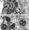
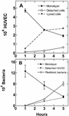
Colonization of the cardiovascular endothelium by viridans group streptococci can result in infective endocarditis and possibly atherosclerosis; however, the mechanisms of pathogenesis are poorly understood. We investigated the ability of selected oral streptococci to infect monolayers of human umbilical vein endothelial cells (HUVEC) in 50% human plasma and to produce cytotoxicity. Planktonic Streptococcus gordonii CH1 killed HUVEC over a 5-h period by peroxidogenesis (alpha-hemolysin) and by acidogenesis but not by production of protein exotoxins. HUVEC were protected fully by addition of supplemental buffers and bovine liver catalase to the culture medium. Streptococci were also found to invade HUVEC by an endocytic mechanism that was dependent on polymerization of actin microfilaments and on a functional cytoskeleton, as indicated by inhibition with cytochalasin D and nocodazole. Electron microscopy revealed streptococci attached to HUVEC surfaces via numerous fibrillar structures and bacteria in membrane-encased cytoplasmic vacuoles. Following invasion by S. gordonii CH1, HUVEC monolayers showed 63% cell lysis over 4 h, releasing 64% of the total intracellular bacteria into the culture medium; however, the bacteria did not multiply during this time. The ability to invade HUVEC was exhibited by selected strains of S. gordonii, S. sanguis, S. mutans, S. mitis, and S. oralis but only weakly by S. salivarius. Comparison of isogenic pairs of S. gordonii revealed a requirement for several surface proteins for maximum host cell invasion: glucosyltransferase, the sialic acid-binding protein Hsa, and the hydrophobicity/coaggregation proteins CshA and CshB. Deletion of genes for the antigen I/II adhesins, SspA and SspB, did not affect invasion. We hypothesize that peroxidogenesis and invasion of the cardiovascular endothelium by viridans group streptococci are integral events in the pathogenesis of infective endocarditis and atherosclerosis.
Figures


Similar articles
-
Invasion of human aortic endothelial cells by oral viridans group streptococci and induction of inflammatory cytokine production.Mol Oral Microbiol. 2011 Feb;26(1):78-88. doi: 10.1111/j.2041-1014.2010.00597.x. Epub 2010 Dec 3. Mol Oral Microbiol. 2011. PMID: 21214874
-
Glucosyltransferase mediates adhesion of Streptococcus gordonii to human endothelial cells in vitro.Infect Immun. 1994 Jun;62(6):2187-94. doi: 10.1128/iai.62.6.2187-2194.1994. Infect Immun. 1994. PMID: 8188339 Free PMC article.
-
Multiple adhesin proteins on the cell surface of Streptococcus gordonii are involved in adhesion to human fibronectin.Microbiology (Reading). 2009 Nov;155(Pt 11):3572-3580. doi: 10.1099/mic.0.032078-0. Epub 2009 Aug 6. Microbiology (Reading). 2009. PMID: 19661180 Free PMC article.
-
Functions of cell surface-anchored antigen I/II family and Hsa polypeptides in interactions of Streptococcus gordonii with host receptors.Infect Immun. 2005 Oct;73(10):6629-38. doi: 10.1128/IAI.73.10.6629-6638.2005. Infect Immun. 2005. PMID: 16177339 Free PMC article.
-
Putative pathogenic factors underlying Streptococcus oralis opportunistic infections.J Microbiol Immunol Infect. 2024 Sep 6:S1684-1182(24)00159-2. doi: 10.1016/j.jmii.2024.09.001. Online ahead of print. J Microbiol Immunol Infect. 2024. PMID: 39261123 Review.
Cited by
-
The group B streptococcal serine-rich repeat 1 glycoprotein mediates penetration of the blood-brain barrier.J Infect Dis. 2009 May 15;199(10):1479-87. doi: 10.1086/598217. J Infect Dis. 2009. PMID: 19392623 Free PMC article.
-
Group B streptococcal serine-rich repeat proteins promote interaction with fibrinogen and vaginal colonization.J Infect Dis. 2014 Sep 15;210(6):982-91. doi: 10.1093/infdis/jiu151. Epub 2014 Mar 11. J Infect Dis. 2014. PMID: 24620021 Free PMC article.
-
Streptococcus mitis and Gemella haemolysans were simultaneously found in atherosclerotic and oral plaques of elderly without periodontitis-a pilot study.Clin Oral Investig. 2017 Jan;21(1):447-452. doi: 10.1007/s00784-016-1811-6. Epub 2016 Apr 2. Clin Oral Investig. 2017. PMID: 27037569
-
The oral microbiota and cardiometabolic health: A comprehensive review and emerging insights.Front Immunol. 2022 Nov 18;13:1010368. doi: 10.3389/fimmu.2022.1010368. eCollection 2022. Front Immunol. 2022. PMID: 36466857 Free PMC article. Review.
-
Use of phylogenetic and phenotypic analyses to identify nonhemolytic streptococci isolated from bacteremic patients.J Clin Microbiol. 2005 Dec;43(12):6073-85. doi: 10.1128/JCM.43.12.6073-6085.2005. J Clin Microbiol. 2005. PMID: 16333101 Free PMC article.
References
-
- Baddour, L. M., G. D. Christensen, J. H. Lowrance, and W. A. Simpson. 1989. Pathogenesis of Experimental endocarditis. Rev. Infect. Dis. 11:452-463. - PubMed
Publication types
MeSH terms
Substances
Grants and funding
LinkOut - more resources
Full Text Sources

