. 2003 Apr 1;17(7):896-911.
doi: 10.1101/gad.252103.

Human Sin3 deacetylase and trithorax-related Set1/Ash2 histone H3-K4 methyltransferase are tethered together selectively by the cell-proliferation factor HCF-1

Affiliations

Human Sin3 deacetylase and trithorax-related Set1/Ash2 histone H3-K4 methyltransferase are tethered together selectively by the cell-proliferation factor HCF-1

Joanna Wysocka et al. Genes Dev. .

Abstract

The abundant and chromatin-associated protein HCF-1 is a critical player in mammalian cell proliferation as well as herpes simplex virus (HSV) transcription. We show here that separate regions of HCF-1 critical for its role in cell proliferation associate with the Sin3 histone deacetylase (HDAC) and a previously uncharacterized human trithorax-related Set1/Ash2 histone methyltransferase (HMT). The Set1/Ash2 HMT methylates histone H3 at Lys 4 (K4), but not if the neighboring K9 residue is already methylated. HCF-1 tethers the Sin3 and Set1/Ash2 transcriptional regulatory complexes together even though they are generally associated with opposite transcriptional outcomes: repression and activation of transcription, respectively. Nevertheless, this tethering is context-dependent because the transcriptional activator VP16 selectively binds HCF-1 associated with the Set1/Ash2 HMT complex in the absence of the Sin3 HDAC complex. These results suggest that HCF-1 can broadly regulate transcription, both positively and negatively, through selective modulation of chromatin structure.


Figures
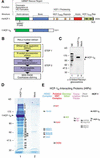
Figure 1
Figure 1
Identification of the HCF-1-interacting proteins (HIPs). (A) Overall structures of HCF-1 and f-HCF-1N. (Upper diagram) Human (Hs) HCF-1. Above are shown structural or sequence elements and functional regions. Below, the position of the tsBN67-cell P134S missense mutation is indicated. (Lower diagram) The Flag-epitope-tagged HCF-1N subunit used in this study. VIC, VP16-induced complex; HCF-1PRO, HCF-1 proteolytic processing repeats; HCF-1Fn3 Rep., HCF-1 fibronectin type 3 repeats. (B) Two-step purification scheme for f-HCF-1N-associated polypeptides. (C) f-HCF-1N is modified by O-linked N-acetyl-glucosamine residues and is a major glycoprotein in the HIPs purification. Two-step purified f-HCF-1N or mock samples were analyzed by immunoblotting with an anti-O-linked N-acetyl-glucosamine antibody. The position of the f-HCF-1N subunit is indicated by the arrowhead. A weak doublet, displaying the correct migration for Sp1 polypeptides, is indicated by the two dots. NE, HeLa nuclear extract. (D) f-HCF-1N-associated proteins were purified from 2.4 × 1010 f-HCF-1N or mock HeLa cells by the two-step procedure outlined. Polypeptides were resolved by SDS-PAGE and stained with Coomassie blue. The positions of the molecular weight markers and f-HCF-1N species are indicated on the left. The 25 additional bands present in the f-HCF-1N-purified material are labeled 1–25 and were analyzed by mass spectrometry. Only two polypeptides could be visualized in the mock-purified sample, and they were subsequently identified as Hsp70 (minor species) and nuclear lamin (major species). (E) Identity of a subset of HIPs. Sixteen out of 33 unique polypeptides identified by HIP mass spectrometry analysis are grouped into six categories: members of the Sin3 HDAC complex (blue), human homologs of the Saccharomyces cerevisiae Set1/Ash2 HMT complex (red), O-linked N-acetyl-glucosamine transferase (OGT; green), transcription factor Sp1 (purple), heat-shock proteins (gray), and endogenous HCF-1C subunits (black). The number to the left of each species represents the stained band from which each protein originates.
Figure 2
Figure 2
Association of the f-HCF-1N subunit with endogenous HCF-1C subunits, Sp1, OGT, and the Sin3 HDAC complex, and histone deacetylase activity. (A) Two-step f-HCF-1N (lane 2) and mock (lane 3) purified samples were analyzed by immunoblotting with the antibodies against the HCF-1C subunit (panel a), Sp1 (panel b), and GABPβ (panel c). Labeled arrowheads identify polypeptides of interest. NE, HeLa nuclear extract (lane 1) used as a positive control. (B) The f-HCF-1N subunit specifically associates with the Sin3 HDAC complex. Samples as in A were analyzed by immunoblotting with antibodies against OGT (panel a), Sin3A, Sin3B, HDAC1, HDAC2, and Sds3 components of the Sin3 HDAC complex (panels bf), a component of the NuRD complex Mi-2 (panel g), and the HDAC1/2-related class I histone deacetylase HDAC3 (panel h). Labeled arrowheads identify polypeptides of interest. NE, HeLa nuclear extract (lane 1) corresponding to 5% of the samples in lanes 2 and 3 was used as a positive control. (C) HCF-1 associates with HDAC activity. Two-step purified f-HCF-1N and mock-purified samples were added to reactions with [3H]-acetylated histone H4 peptide in the presence or absence of deacetylase inhibitor. Counts above the background CPM released from the acetylated peptide incubated alone are shown. The average of three experiments is shown.
Figure 3
Figure 3
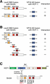
Association of Sin3A PAH1 with the HCF-1 Basic region in a yeast two-hybrid assay. (A) The LexA DNA-binding domain fused to mouse Sin3A amino acids 119–1219 (PAH1-E) was used as a bait to screen a library of mouse embryo cDNAs fused to the VP16 transactivation domain. Four identical positive clones encoding a portion of the HCF-1 Basic region were identified. (B) Directed two-hybrid assays were performed to map the Sin3A-interaction domain of HCF-1 Basic region using LexA fusions containing different regions of Sin3A: PAH1, PAH2, PAH3, PAH4, and PAH3-E (see Materials and Methods). (C) Schematic representations of HCF-1 and Sin3A are shown. The HCF-1 Sp1-interaction region is indicated above the diagram. Four paired amphipatic PAH1–4 helices of Sin3A are depicted. Regions of Sin3A involved in association with various protein partners are shown below the Sin3A diagram. Regions of HCF-1 and Sin3A that interact in the yeast two-hybrid assay are indicated.
Figure 4
Figure 4
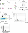
Association of HCF-1 with the human homologs of components of the Saccharomyces cerevisiae Set1/Ash2 HMT complex and HMT activity. (A) Similarity between components of the S. cerevisiae Set1/Ash2 complex and human proteins identified among the HIPs. Schematic representations of yeast Set1/Ash2 complex proteins, and related HIPs (hSet1, accession no. gi 6683126; hAsh2, accession no. gi 4757790; and WDR5, accession no. gi 16554627) are shown on the left and right, respectively. Structural domains and motifs are shown in each protein and defined by the key on the bottom right. RRM, RNA recognition motif; SET, Set domain; postSET, Set domain-associated cysteine-rich motif; HBM, HCF-1-binding motif; PHD, PHD zinc-finger motif; SPRY, domain in SPla and the RYanodine receptor; WD40, WD40 repeats; RIIa, protein kinase A regulatory subunit dimerization domain motif. (B) Two-step purified f-HCF-1N (lane 2) and mock (lane 3) samples were analyzed by immunoblot with anti-human Set1 (panel a), Ash2 (panel b), WDR5 (panel c), and SUV39H1 (panel d) antibodies. Labeled arrowheads indicate polypeptides of interest; NE, HeLa nuclear extract (lane 1) corresponding to 5% of the samples in lanes 1 and 2 was used as a positive control. (C) HCF-1 associates with HMT activity. Two-step f-HCF-1N and mock HeLa-cell purified samples were incubated with core histones or recombinant histone H3 in the presence of [3H]AdoMet. Proteins were resolved by 15% SDS-PAGE and examined by Coomassie blue staining (lower panel) or fluorography (upper panel). The positions of histones are indicated on the right. NE, HeLa nuclear extract used as a positive control. (D) Histone H3-K4 specificity of human Set1/Ash2 HMT. Recombinant histone H3 was in vitro methylated using f-HCF-1N two-step purified material and subjected to 16 Edman degradation cycles. The radioactivity released in each cycle is indicated. (E) Interplay between K9 methylation and K4 methylation by Set1/Ash2 HMT. Two-step f-HCF-1N and mock-purified samples were incubated with a histone H3 peptide corresponding to amino acids 1–15, or the corresponding peptide trimethylated at K4 or K9. 3H incorporation for each substrate peptide was defined as the difference in 3H CPM obtained with the f-HCF-1N and mock samples. The average of five experiments is shown.
Figure 5
Figure 5
Independent Sin3 HDAC and Set1/Ash2 HMT association with separate functional regions of the HCF-1N subunit. (A) A schematic of f-HCF-1N and f-HCF-1Kelch is shown with Set1/Ash2- and Sin3-interaction regions depicted above the f-HCF-1N diagram. (B) Anti-Flag immunoprecipitations were performed from cells expressing f-HCF-1N, f-HCF-1Kelch, or transduced with the empty vector, and analyzed by immunoblotting with anti-HCF-1N (panel a), anti-Sin3 (panel b), and anti-Ash2 (panel c) antibodies. Samples were normalized to the cell equivalents. *, nonspecific anti-Ash2 cross-reacting species. (C) HCF-1N association with Sin3 and Ash2 is DNA-independent. Two-step purification was performed in parallel from f-HCF-1N and empty vector-transduced cells either in the presence (lanes 1,2) or in the absence (lanes 3,4) of 100 μg/mL ethidium bromide (see Materials and Methods). Purified material was analyzed by immunoblotting with anti-Sin3A (panel a) and anti-Ash2 (panel b) antibodies.
Figure 5
Figure 5
Independent Sin3 HDAC and Set1/Ash2 HMT association with separate functional regions of the HCF-1N subunit. (A) A schematic of f-HCF-1N and f-HCF-1Kelch is shown with Set1/Ash2- and Sin3-interaction regions depicted above the f-HCF-1N diagram. (B) Anti-Flag immunoprecipitations were performed from cells expressing f-HCF-1N, f-HCF-1Kelch, or transduced with the empty vector, and analyzed by immunoblotting with anti-HCF-1N (panel a), anti-Sin3 (panel b), and anti-Ash2 (panel c) antibodies. Samples were normalized to the cell equivalents. *, nonspecific anti-Ash2 cross-reacting species. (C) HCF-1N association with Sin3 and Ash2 is DNA-independent. Two-step purification was performed in parallel from f-HCF-1N and empty vector-transduced cells either in the presence (lanes 1,2) or in the absence (lanes 3,4) of 100 μg/mL ethidium bromide (see Materials and Methods). Purified material was analyzed by immunoblotting with anti-Sin3A (panel a) and anti-Ash2 (panel b) antibodies.
Figure 6
Figure 6
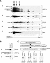
Sin3 HDAC and Set1/Ash2 HMT complex components coexist with HCF-1 in high-molecular-weight complexes. (A) Cosedimentation of HCF-1N, Sin3A, and Ash2 on glycerol gradients. HCF-1-enriched samples were prepared from cells synthesizing f-HCF-1N by the WGA purification step and applied to a 25%–50% glycerol gradient (see Materials and Methods). Fractions were analyzed by immunoblotting with antibodies recognizing endogenous HCF-1C (panel a), ectopic f-HCF-1N (panel b), Sin3A (panel c), Ash2 (panel d), and Sp1 (panel e). The position of molecular weight markers applied to a parallel gradient is indicated at the top. (B) Sin3A, endogenous HCF-1, and Ash2 copurify in nontranduced cells. Anti-Sin3A immunoprecipitation (lane 1) and control immunoprecipitation (lane 2) with a corresponding amount of the irrelevant antibody of the same isotype (12CA5) were performed from nontransduced HeLa cells. Immunoprecipitates were analyzed by immunoblotting with anti-Sin3A (panel a), anti-HCF-1 (panel b), and anti-Ash2 (panel c) antibodies. (C) Sin3A and Ash2 can associate with HCF-1 simultaneously. (Top) Purification scheme for f-HCF-1N/Sin3A complexes from f-HCF-1N/HA-VP16ΔC HeLa cells. Corresponding amounts of the two-step-purified samples from f-HCF-1N/HA-VP16ΔC and HA-VP16ΔC only HeLa cells fractionated by anti-Sin3A immunoprecipitation were analyzed by immunoblotting with Sin3A (lower panel, lanes 1,2), anti f-HCF-1N (lanes 3,4), Ash2 (lanes 5,6), and VP16 (lanes 7,8) antibodies.
Figure 7
Figure 7
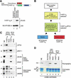
Preferential VP16 association with f-HCF-1N–Set1/Ash2 HMT over f-HCF-1N–Sin3 HDAC complexes. (A) A significant portion of HA-VP16ΔC is bound to chromatin in the f-HCF-1N/VP16ΔC cells. A schematic of HA-VP16ΔC and f-HCF-1N is shown. f-HCF-1N/HA-VP16ΔC HeLa cells were fractionated into cytosolic/soluble nuclear and chromatin-associated protein fractions and probed with anti-Flag (upper panel) and anti-HA (lower panel) epitope antibodies. (B) Schematic of the VP16-bound and VP16-free f-HCF-1N complexes fractionation. (C) Two-step f-HCF-1N and mock-purified samples from f-HCF-1N/HA-VP16ΔC and HA-VP16ΔC only cells, were fractionated by anti-HA coimmunoprecipitation into a VP16-bound f-HCF-1N fraction (pellet) and VP16-free f-HCF-1N fraction (supernatant). Samples were analyzed by immunoblotting with VP16 (panel a), HCF-1 (αN18; panel b), Sin3A (panel c), Sds3 (panel d), Ash2 (panel e), and WDR5 (panel f) antibodies. (D) VP16-bound HCF-1 complexes contain HMT activity. VP16-bound and VP16-free f-HCF-1N complexes were isolated as for C, except that VP16-bound complexes were eluted from the beads with excess of HA peptide. Fractions were then normalized to the amount of Ash2 present (threefold more of VP16-bound than VP16-free fraction was used, indicated by 3×) and assayed for the HMT activity using core histones as a substrate. Proteins were resolved by 15% SDS-PAGE and examined by Coomassie blue staining (lower panel) or fluorography (upper panel). The positions of the histones are indicated on the left. NE, HeLa nuclear extract (10-fold less than used in Fig. 4C) was used as a positive control.

Similar articles

Cited by

References

    1. Alland L, David G, Shen-Li H, Potes J, Muhle R, Lee HC, Hou H, Jr, Chen K, DePinho RA. Identification of mammalian Sds3 as an integral component of the Sin3/histone deacetylase corepressor complex. Mol Cell Biol. 2002;22:2743–2750. - PMC - PubMed
    1. Bernstein BE, Humphrey EL, Erlich RL, Schneider R, Bouman P, Liu JS, Kouzarides T, Schreiber SL. Methylation of histone H3 Lys 4 in coding regions of active genes. Proc Natl Acad Sci. 2002;99:8695–8700. - PMC - PubMed
    1. Briggs SD, Bryk M, Strahl BD, Cheung WL, Davie JK, Dent SY, Winston F, Allis CD. Histone H3 lysine 4 methylation is mediated by Set1 and required for cell growth and rDNA silencing in Saccharomyces cerevisiae. Genes & Dev. 2001;15:3286–3295. - PMC - PubMed
    1. Bryk M, Briggs SD, Strahl BD, Curcio MJ, Allis CD, Winston F. Evidence that Set1, a factor required for methylation of histone H3, regulates rDNA silencing in S. cerevisiae by a Sir2-independent mechanism. Curr Biol. 2002;12:165–170. - PubMed
    1. Dignam JD, Lebovitz RM, Roeder RG. Accurate transcription initiation by RNA polymerase II in a soluble extract from isolated mammalian nuclei. Nucleic Acids Res. 1983;11:1475–1489. - PMC - PubMed

Publication types

MeSH terms