. 2003 Apr;23(7):2451-62.
doi: 10.1128/MCB.23.7.2451-2462.2003.

Involvement of poly(ADP-Ribose) polymerase 1 and poly(ADP-Ribosyl)ation in regulation of centrosome function

Affiliations

Involvement of poly(ADP-Ribose) polymerase 1 and poly(ADP-Ribosyl)ation in regulation of centrosome function

Masayuki Kanai et al. Mol Cell Biol. 2003 Apr.

Abstract

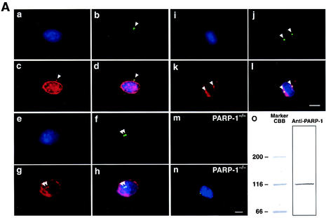
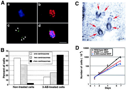
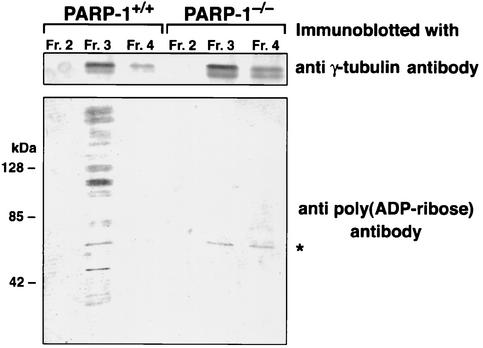
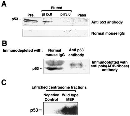
The regulatory mechanism of centrosome function is crucial to the accurate transmission of chromosomes to the daughter cells in mitosis. Recent findings on the posttranslational modifications of many centrosomal proteins led us to speculate that these modifications might be involved in centrosome behavior. Poly(ADP-ribose) polymerase 1 (PARP-1) catalyzes poly(ADP-ribosyl)ation to various proteins. We show here that PARP-1 localizes to centrosomes and catalyzes poly(ADP-ribosyl)ation of centrosomal proteins. Moreover, centrosome hyperamplification is frequently observed with PARP inhibitor, as well as in PARP-1-null cells. Thus, it is possible that chromosomal instability known in PARP-1-null cells can be attributed to the centrosomal dysfunction. P53 tumor suppressor protein has been also shown to be localized at centrosomes and to be involved in the regulation of centrosome duplication and monitoring of the chromosomal stability. We found that centrosomal p53 is poly(ADP-ribosyl)ated in vivo and centrosomal PARP-1 directly catalyzes poly(ADP-ribosyl)ation of p53 in vitro. These results indicate that PARP-1 and PARP-1-mediated poly(ADP-ribosyl)ation of centrosomal proteins are involved in the regulation of centrosome function.


Figures

FIG. 1.
FIG. 1.
(A) PARP-1 physically associates with centrosomes throughout the cell cycle. Exponentially growing wild-type MEFs were coimmunostained with anti-γ-tubulin monoclonal antibodies (panels b, f, and j, green) and anti-PARP-1 polyclonal antibodies (panels c, g, and k, red). Cells were also counterstained with DAPI (panels a, e, and i, blue). Panels d, h, and l show the overlay images. Panels a to d show a cell with unduplicated centrosome. Panels e to h show a cell with duplicated centrosomes. Panels i to l show a mitotic cell. Arrowheads point to centrosomes. Panels m and n show that negative immunostaining of PARP-1−/− MEFs by anti-PARP-1 antibody. Exponentially growing PARP-1−/− MEFs were coimmunostained with anti-PARP-1 polyclonal (panel m, red) and anti-γ-tubulin polyclonal (panel n, green) antibodies. Cells were also counterstained with DAPI (panel n, blue). Scale bar, 10 μm. Wild-type MEFs were lysed in RIPA buffer, resolved by SDS-PAGE, and immunoblotted with anti-PARP-1 antibody (panel o). (B) Centrosomal proteins are poly(ADP-ribosyl)ated. Exponentially growing wild-type MEFs were coimmunostained with anti-γ-tubulin polyclonal (panels b, f, and j, red) and anti-poly(ADP-ribose) monoclonal (panels c, g, and k, green) antibodies. Cells were also counterstained with DAPI (panels a, e, and i, blue). Panels d, h, and l show the overlay images. Panels a to d show a cell with unduplicated centrosome. Panels e to h show a cell with duplicated centrosomes. Panels I to l show a mitotic cell. The arrowheads point to centrosomes. Panels m and n show the immunostaining of PARP-1−/− MEFs by anti-poly(ADP-ribose) antibody. Exponentially growing PARP-1−/− MEFs were coimmunostained with anti-poly(ADP-ribose) monoclonal (panel m, green) and anti-γ-tubulin polyclonal (panel n, red) antibodies. Cells were also counterstained with DAPI (panel n, blue). Scale bar, 10 μm.
FIG. 1.
FIG. 1.
(A) PARP-1 physically associates with centrosomes throughout the cell cycle. Exponentially growing wild-type MEFs were coimmunostained with anti-γ-tubulin monoclonal antibodies (panels b, f, and j, green) and anti-PARP-1 polyclonal antibodies (panels c, g, and k, red). Cells were also counterstained with DAPI (panels a, e, and i, blue). Panels d, h, and l show the overlay images. Panels a to d show a cell with unduplicated centrosome. Panels e to h show a cell with duplicated centrosomes. Panels i to l show a mitotic cell. Arrowheads point to centrosomes. Panels m and n show that negative immunostaining of PARP-1−/− MEFs by anti-PARP-1 antibody. Exponentially growing PARP-1−/− MEFs were coimmunostained with anti-PARP-1 polyclonal (panel m, red) and anti-γ-tubulin polyclonal (panel n, green) antibodies. Cells were also counterstained with DAPI (panel n, blue). Scale bar, 10 μm. Wild-type MEFs were lysed in RIPA buffer, resolved by SDS-PAGE, and immunoblotted with anti-PARP-1 antibody (panel o). (B) Centrosomal proteins are poly(ADP-ribosyl)ated. Exponentially growing wild-type MEFs were coimmunostained with anti-γ-tubulin polyclonal (panels b, f, and j, red) and anti-poly(ADP-ribose) monoclonal (panels c, g, and k, green) antibodies. Cells were also counterstained with DAPI (panels a, e, and i, blue). Panels d, h, and l show the overlay images. Panels a to d show a cell with unduplicated centrosome. Panels e to h show a cell with duplicated centrosomes. Panels I to l show a mitotic cell. The arrowheads point to centrosomes. Panels m and n show the immunostaining of PARP-1−/− MEFs by anti-poly(ADP-ribose) antibody. Exponentially growing PARP-1−/− MEFs were coimmunostained with anti-poly(ADP-ribose) monoclonal (panel m, green) and anti-γ-tubulin polyclonal (panel n, red) antibodies. Cells were also counterstained with DAPI (panel n, blue). Scale bar, 10 μm.
FIG. 2.
FIG. 2.
Inhibition of PARP induces abnormal centrosome amplification. (A) Representative immunostaining of abnormally amplified centrosomes in 3-AB-treated MEFs. Wild-type MEFs were cultured in the presence of 7 mM 3-AB for 1 week. Cells were coimmunostained for anti-γ-tubulin polyclonal (panel c, green) and anti-α-tubulin monoclonal (panel b, red) antibodies. Cells were also counterstained with DAPI (panel a, blue). Panel d shows the overlay images. (B) Statistical analysis of centrosome hyperamplification in 3-AB-treated MEFs. The number of centrosomes per cell (i.e., 1, 2, or >2 centrosomes) was determined by coimmunostaining of α-tubulin and γ-tubulin of cells that were cold treated and briefly extracted before fixation. For the analysis, >200 cells were examined. (C) Electron micrographs of thin sections in 3-AB-treated cells. The arrows indicate the centriole. Scale bar, 0.2 μm. (D) Kinetic analysis of cell growth. Wild-type MEFs, PARP-1−/− MEFs, and 3-AB-treated cells were treated with trypsin, negatively stained with 0.4% trypan blue, and counted on a hemacytometer.
FIG. 3.
FIG. 3.
Centrosome hyperamplification in PARP-1−/− MEFs. (A) Exponentially growing PARP-1−/− and PARP-1+/+ MEFs were immunostained with anti-γ-tubulin polyclonal antibody (red). Cells were also counterstained with DAPI (blue). Panels a and b are PARP-1+/+ MEFs, and panels c and d are PARP-1−/− MEFs. Panels a and c show mitotic cells, whereas panels b and d show interphase cells. The arrowheads point to centrosomes, and an arrow indicates aggregated centrosomes. Scale bar, 10 μm. (B) Statistical analysis of centrosome hyperamplification in PARP-1−/− MEFs. The number of centrosomes per cell (i.e., 1, 2, or >2 centrosomes) was determined by immunostaining of γ-tubulin. For the analysis, >200 cells were examined. Bars represent the average ± the standard error calculated from three independent measurements. (C) PARP-1+/+ and PARP-1−/− primary MEFs were immunostained with anti-γ-tubulin polyclonal (green) and/or α-tubulin monoclonal (red) antibodies. Cells were counterstainined with DAPI (blue). Representative images in panels a and b show the mitotic cells. Scale bar, 10 μm. (D) Quantification of centrosome hyperamplification in PARP-1−/− PMEFs. The number of centrosomes per cell (1, 2, or >2 centrosomes) was determined by immunostaining of γ-tubulin. For the analysis, >200 cells were examined.
FIG. 4.
FIG. 4.
(A) Uncoupling of initiation of DNA and centrosome duplication in PARP-1−/− MEFs. PARP-1+/+ and PARP-1−/− MEFs were serum starved for 36 h, followed by serum stimulation with the medium containing 20% FBS and BrdU. At indicated time points, cells were briefly extracted, fixed, and coimmunostained with anti-BrdU monoclonal and anti-γ-tubulin polyclonal antibodies. The number of centrosomes per cell and BrdU incorporation were scored by fluorescence microscopy. For each immunostaining, >200 cells were examined. (B) Centrosome reduplication assay. PARP-1+/+ and PARP-1−/− MEFs were incubated for 36 h with Aph, stained with an antibody against γ-tubulin, and analyzed by immunofluorescence microscopy. The number of centrosomes was then counted. (C) Flow cytometric analysis of PARP-1−/− MEFs. Nuclei were prepared and stained with propidium iodide for flow cytometric analysis. In addition to the two major peaks of nuclei at 2N and 4N that appeared in the DNA histograms of PARP-1+/+ MEFs, PARP-1−/− MEFs exhibit peaks at 1N, 3N, and 8N (arrows).
FIG. 5.
FIG. 5.
Hypo-poly(ADP-ribosyl)ation of centrosome in PARP-1−/− MEFs. The cell homogenates derived from exponentially growing PARP-1+/+ and PARP-1−/− MEFs (∼2 × 10⁷ cells) were subjected to a discontinuous sucrose gradient fractionation. The fractions were subjected to immunoblot analysis for γ-tubulin (upper panels) to identify the centrosomal fractions. The fraction most enriched for centrosomes (fraction 3) and neighboring fractions (fractions 2 and 4) were subjected to immunoblot analysis for poly(ADP-ribose) (lower panels). The asterisk indicates a nonspecific band or a protein poly(ADP-ribosyl)ated by other PARP family.
FIG. 6.
FIG. 6.
p53 is poly(ADP-ribosyl)ated in the centrosome in vivo and in vitro. (A) Detection of poly(ADP-ribosyl)ated p53 in centrosome by using an affinity column of anti-poly(ADP-ribose) monoclonal antibody. The centrosome fraction of wild-type MEFs were heat denatured in 10 mM PIPES buffer containing 0.5% SDS at 95°C for 10 min and then diluted with PBS to 0.05% SDS. The denatured centrosomes were applied onto the column of anti-poly(ADP-ribose) antibody. The samples before appliaction to the column (lane 1 [Pre]), the eluates (lane 2 [eluted at pH 5.0] and lane 3 [eluted at pH 3.0]), and a pass-through fraction (lane 4 [Pass]) were resolved by SDS-PAGE and immunoblotted with anti-p53 monoclonal antibody (upper panel) or normal mouse IgG (lower panel). (B) Detection of poly(ADP-ribosyl)ated p53 in centrosomes. The centrosomes isolated from wild-type MEFs were heat denatured as described for panel A and subjected to immunodepletion with normal mouse IgG or anti-p53 monoclonal antibody. The supernatants after precipitation of the antibody-antigen complexes were immunoblotted with anti-poly(ADP-ribose) antibody. (C) In vitro poly(ADP-ribosyl)ation of p53 in the isolated centrosomes. The enriched centrosome fractions were resuspended in the in vitro poly(ADP-ribosyl)ation reaction buffer and were incubated with [32P]NAD. A negative control was subjected to analysis with the enriched centrosome fractions derived from PARP-1−/− MEFs. After the reaction, the centrosomes were heat denatured as described for panel A and subjected to immunoprecipitation with anti-p53 antibody. The immunoprecipitates were resolved by SDS-PAGE and subjected to phospho-imaging analysis.

Similar articles

Cited by

References

    1. Adolph, K. W., and M. K. Song. 1985. Decrease in ADP-ribosylation of HeLa non-histone proteins from interphase to metaphase. Biochemistry 24:345-352. - PubMed
    1. Amstad, P. A., G. Krupitza, and P. A. Cerutti. 1992. Mechanism of c-fos induction by active oxygen. Cancer Res. 52:3952-3960. - PubMed
    1. Balczon, R., L. Bao, W. E. Zimmer, K. Brown, R. P. Zinkowski, and B. R. Brinkley. 1995. Dissociation of centrosome replication events from cycles of DNA synthesis and mitotic division in hydroxyurea-arrested Chinese hamster ovary cells. J. Cell Biol. 130:105-115. - PMC - PubMed
    1. Blair Zajdel, M. E., and G. E. Blair. 1988. The intracellular distribution of the transformation-associated protein p53 in adenovirus-transformed rodent cells. Oncogene 2:579-584. - PubMed
    1. Borel, F., O. D. Lohez, F. B. Lacroix, and R. L. Margolis. 2002. Multiple centrosomes arise from tetraploidy checkpoint failure and mitotic centrosome clusters in p53 and RB pocket protein-compromised cells. Proc. Natl. Acad. Sci. USA 99:9819-9824. - PMC - PubMed

Publication types

MeSH terms