. 2003 Mar 1;23(5):1781-91.
doi: 10.1523/JNEUROSCI.23-05-01781.2003.

Sensory neuron subtypes have unique substratum preference and receptor expression before target innervation

Affiliations

Sensory neuron subtypes have unique substratum preference and receptor expression before target innervation

Wei Guan et al. J Neurosci. .

Abstract

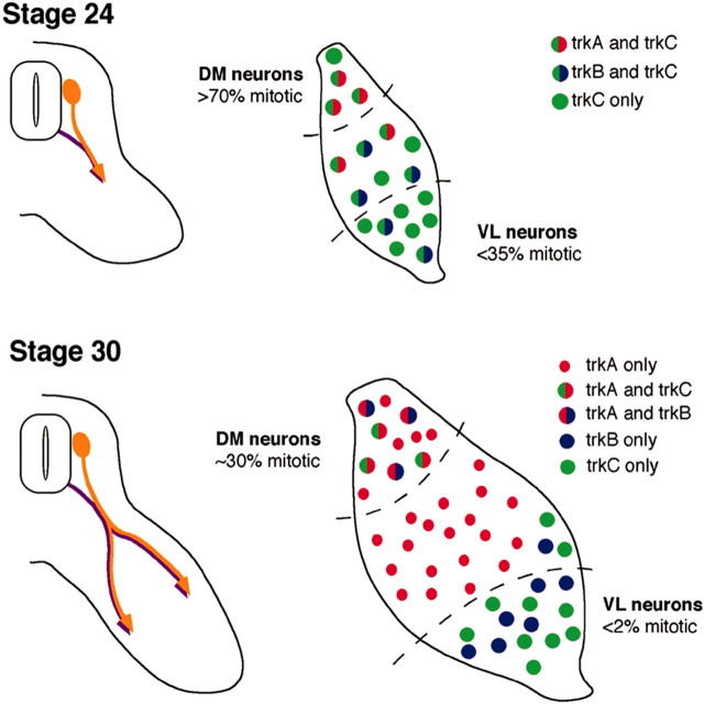
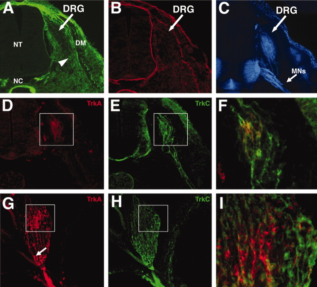
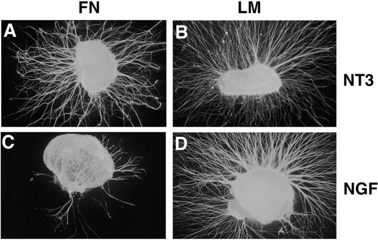
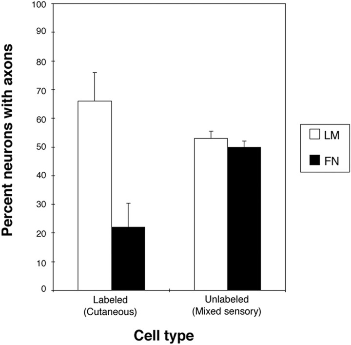
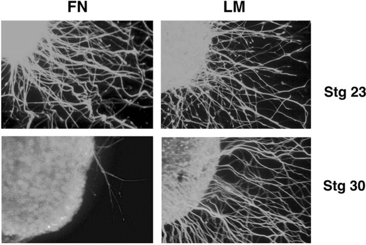
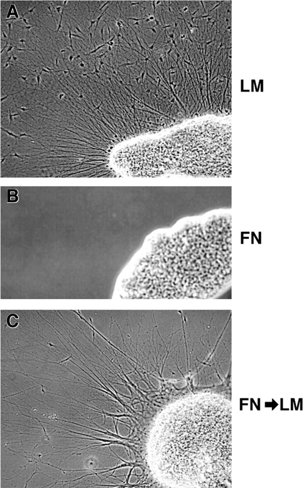
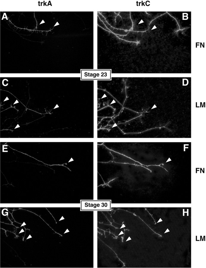
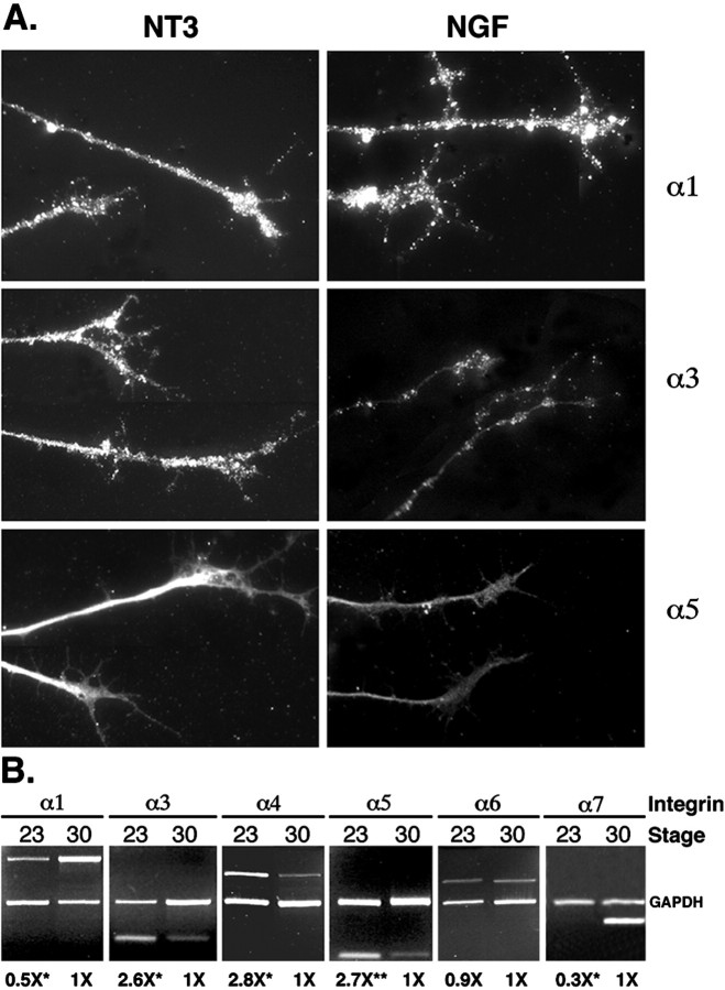
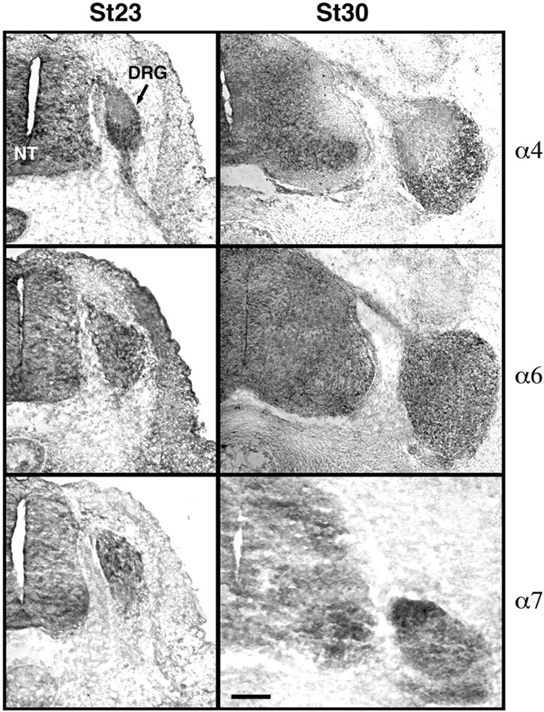
The factors controlling the specification and subsequent differentiation of sensory neurons are poorly understood. Data from embryological manipulations suggest that either sensory neuron fates are specified by the targets they encounter or sensory neurons are considerably more "plastic" with respect to specification than are neurons of the CNS. The prevailing view that sensory neurons are specified late in development is not consistent, however, with the directed outgrowth of sensory neurons to their targets and the characteristic spatial distribution of sensory neuron fates within the peripheral ganglia. To address when in development different classes of sensory neurons can first be distinguished, we investigated the interactions of early dorsal root ganglia neurons with the extracellular matrix before neurite outgrowth to targets. We found that subclasses of sensory neurons in early dorsal root ganglia show different patterns of neurite outgrowth and integrin expression that are predictive of their fates. In the absence of neurotrophins, presumptive proprioceptive neurons extend neurites robustly on both laminin and fibronectin, whereas presumptive cutaneous neurons show a strong preference for laminin. Cutaneous afferents that have innervated targets show a similar strong preference for laminin and show higher levels of integrin alpha7beta1 than do proprioceptive neurons. Finally, presumptive proprioceptive neurons express fibronectin receptors, integrin alpha3beta1, alpha4beta1, and alpha5beta1, at higher levels than do presumptive cutaneous neurons. Our results indicate that subtypes of sensory neurons have unique patterns of neurite outgrowth and receptor expression before target innervation.


Figures

Fig. 1.
Fig. 1.
A schematic diagram of DRG development at stages 23–24 and 30. Diagram summarizes sensory axon extension from the whole DRG (left) (Tosney and Landmesser, 1985a; Hollyday, 1995; Wang and Scott, 2000) and the patterns of mitosis (Carr and Simpson, 1978) and trk expression (Rifkin et al., 2000) in dorsomedial (DM) and ventrolateral (VL) thirds of the DRG (right). At stage 23–24, the neurons are still being born throughout the DRG, although the earliest born ventrolateral neurons have begun extending axons. These early cells are trkC+ and include most proprioceptive neurons as well as cutaneous neurons. By stage 30, ventrolateral cells have exited the cell cycle, and most axons from cells in this region have innervated targets and are neurotrophin dependent. In contrast, the latest born dorsomedial cells are still being generated. Newly born neurons that have not yet extended axons into the periphery are neurotrophin independent. All dorsomedial cells are fated to be cutaneous afferents.
Fig. 2.
Fig. 2.
Sensory neurons expressing trkC arise earlier in development than trkA-expressing neurons and extend axons through a fibronectin-rich environment. Cryostat sections of stage 22 (A–F) and stage 25 (G–I) lumbar DRGs. Dorsal is up. Medial isleft. A, At stage 22, FN was expressed at high levels along the pathways of early DRG axons (arrowheads). B, At stage 22, laminin was expressed mainly in the basement membranes of the neural tube (NT), the notochord (NC), the skin, and the dermamyotome (DM).C, Anti-DMI/GRASP indicates the position of sensory (arrow) and motor axons (MNs) at stage 22. D, At stage 22, trkA was expressed by only a few DRG neurons, and no trkA+ axons are observed. E, The same section shown in D, stained for trkC expression. trkC was expressed in all neurons and ventrally extending axons.F, Enlargement of boxed areas inD and E. trkA and trkC expression overlaid. Most trkA+ neurons at this stage also express trkC.G, Anti-trkA staining at stage 25. trkA+ cell bodies are predominantly located in the dorsomedial quadrant. Numerous trkA+ axons have extended axons that have fasciculated with earlier arising sensory and motor axons. The arrow indicates trkA+ axon bundles in the ventral aspect of the DRG. H, trkC continues to be expressed in ventrolateral neurons. I, Enlargement of boxed areas in G andH. trkA and trkC expression overlaid. Most trkA+ neurons do not express trkC at stage 25.
Fig. 3.
Fig. 3.
NGF- and NT3-responsive neurons show different neurite outgrowth on LM and FN substrata. DRGs from embryonic day 7 (approximately stage 30) were cultured 24 hr in the presence of NT3 (A, B) or NGF (C,D) on fibronectin (A, C) or laminin (B, D). A, NT3-responsive neurons extend neurites robustly on FN substrata.B, NT3-responsive neurons also extend neurites robustly on LM. C, In contrast to NT3-responsive neurons, NGF-responsive neurons are much less likely to extend neurites on FN.D, Extension of NGF-responsive neurites was robust on laminin. Data from at least nine independent experiments were examined. Number of DRGs extending axons and the total number examined in each condition are as follows: NT3–FN, 23 of 32 (72%); NT3–LM, 27 of 30 (90%); NGF–FN, 9 of 27 (33%); and NGF–LM, 29 of 29 (100%). Number of DRGs extending neurites in the NGF–FN condition is significantly different from all other conditions (p < 0.005; Fisher's exact test).
Fig. 4.
Fig. 4.
Retrogradely labeled cutaneous neurons that have innervated targets also show a strong preference for laminin. Quantification of neurite extension from labeled and unlabeled neurons. Unlabeled cells (mixed sensory modalities, including cutaneous) extend equally well on LM and FN substrata. Labeled, cutaneous neurons are three times as likely to extend on LM as on FN. Results from three experiments are presented. Numbers of cells counted are as follows: labeled on LM, 207; labeled on FN, 303; unlabeled on LM, 102; and unlabeled on FN, 102. Percentage of labeled neurons extending on LM and FN are statistically different from each other and from control (unlabeled) neurons (p < 0.001; χ2 test).
Fig. 5.
Fig. 5.
Substratum preferences persist in the absence of exogenous neurotrophins. DRGs from stage 23 and stage 30 embryos are cultured 24 hr in the absence of neurotrophins on either fibronectin or laminin. At stage 23 (E3.5–E4), the majority of the neurites extending are trkC+ (see Figs. 1, 2, 7, Table 3) and fated to be proprioceptive. At this stage, neurites grew robustly on both LM and FN in the absence of neurotrophins. At stage 30 (E6.5), most neurons undergoing initial axonogenesis are fated to be cutaneous. None of the DRGs cultured on FN at this age extended neurites long enough to pass through the limit of non-neuronal cells (see Materials and Methods). In contrast, DRGs cultured on LM robustly extended neurites in the absence of neurotrophins at stage 30. For all conditions, at least three independent experiments were examined. Number of DRGs extending axons out of the total number examined in each condition are as follows: stage 23–FN, 10 of 12 (83%); stage 23–LM, 9 of 10 (90%); stage 30–FN, 0 of 25 (0%); and stage 30–LM, 20 of 26 (77%). Number of DRGs extending axons on fibronectin at stage 30 is significantly different from all other conditions (p < 0.001; Fisher's exact test).
Fig. 6.
Fig. 6.
Sympathetic neurites extend preferentially on laminin. Lumbosacral sympathetic chain ganglia were dissected from stage 35/36 (E9) embryos and cultured on either laminin (A) or fibronectin (B) for 24 hr. Neurites extended only on laminin, regardless of neurotrophin treatment at the time of culture (Table 2). C, Failure to extend on fibronectin did not reflect loss of ability to extend axons; ganglia removed from fibronectin substrata after the initial 24 hr, transferred to laminin substrata, and cultured for an additional 24 hr extended axons robustly.
Fig. 7.
Fig. 7.
trkA+ neurites from stage 23 and all neurites from stage 30 prefer laminin. trkA and trkC double labeling of DRG neurons cultured in the absence of neurotrophins on FN (A,B, E, F) and LM (C, D, G,H). At stage 23 (A–D), most neurotrophin-independent neurites express trkC only. trkC+ neurites extend equally well on LM and FN. trkA-expressing neurites (arrowheads) are few and prefer LM over FN (see Table3). At stage 30 (E–H), there are more trkA+ neurites (arrowheads), and more neurites extend out on LM than on FN. Stage 30 neurites show a strong preference for LM (see Table 3). Arrowheads indicate neurites expressing both trkA and trkC.
Fig. 8.
Fig. 8.
Differences in integrin expression are seen between different classes of sensory neurons. A, Stage 30 neurons cultured in the presence of NGF or NT3 and stained for integrin α1β1, α3β1, and α5β1. NT3- and NGF-responsive neurons in culture express similar levels of the laminin receptor integrin α1. In contrast, NT3-responsive neurons express much higher levels of the fibronectin receptors integrin α3 and α5. DRGs stained for α1 and α3 were cultured on laminin, and those stained for α5β1 were cultured on fibronectin. Exposure for α3–NGF is twice as long as exposure for α3–NT3 attributable to very weak staining of NGF cultures. All other exposures are identical across the two conditions. B, PCR analysis of integrin mRNA levels from stage 23 and 30 cultured without neurotrophins. Integrins α1, α3, α4, α5, α6, and α7 are expressed at both stages. Integrins α3, α4, and α5 are expressed at higher levels in stage 23 DRGs, whereas α1 and α7 levels are significantly lower at this stage. Average relative levels (normalized to stage 30) of amplified integrin mRNA from at least three independent experiments are given below the lanes. The 95% confidence intervals for stage 23 ratios are as follows: α1, 0.7–0.4; α3, 3.4–2.0; α4, 4.5–1.7; α5, 3.2–2.0; α6, 0.9–0.8; and α7, 0.4–0.2. *p < 0.05 indicates significantly different from 1 (t test); **p < 0.01 indicates significantly different from 1 (t test).
Fig. 9.
Fig. 9.
In situ hybridization for integrins α4, α6, and α7 at stage 23 and stage 30. Integrin α4 message is localized to the ventral third of the DRG at both stages, whereas integrin α6 is more evenly distributed in cells scattered throughout the DRG. Integrin α7 is expressed in only a few cells at stage 23 and is localized predominantly dorsally by stage 30, consistent with the PCR analysis of mRNA expression (Table 4, Fig. 8). Dorsal isup, and medial to the left.NT, Neural tube. Scale bar, 100 μm.

Similar articles

Cited by

References

    1. Adams DH, Scott SA. Response of “naive” cutaneous and muscle afferents to potential targets in vitro. Dev Biol. 1998;203:210–220. - PubMed
    1. Anderson DJ. Lineages and transcription factors in the specification of vertebrate primary sensory neurons. Curr Opin Neurobiol. 1999;9:517–524. - PubMed
    1. Aumailley M, Rousselle P. Laminins of the dermo-epidermal junction. Matrix Biol. 1999;18:19–28. - PubMed
    1. Backstrom A, Soderstrom S, Kylberg A, Ebendal T. Molecular cloning of the chicken trkA and its expression in early peripheral ganglia. J Neurosci Res. 1996;46:67–81. - PubMed
    1. Baker CV, Bronner-Fraser M. Establishing neuronal identity in vertebrate neurogenic placodes. Development. 2000;127:3045–3056. - PubMed

Publication types

MeSH terms

LinkOut - more resources