. 2003 Feb;14(2):611-24.
doi: 10.1091/mbc.e02-08-0528.

Raft-mediated trafficking of apical resident proteins occurs in both direct and transcytotic pathways in polarized hepatic cells: role of distinct lipid microdomains

Affiliations

Raft-mediated trafficking of apical resident proteins occurs in both direct and transcytotic pathways in polarized hepatic cells: role of distinct lipid microdomains

Tounsia Aït Slimane et al. Mol Biol Cell. 2003 Feb.

Abstract

In polarized hepatic cells, pathways and molecular principles mediating the flow of resident apical bile canalicular proteins have not yet been resolved. Herein, we have investigated apical trafficking of a glycosylphosphatidylinositol-linked and two single transmembrane domain proteins on the one hand, and two polytopic proteins on the other in polarized HepG2 cells. We demonstrate that the former arrive at the bile canalicular membrane via the indirect transcytotic pathway, whereas the polytopic proteins reach the apical membrane directly, after Golgi exit. Most importantly, cholesterol-based lipid microdomains ("rafts") are operating in either pathway, and protein sorting into such domains occurs in the biosynthetic pathway, largely in the Golgi. Interestingly, rafts involved in the direct pathway are Lubrol WX insoluble but Triton X-100 soluble, whereas rafts in the indirect pathway are both Lubrol WX and Triton X-100 insoluble. Moreover, whereas cholesterol depletion alters raft-detergent insolubility in the indirect pathway without affecting apical sorting, protein missorting occurs in the direct pathway without affecting raft insolubility. The data implicate cholesterol as a traffic direction-determining parameter in the direct apical pathway. Furthermore, raft-cargo likely distinguishing single vs. multispanning membrane anchors, rather than rafts per se (co)determine the sorting pathway.


Figures

Figure 1
Figure 1
GPI-GFP is localized at the BC membrane of HepG2 cells, reached by the indirect pathway. (A) GPI-GFP–transfected cells were grown on coverslips, fixed, permeabilized, and stained with the anti-GFP antibody and secondary Cy5-conjugated antibody. Confocal microscopy shows that at steady state the GFP fluorescence preferentially localizes at the BC surface (a, asterisk), but is also detected at the BL membrane. Staining with anti-GFP antibody confirms this distribution (b). Nuclei (N) are stained with propidium iodide. (B) BL-to-AP transcytosis of GPI-GFP and DPPIV was monitored by an antibody-immunoassay. GPI-GFP–expressing cells were incubated for 30 min at 0°C with anti-GFP (a–d) or anti-DPPIV antibodies (a′–d′). Immediately (a and a′) or after 30 (b and b′), 60 (c and c′), or 180 min (d and d′) at 37°C, cells were fixed and permeabilized. Primary antibodies were visualized with secondary TRITC-(GFP)– or Cy3-(DPPIV)–conjugated antibodies. For each time point, a minimum of 50 BC structures was analyzed in three independent experiments. BCs are marked by asterisks; arrows (b, c, b′, and c′) point to GPI-GFP– and DPPIV-positive vesicles detected during internalization of both antibody-conjugated proteins. Bar, 10 μm.
Figure 2
Figure 2
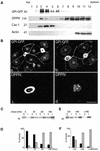
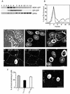
GPI-GFP and DPPIV are recruited in TX-100–insoluble microdomains. (A) GPI-GFP–expressing cells were lysed in TNE/TX-100 buffer at 4°C and run through a 5–40% sucrose gradient. For DPPIV analysis, HepG2 cells were 35S-labeled for 6 h, and after lysis fractionated on a similar gradient. Cav-1, stably expressed in HepG2 cells, was analyzed in parallel as a marker to indicate DRM fractions. Actin was used as a negative control. Fractions of 1 ml were collected from top to bottom after centrifugation to equilibrium. GPI-GFP, Cav-1, and actin were TCA precipitated, whereas labeled DPPIV was immunoprecipitated. (B) GPI-GFP–expressing cells were fixed and analyzed for the presence of GPI-GFP and DPPIV either directly (a and c) or after in situ extraction with 1% TX-100 (b and d). *, BC. Bar, 10 μm. (C) GPI-GFP–expressing cells were pulse chased with [35S]methionine for 10 min, followed by incubation in chase medium for the indicated times. After extraction in TNE/TX-100 buffer at 4°C, both the soluble (S; supernatant) and insoluble (I; pellet) fractions were collected by centrifugation, immunoprecipitated, and analyzed by SDS-PAGE. Quantification of three independent experiments is shown in D as mean ± SD. Black bars represent soluble protein and hatched bars represent insoluble protein. (E) Pulse chase analysis of GPI-GFP after a temperature block. After a short pulse (10 min), GPI-GFP–expressing cells were chased for 120 min at 20°C and then warmed to 37°C and chased for another 60 and 180 min. Quantification of three independent experiments is shown in F as mean ± SD. Black bars represent soluble protein and hatched bars represent insoluble protein.
Figure 3
Figure 3
MDR1-GFP is localized at the BC surface of HepG2 cells. (A) MDR1-GFP–transfected cells were biosynthetically labeled for 10 min with [35S]methionine and chased for the indicated times. Samples were immunoprecipitated with pAb against GFP and analyzed on SDS-PAGE. In B and C, transfected HepG2 cells were directly visualized for GFP fluorescence localization (a and a′), or examined after staining with either anti-GFP (B, b) or anti-MDR1 (C, b′). Nuclei (N) stained with propidium iodide. *, BC. Bar, 10 μm.
Figure 4
Figure 4
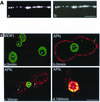
Direct transport of MDR1-GFP in HepG2 cells as detected by mAb 4E3. (A) MDCK cells stably transfected with MDR1-GFP were grown on Transwell filters, fixed, and visualized directly (a) or after staining with mAb 4E3 added to both the AP and BL sides (b). The XZ sections show that MDR1-GFP is restricted to the AP surface of MDCK cells. (B) BL-to-AP-transcytosis of MDR1-GFP and APN in HepG2 cells. MDR1-GFP–expressing cells were incubated for 30 min at 0°C with mAb 4E3 (a) or anti-APN antibody (b–d). After washing, cells were either fixed and permeabilized immediately (a and b) or warmed to 37°C for 30 min (c) and 180 min (d), and stained with secondary Cy3- (MDR1) or TRITC-(APN)-conjugated antibodies. *, BC, arrows (c) point to APN-positive vesicles detected during internalization of the antibody-conjugated complex. Bar, 10 μm.
Figure 5
Figure 5
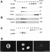
MDR1-GFP localizes to LubWX-insoluble microdomains and is solubilized in TX-100. (A) MDR1-GFP–expressing HepG2 cells were lysed either in TNE/TX-100 buffer or TNE/LubWX buffer at 4°C and analyzed by flotation on sucrose gradient. For details, see legend to Figure 2. Cav-1 and actin were analyzed in parallel as positive and negative controls, respectively. (B) Insolubility of MDR1-GFP in LubWX is not due to interaction with cytoskeletal elements. MDR1-GFP–expressing cells were lysed in 1% LubWX at 4°C in the presence of 250 mM (NH4)2SO4 [(+NH4)2SO4] or incubated for 1 h at 37°C in the presence of 10 μg/ml cytochalasin D (+CytD) followed by lysis in 1% LubWX at 4°C. (C) A similar analysis as in A was carried out in MDR1-GFP–transfected MDCK cells. (D) Analysis of MDR1-GFP association with lipid microdomains after in situ extraction with detergents. MDR1-GFP–expressing cells were either fixed directly (a) or extracted with 1% LubWX (b) or 1% TX 100 (c) before fixation.
Figure 6
Figure 6
Kinetics of MDR1-GFP in LubWX-resistant domains revealed by pulse-chase analysis. MDR1-GFP–expressing cells were 35S-labeled for 10 min, followed by incubation in chase medium for the indicated times. Cells were extracted either in TNE/LubWX buffer (A) or in TNE/TX-100 buffer (C) at 4°C, and processed as in Figure 2C. In B, quantification of three independent experiments, after extraction with LubWX, is shown as mean ± SD. Black bars represent soluble protein and hatched bars represent insoluble protein.
Figure 7
Figure 7
ATP7B is soluble in TX-100 but insoluble in LubWX. (A) Endogenous ATP7B, present in HepG2 cells, was analyzed for lipid microdomain association on sucrose density gradients after lysis of the cells either in TNE/TX-100 or TNE/LubWX at 4°C, in the absence of copper or after treatment for 4 h with 20 μM CuSO4 (+Cu2+). (B) Flotation of GPI-GFP and DPPIV in sucrose gradients after extraction with LubWX.
Figure 8
Figure 8
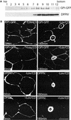
Cholesterol removal renders GPI-GFP and DPPIV less TX-100 insoluble without affecting their AP targeting. (A) GPI-GFP–expressing cells were grown in the presence of Lov/CD as described under MATERIALS AND METHODS. Cells were then lysed in TNE/TX-100 buffer and analyzed by flotation on sucrose gradient. (B) GPI-GFP–expressing cells were grown in the absence (−Lov/CD) or presence (+Lov/CD) of Lov/CD. The dynamics of GPI-GFP and DPPIV was investigated using the trafficking antibody assay as in Figure 1B. *, BC. Bar, 10 μm.
Figure 9
Figure 9
Cholesterol depletion causes missorting of MDR1-GFP without affecting its insolubility in LubWX. (A) MDR1-GFP– and GPI-GFP–transfected cells were treated as in Figure 8, and then lysed in TNE/LubWX buffer and processed as in Figure 8A. (B) HepG 2 cells were incubated with [3H]cholesterol for 24 h at 37°C and subsequently washed and solubilized with either TX-100 or LubWX buffer. The lysates were then analyzed by sucrose gradient ultracentrifugation. Gradient fractions were collected from the top of the tube and [3H]cholesterol was measured by liquid scintillation counting. The results indicate the percentage of total [3H]cholesterol determined in each fraction. ○, LubWX; ●, TX-100. (C) MDR1-GFP–expressing cells were treated with (+Lov/CD) or without (−Lov/CD) Lov/CD, fixed, permeabilized, and examined by confocal microscopy. A phase contrast image of Lov/CD-treated cells is shown at the top, left. (D) MDR1-GFP–expressing cells treated with Lov/CD were incubated for 30 min at 0°C with SM-BODIPY and subsequently analyzed by confocal microscopy. (E) MDR1-GFP–expressing cells were incubated in the presence of cycloheximide for 60 min and subsequently treated with Lov/CD in the continuous presence of cycloheximide. After fixation and permeabilization, the cells were examined by confocal microscopy. Nuclei (N) are stained with propidium iodide. (F) The cholesterol concentration of control, cholesterol-depleted, and cholesterol-repleted cells was determined (see MATERIALS AND METHODS for details). Data were obtained from three independent experiments. (G) Cholesterol-depleted cells were grown in the presence of CD/Chol complexes as described under MATERIALS AND METHODS and subsequently examined for the localization of MDR1-GFP. *, BC. Bar, 10 μm.

Similar articles

Cited by

References

    1. Bartles JR, Feracci HM, Stieger B, Hubbard AL. Biogenesis of the rat hepatocyte plasma membrane in vivo: comparison of the pathways taken by apical and basolateral proteins using subcellular fractionation. J Cell Biol. 1987;105:1241–1251. - PMC - PubMed
    1. Bastaki M, Braiterman LT, Johns DC, Chen YH, Hubbard AL. Absence of direct delivery for single transmembrane apical proteins or their “secretory” forms in polarized hepatic cells. Mol Biol Cell. 2002;13:225–237. - PMC - PubMed
    1. Brown DA, Crise B, Rose JK. Mechanism of membrane anchoring affects polarized expression of two proteins in MDCK cells. Science. 1989;245:1499–1501. - PubMed
    1. Brown DA, Rose JK. Sorting of GPI-anchored proteins to glycolipid-enriched membrane subdomains during transport to the apical cell surface. Cell. 1992;68:533–544. - PubMed
    1. Bull PC, Thomas GR, Rommens JM, Forbes JR, Cox DW. The Wilson disease gene is a putative copper transporting P-type ATPase similar to the Menkes gene. Nat Genet. 1993;5:327–337. - PubMed

MeSH terms

LinkOut - more resources