. 2003 Mar 15;547(Pt 3):761-74.
doi: 10.1113/jphysiol.2002.035683. Epub 2003 Jan 24.

Ryanodine stores and calcium regulation in the inner segments of salamander rods and cones

Affiliations

Ryanodine stores and calcium regulation in the inner segments of salamander rods and cones

David Krizaj et al. J Physiol. .

Abstract

Despite the prominent role played by intracellular Ca2+ stores in the regulation of neuronal Ca2+ homeostasis and in invertebrate photoreception, little is known about their contribution to the control of free Ca2+ concentration ([Ca2+]i) in the inner segments of vertebrate photoreceptors. Previously, caffeine-sensitive intracellular Ca2+ stores were shown to play a role in regulating glutamate release from photoreceptors. To understand the properties of these intracellular stores better we used pharmacological approaches that alter the dynamics of storage and release of Ca2+ from intracellular compartments. Caffeine evoked readily discernible changes in [Ca2+]i in the inner segments of rods, but not cones. Caffeine-evoked Ca2+ responses in cone inner segments were unmasked in the presence of inhibitors of the plasma membrane Ca2+ ATPases (PMCAs) and mitochondrial Ca2+ sequestration. Caffeine-evoked responses were blocked by ryanodine, a selective blocker of Ca2+ release and by cyclopiazonic acid, a blocker of Ca2+ sequestration into the endoplasmic reticulum. These two inhibitors also substantially reduced the amplitude of depolarization-evoked [Ca2+]i increases, providing evidence for Ca2+-induced Ca2+ release (CICR) in rods and cones. The magnitude and kinetics of caffeine-evoked Ca2+ elevation depended on the basal [Ca2+]i, PMCA activity and on mitochondrial function. These results reveal an intimate interaction between the endoplasmic reticulum, voltage-gated Ca2+ channels, PMCAs and mitochondrial Ca2+ stores in photoreceptor inner segments, and suggest a role for CICR in the regulation of synaptic transmission.


Figures
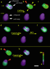
Figure 1
Figure 1. Caffeine evokes an increase in free [Ca2+]i in the inner segment (IS) of rods but not cones
a, simultaneous [Ca2+] measurement from salamander rod and cone ISs loaded with the Ca2+ indicator fura-2. Note that due to limited loading of the fura-2 ester into the small outer segment (OS) cytoplasmic volume, the OS Ca2+ signal is not detected at the 340/380 nm excitation used in this particular experiment. The cell body and the ellipsoid of the rod and cone ISs, respectively, are marked with white arrows. b, 10 mm caffeine evoked an increase in [Ca2+]i in the rod IS but not the cone IS. The [Ca2+]i increase was prominent in the cell body (yellow arrowheads) and modest in the ellipsoid (red arrowhead). e, caffeine washout. f, subsequent exposure to 20 mm KCl raised [Ca2+]i in both cells. The increase was again less pronounced in the ellipsoid regions of both cells (red arrowheads in g). h, [Ca2+]i returned to baseline levels 2 min following KCL washout. Scale bar = 20 μm.
Figure 2
Figure 2. Time course of caffeine effects on intracellular free [Ca2+] in rod and cone ISs
A, simultaneous measurement of [Ca2+] from salamander rod and cone ISs using a CCD camera. Cells were loaded with fura-2 AM and stimulated with brief puffs of 90 mm KCl from a nearby pipette (indicated by the asterisks) followed by superfusion with 10 mm caffeine (indicated by the horizontal bar). Caffeine evoked a transient [Ca2+]i increase in the rod, but not cone, IS. B, simultaneous line-scan measurement of Ca2+ signals from salamander rod and cone ISs loaded with fluo-4 AM. Caffeine (10 mm) evoked a large fluorescence increase in the rod IS but did not change the fluorescence in the cone. C, caffeine triggered an increase in [Ca2+]i in a subset of cone ISs. Simultaneous recording of fluo-4 fluorescence from rod and cone IS. Caffeine (10 mm) triggered a relatively rapid large-amplitude increase in the rod Ca2+ signal. Concomitantly, a small Ca2+ signal with a slow rise time was observed in the cone IS. a.u. = arbitrary units.
Figure 3
Figure 3. Simultaneous [Ca2+]i measurement from an IS and OS of a rod
A, rod loaded with fura-2 AM. The cell was depolarized by 20 mm KCl to maximize store loading. Caffeine (10 mm) transiently elevated [Ca2+]i in the IS, but not the OS. B, confocal image of a dissociated rod photoreceptor loaded with 5 μm fluo-4. Prominent fluo-4 fluorescence is observed in the IS of the cell, whereas the OS signal is weak. C, line-scan across the IS (top) and the OS (bottom) of the cell shown in B. The abscissa represents the time axis. Exposure to 10 mm caffeine caused a large saturating increase in the fluo-4 signal in the IS but caused no fluorescence change in the OS. Note that the caffeine-evoked Ca2+ signal is confined to the IS.
Figure 4
Figure 4. Plasma membrane Ca2+ ATPases (PMCAs) shape the decay of caffeine-evoked [Ca2+]i transients
A rod IS was superfused continually with 1 mm La3+, a blocker of PMCAs, and then stimulated with steps of 10 mm caffeine. In the presence of La3+, caffeine evoked sustained [Ca2+]i elevations. Following caffeine removal, [Ca2+]i returned to baseline in the continued presence of La3+.
Figure 5
Figure 5. Inhibition of PMCAs unmasks a caffeine-evoked [Ca2+]i response in the cone IS
A cone IS that was stimulated with caffeine in control saline showed no [Ca2+]i response. Subsequently, putative caffeine-sensitive stores were replenished with a step of 20 mm KCl and the solution was switched to 1 mm La3+. In the presence of La3+, caffeine evoked a sustained increase in [Ca2+]i.
Figure 6
Figure 6. Ryanodine receptors (RyRs) are localized to photoreceptor ISs
Confocal fluorescence images of retinal cells immunostained with antisera against RyRs and SV2. A and E, Nomarski images of rod and double cone ISs dissociated from the salamander retina. B and F, RyR immunofluorescence is prominent in the synaptic terminal and the ellipsoid region. A moderate RyR signal is also observed in the plasma membrane surrounding the perikaryon. C, G and K, SV2 immunolabels synaptic terminals of retinal neurons. D and H, RyR and SV2 signals colocalize in the synaptic terminals of the rod and the cone. I, Nomarski image from a salamander retinal section. J, the RyR antibody labels photoreceptor perikarya and synaptic terminals. A prominent signal is also observed in Müller cell bodies and processes (arrowheads), in ganglion cell bodies and in synaptic processes in the inner plexiform layer (IPL). Little staining is seen in photoreceptor OSs and in bipolar cell bodies. L, RyR and SV2 colocalize in synaptic processes of the outer plexiform layer (OPL) and the IPL. ONL, outer nuclear layer; INL, inner nuclear layer; GCL, ganglion cell layer. Scale bars are 10 μm in AH and 50 μm in IL.
Figure 7
Figure 7. Ca2+ is sequestered into intracellular stores by sarcoplasmic-endoplasmic Ca2+ ATPases
Simultaneous recordings from two rod ISs. A puffer pipette containing 50 mm caffeine was positioned close to rod 1; rod 2 was located at the opposite side of the coverslip. Brief puffs of caffeine evoked transient increases in [Ca2+]i in rod 1, but had no effect on rod 2. In both cells, cyclopiazonic acid (CPA) by itself evoked a slow elevation in [Ca2+]i by several tens of nanomoles.
Figure 8
Figure 8. The magnitude of caffeine-evoked Ca2+ release depends on the magnitude of the conditioning depolarization
A rod photoreceptor IS exposed to conditioning steps of high K+ followed by superfusion with 10 mm caffeine. Each asterisk represents a single 128 ms KCl puff; bars denote superfusion. Increasing the duration of the conditioning [Ca2+]i step caused a subsequent increase in the caffeine-evoked [Ca2+]i increase. The caffeine-evoked responses saturated when the conditioning free [Ca2+]i reached ∼500 nm.
Figure 9
Figure 9. Ca2+-induced Ca2+ release contributes to depolarization-evoked [Ca2+]i increases in both rod and cone ISs
A, a rod IS in which [Ca2+]i was raised periodically with 128 ms puffs of 90 mm KCl. Superfusion with 10 mm caffeine itself raised [Ca2+]i by ∼100 nm. However, the magnitude of the subsequent KCl puff was substantially reduced in the presence of caffeine. B, a rod IS in which exposure to CPA slightly elevated the [Ca2+]i baseline and significantly reduced the magnitude of KCl-evoked [Ca2+]i transients. Following recovery, caffeine itself evoked a normal [Ca2+]i response. C, a rod IS in which 20 μm ryanodine irreversibly decreased the magnitude of KCl-evoked [Ca2+]i transients. D, a cone IS in which CPA caused a transient elevation of [Ca2+]i and reversibly reduced the magnitude of KCl-evoked transients. Note that whereas KCl-evoked responses recovered following CPA washout, subsequent superfusion with 10 mm caffeine did not result in elevation of [Ca2+]i.
Figure 11
Figure 11. Ryanodine-sensitive Ca2+ stores communicate with the IS mitochondria in rod and cone ISs
A, a rod IS loaded with fura-2 AM. The rod was depolarized by 20 mm KCl throughout the experiment. Caffeine (10 mm) evoked a transient [Ca2+]i increase followed by an undershoot. p-Trifluoromethoxy-phenyl hydrazone (FCCP, 1 μm) itself caused an increase in [Ca2+]i and a fivefold potentiation in the magnitude of the caffeine-evoked [Ca2+]. B, line-scan recording from a cone IS loaded with fluo-4. No response to caffeine was observed in control saline. FCCP (2 μm) by itself caused a large increase in the Ca2+ signal in the cone IS. In the presence of FCCP, caffeine evoked a significant elevation in [Ca2+]i.
Figure 10
Figure 10. Photoreceptors possess multiple types of internal Ca2+ stores
Ryanodine stores were depleted via a prolonged exposure to caffeine in 0 Ca2+ supplemented with 3 mm EGTA. Subsequent exposure to ionomycin caused an additional elevation of Ca2+ by ∼800 nm, consistent with the presence of caffeine-insensitive stores in the IS.

Similar articles

Cited by

References

    1. Akaike N, Sadoshima J. Caffeine affects four different ionic currents in the bull-frog sympathetic neurone. J Physiol. 1989;412:221–244. - PMC - PubMed
    1. Babcock DF, Herrington J, Goodwin PC, Park YB, Hille B. Mitochondrial participation in the intracellular Ca2+ network. J Cell Biol. 1997;136:833–844. - PMC - PubMed
    1. Baumann O, Walz B. Endoplasmic reticulum of animal cells and its organization into structural and functional domains. Int Rev Cytol. 2001;205:149–214. - PubMed
    1. Berger ER. Subsurface membranes in paired cone photoreceptor inner segments of adult and neonatal Lebistes retinae. J Ultrastruct Res. 1967;17:220–232. - PubMed
    1. Berridge MJ, Lipp P, Bootman MD. The versatility and universality of calcium signalling. Nat Rev Mol Cell Biol. 2000;1:11–21. - PubMed

Publication types

MeSH terms

LinkOut - more resources