. 2003 Feb;185(4):1299-315.
doi: 10.1128/JB.185.4.1299-1315.2003.

Genetic and biochemical analysis of phosphatase activity of Escherichia coli NRII (NtrB) and its regulation by the PII signal transduction protein

Affiliations

Genetic and biochemical analysis of phosphatase activity of Escherichia coli NRII (NtrB) and its regulation by the PII signal transduction protein

Augen A Pioszak et al. J Bacteriol. 2003 Feb.

Abstract

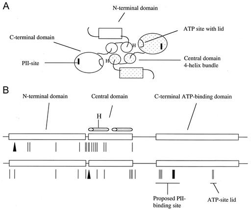
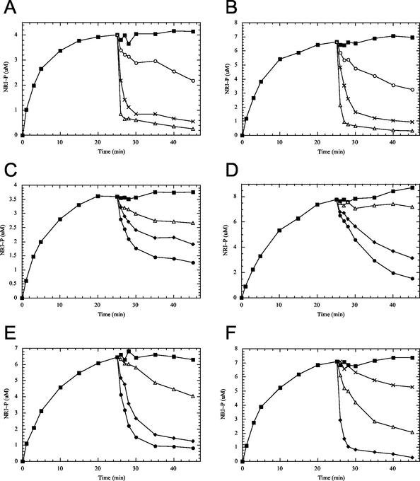
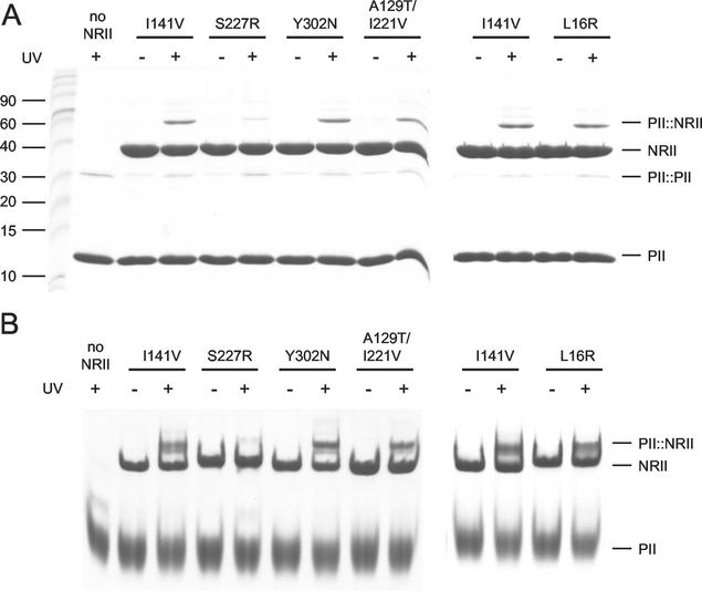
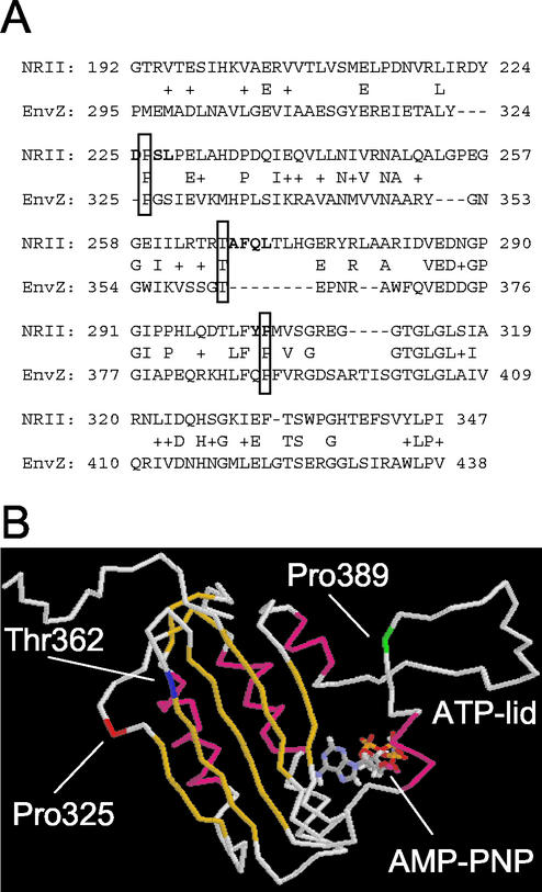
Mutant forms of Escherichia coli NRII (NtrB) were isolated that retained wild-type NRII kinase activity but were defective in the PII-activated phosphatase activity of NRII. Mutant strains were selected as mimicking the phenotype of a strain (strain BK) that lacks both of the related PII and GlnK signal transduction proteins and thus has no mechanism for activation of the NRII phosphatase activity. The selection and screening procedure resulted in the isolation of numerous mutants that phenotypically resembled strain BK to various extents. Mutations mapped to the glnL (ntrB) gene encoding NRII and were obtained in all three domains of NRII. Two distinct regions of the C-terminal, ATP-binding domain were identified by clusters of mutations. One cluster, including the Y302N mutation, altered a lid that sits over the ATP-binding site of NRII. The other cluster, including the S227R mutation, defined a small surface on the "back" or opposite side of this domain. The S227R and Y302N proteins were purified, along with the A129T (NRII2302) protein, which has reduced phosphatase activity due to a mutation in the central domain of NRII, and the L16R protein, which has a mutation in the N-terminal domain of NRII. The S227R, Y302N, and L16R proteins were specifically defective in the PII-activated phosphatase activity of NRII. Wild-type NRII, Y302N, A129T, and L16R proteins bound to PII, while the S227R protein was defective in binding PII. This suggests that the PII-binding site maps to the "back" of the C-terminal domain and that mutation of the ATP-lid, central domain, and N-terminal domain altered functions necessary for the phosphatase activity after PII binding.


Figures

FIG. 1.
FIG. 1.
Mutations affecting the phosphatase activity of NRII map to all domains of the protein. (A) Model for the domain organization of the NRII dimer based on structural information available for other two-component system transmitter proteins and biochemical studies of NRII. Each NRII subunit is composed of three domains: an unconserved N-terminal domain involved in intramolecular signal transduction, and central and C-terminal domains that together compose the conserved transmitter module. The central domain is involved in dimerization and probably forms half of a four-helix bundle containing His139, the site of autophosphorylation. The C-terminal domain is the ATP-binding kinase domain that directly interacts with PII. The dimer is shown viewed down the four-helix bundle. (B) Linear depiction of the domain structure of the 349-amino-acid NRII showing the distribution of mutations affecting the phosphatase activity. The top drawing shows the mutations obtained in the 1992 study (5), while the bottom drawing shows the mutations obtained in this study. The lines indicate amino acid substitutions, the bar indicates a deletion, and the triangles indicate insertions. The figure is roughly to scale.
FIG. 2.
FIG. 2.
Growth phenotypes of glnL* alleles. Strains containing representative glnL* alleles in a wild-type or nac background were streaked for single colonies on glucose-ammonia-glutamine-tryptophan minimal medium. Growth was carried out for 42 h at 37°C. The strains were as follows, with relevant genotypes in brackets: 1, YMC10φ [wild type]; 2, BKgφ [ΔglnBΩGmrΔglnK1]; 3, L*(L16R)φ [glnL (L16R)]; 4, L*(L154R)φ [glnL (L154R)]; 5, L*(S227R)φ [glnL (S227R)]; 6, L*(Y302N)φ [glnL (Y302N)]; 7, Nφ [nac::Camr]; 8, BKgNφ [ΔglnBΩGmrΔglnK1 nac::Camr]; 9, L*(L16R)Nφ [glnL (L16R) nac::Camr]; 10, L*(L154R)Nφ [glnL (L154R) nac::Camr]; 11, L*(S227R)Nφ [glnL (S227R) nac::Camr]; 12, L*(Y302N)Nφ [glnL (Y302N) nac::Camr].
FIG. 3.
FIG. 3.
Gel analysis of the autophosphorylation activities of NRII mutants. (A) Nondenaturing polyacrylamide gel electrophoresis of autophosphorylation reaction mixtures. NRII (2 μM) or mutant forms of NRII were incubated on ice for 20 min in reaction mixtures containing 0.5 mM ATP or as indicated, and autophosphorylation reactions were performed as described in Materials and Methods. Reactions were stopped by addition of 50 mM EDTA, and the mixtures were analyzed on 10% nondenaturing polyacrylamide gels stained with Coomassie brilliant blue R-250. The panel shows results from three separate gels. (B) Urea-polyacrylamide gel electrophoresis of autophosphorylation reactions. The indicated NRII proteins (2 μM) were incubated on ice for 30 min in reaction mixtures containing 0, 0.02, 0.1, 0.5, or 2 mM ATP (left to right), as described in Materials and Methods. Reactions were stopped by addition of 4 M urea, and the mixtures were analyzed on 10% polyacrylamide-6 M urea gels stained with Coomassie brilliant blue R-250. The panel shows results from three separate gels.
FIG. 4.
FIG. 4.
Effect of PII on the autophosphorylation activities of NRII mutants. NRII (2 μM) or mutant forms of NRII were incubated on ice in reaction mixtures containing 50 mM Tris-HCl (pH 7.5), 10 mM MgCl2, 100 mM KCl, 0.5 mg of BSA per ml, 50 μM 2-ketoglutarate, and 0.5 mM [γ-32P]ATP in the absence (▪) or presence (○) of 12 μM PII. At various times, aliquots were spotted onto nitrocellulose filters and analyzed as described in Materials and Methods. (A) NRII; (B) NRII (I141V); (C) NRII (S227R); (D) NRII (Y302N); (E) NRII (L16R); (F) NRII (A129T).
FIG. 5.
FIG. 5.
Kinase and phosphatase activities of NRII mutants. Reaction mixtures containing 30 μM NRI-N, 0.3 μM NRII (or mutant NRII), 50 μM 2-ketoglutarate, and 0.5 mM [γ-32P]ATP were incubated at 25°C. After 25 min, the reaction mixtures were split into tubes containing buffer (▪), 0.075 μM PII (○), 0.15 μM PII (×), 0.30 μM PII (▵), 1.0 μM PII (⧫), or 3.0 μM PII (•). At the indicated times, aliquots were spotted onto nitrocellulose filters and analyzed as described in Materials and Methods. (A) NRII; (B) NRII (I141V); (C) NRII (S227R); (D) NRII (Y302N); (E) NRII (L16R); (F) NRII (A129T).
FIG. 6.
FIG. 6.
Cross-linking of PII to NRII mutants. PII (E44C/C73S)-TFPAM-3 (9 μM) was incubated with the indicated NRII protein (4.5 μM) in reaction mixtures containing 50 μM 2-ketoglutarate and 0.5 mM ATP as described in Materials and Methods. Cross-linking was initiated by exposure to UV light for 20 min as indicated. (A) Reactions were stopped by addition of SDS gel-loading buffer, and the mixtures were analyzed on SDS-15% polyacrylamide gels. Molecular mass markers are indicated in kilodaltons. (B) Alternatively, reactions were stopped by addition of 120 mM glycine (pH 9.0) and the mixtures were analyzed on 10% nondenaturing polyacrylamide gels. For both panels, the gels were stained with Coomassie brilliant blue R-250.
FIG. 7.
FIG. 7.
Gel filtration assay for PII binding to NRII mutants. PII (48 μM) and the indicated NRII protein (12 μM) were mixed on ice in reaction mixtures containing 1 mM MgCl2, 50 μM free 2-ketoglutarate, and 0.5 mM free ATP, as indicated in Materials and Methods. The bulk of the reaction mixture was loaded onto a Sephadex G-100 column equilibrated in buffer containing 1 mM MgCl2, 50 μM 2-ketoglutarate, and 0.5 mM ATP and eluted at 4°C. Fractions were collected and analyzed on SDS-15% polyacrylamide gels stained with Coomassie brilliant blue R-250. Fraction numbers are indicated above the lanes, and IN indicates the column input. (A) NRII (I141V); (B) NRII (S227R); (C) NRII (Y302N); (D) NRII (A129T/I221V); (E) NRII (L16R).
FIG. 8.
FIG. 8.
Structure of the kinase domain of EnvZ modeled to show the approximate positions of the mutations obtained in NRII. (A) Amino acid sequence alignment of the kinase domains of NRII and EnvZ. The alignment was generated using a basic BLAST search of the E. coli genome with full-length NRII (accession number AAC76866) as the query (1). The residues shown in bold indicate the positions of the mutations obtained in the kinase domain of NRII. Shown boxed are conserved residues that were used as “anchor” points to represent positions of mutations obtained in NRII. Pro226 (NRII)/Pro325 (EnvZ) represents the cluster of mutations obtained at positions 225 to 228, Thr266 (NRII)/Thr362 (EnvZ) represents the deletion of residues 267 to 270, and Pro303 (NRII)/Pro389 (EnvZ) represents the cluster of mutations obtained at positions 302 to 303. (B) Nuclear magnetic resonance imaging structure of the kinase domain of EnvZ (52) shown as an alpha-carbon trace. Alpha-helices are shown in magenta, and beta-strands are shown in yellow. The AMP-PNP molecule is shown in stick representation with CPK coloring. The positions of Pro325 (representing the 225-to-228 cluster), Thr362 (representing the deletion), and Pro389 (representing the 302-to-303 cluster) are shown in red, blue, and green, respectively. The figure was generated using RasMol.

Similar articles

Cited by

References

    1. Altschul, S. F., T. L. Madden, A. A. Schaffer, J. Zhang, Z. Zhang, W. Miller, and D. J. Lipman. 1997. Gapped BLAST and PSI-BLAST: a new generation of protein database search programs. Nucleic Acids Res. 25:3389-3402. - PMC - PubMed
    1. Atkinson, M. R., T. A. Blauwkamp, and A. J. Ninfa. 2002. Context-dependent functions of the PII and GlnK signal transduction proteins in Escherichia coli. J. Bacteriol. 184:5364-5375. - PMC - PubMed
    1. Atkinson, M. R., T. A. Blauwkamp, V. Bondarenko, V. Studitsky, and A. J. Ninfa. 2002. Activation of the glnA, glnK, and nac promoters as Escherichia coli undergoes the transition from nitrogen-excess growth to nitrogen starvation. J. Bacteriol. 184:5358-5363. - PMC - PubMed
    1. Atkinson, M. R., E. S. Kamberov, R. L. Weiss, and A. J. Ninfa. 1994. Reversible uridylylation of the Escherichia coli PII signal transduction protein regulates its ability to stimulate the dephosphorylation of the transcription factor nitrogen regulator I (NRI or NtrC). J. Biol. Chem. 269:28288-28293. - PubMed
    1. Atkinson, M. R., and A. J. Ninfa. 1992. Characterization of mutations in the glnL gene of Escherichia coli affecting nitrogen regulation. J. Bacteriol. 174:4538-4548. - PMC - PubMed

Publication types

MeSH terms

Substances

LinkOut - more resources