. 2003 Feb 3;160(3):341-53.
doi: 10.1083/jcb.200211048. Epub 2003 Jan 27.

Rae1 is an essential mitotic checkpoint regulator that cooperates with Bub3 to prevent chromosome missegregation

Affiliations

Rae1 is an essential mitotic checkpoint regulator that cooperates with Bub3 to prevent chromosome missegregation

J Ramesh Babu et al. J Cell Biol. .

Abstract

The WD-repeat proteins Rae1 and Bub3 show extensive sequence homology, indicative of functional similarity. However, previous studies have suggested that Rae1 is involved in the mRNA export pathway and Bub3 in the mitotic checkpoint. To determine the in vivo roles of Rae1 and Bub3 in mammals, we generated knockout mice that have these genes deleted individually or in combination. Here we show that haplo-insufficiency of either Rae1 or Bub3 results in a similar phenotype involving mitotic checkpoint defects and chromosome missegregation. We also show that overexpression of Rae1 can correct for Rae1 haplo-insufficiency and, surprisingly, Bub3 haplo-insufficiency. Rae1-null and Bub3-null mice are embryonic lethal, although cells from these mice did not have a detectable defect in nuclear export of mRNA. Unlike null mice, compound haplo-insufficient Rae1/Bub3 mice are viable. However, cells from these mice exhibit much greater rates of premature sister chromatid separation and chromosome missegregation than single haplo-insufficient cells. Finally, we show that mice with mitotic checkpoint defects are more susceptible to dimethylbenzanthrene-induced tumorigenesis than wild-type mice. Thus, our data demonstrate a novel function for Rae1 and characterize Rae1 and Bub3 as related proteins with essential, overlapping, and cooperating roles in the mitotic checkpoint.


Figures
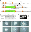
Figure 1.
Figure 1.
Targeted disruption of the mouse Rae1 gene. (A) Indicated are part of the endogenous mouse (m)Rae1 gene (top), the targeting vector (middle), and the disrupted Rae1 allele (bottom). BamHI restriction sites and the 3′ DNA probe (solid bar) used for Southern blot identification of wild-type (WT) and knockout (KO) Rae1 alleles are shown. (B) Southern blot of genomic ES cell DNA, digested with BamH1 and hybridized to a 3′ external probe, revealing the expected 10.2-kb wild-type and 8.5-kb mutant fragments. (C) PCR genotyping of E3.5 embryos from a Rae1+/− intercross showing wild-type and knockout allele-specific amplification products of respectively 516 and 650 bp. (D) In vitro growth of representative Rae1+/+ and Rae1−/− embryos. Embryos from heterozygous crosses were recovered at E3.5 and grown on multi-well slides in DME/15% FCS. They were inspected daily and photographed at 48 (E5.5), 72 (E6.5), and 96 h (E7.5) after seeding. I, ICM; T, trophectoderm cells.
Figure 2.
Figure 2.
Rae1 is not essential for nuclear export of mRNA. (A) Overview of the experimental design. Blastocysts from intercrosses of Rae1+/− mice were cultured for ∼4–5 d and then analyzed by immunostaining or in situ hybridization. (B–C') Double staining of E8.5 embryonic outgrowths with a polyclonal antibody against mouse Rae1(188–368) (Pritchard et al., 1999) and monoclonal antibody mAb414, a marker of the NPC (Wu et al., 2001). Shown are representative high-resolution images of trophectoderm cells. (D and E) Immunostaining of E8.5 embryonic outgrowths with a polyclonal antibody against Nup98(151–224) (Wu et al., 2001). Shown are high-resolution images of trophectoderm cells. (F and G) Localization of poly(A)+ RNA in trophectoderm cells from E8.5 Rae1+/+ and Rae1−/− outgrowths. A FITC–oligo(dT)50 probe was used for visualization of poly(A)+ by in situ hybridization. (H and I) Trophoblast cells stained with a polyclonal antibody against human Tap (Braun et al., 1999).
Figure 3.
Figure 3.
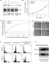
The mitotic checkpoint requires a full complement of Rae1. (A) Analysis of Rae1+/+ and Rae1+/− MEF lines for Rae1, Bub3, and Mad2 protein levels by Western blotting (4–20% polyacrylamide gel). 100 μg of total protein extract from each MEF cell line was used in the analysis. For probes, we used a rabbit antibody against mouse Rae1(188–368), a rabbit antibody against mouse Bub3(145–276), and a mouse monoclonal antibody against human Mad2 that recognizes mouse Mad2 (BD Biosciences). (B) Growth curves of primary MEFs. Data shown are means and standard deviations derived from three Rae1+/+ and three Rae1+/− MEF lines. (C) Mitotic index of nocodazole-treated Rae1+/− and Rae1+/+ MEF cell lines (n = 3 for each genotype). (D) Representative phase contrast images of Rae1+/+ and Rae1+/− MEF cultures before and after 4 h of nocodazole exposure. (E) Representative FACS® profiles of propidium-stained unsynchronized Rae1+/+ (panels 1–3) and Rae1+/− MEFs (panels 4–6). Durations of nocodazole treatment (80 ng/ml) and recovery intervals are indicated. (F) Cyclin B–associated Cdc2 kinase activity of synchronized MEF cells at indicated time points after release into nocodazole.
Figure 4.
Figure 4.
Rae1 haplo-insufficient cells exhibit increased chromosomal instability. (A) Analysis of the percentage of aneuploid cells in MEF cultures at passage 5. Values are from three independent Rae1+/+ and three independent Rae1+/− MEF cultures. 50 metaphases were counted for each MEF line. (B) Distribution of chromosome numbers of wild-type and Rae1 haplo-insufficient MEF lines. Data shown are combined values from three independent MEF lines per genotype.
Figure 5.
Figure 5.
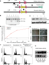
Disruption of a single Bub3 allele leads to mitotic checkpoint dysfunction. (A) Targeted inactivation of the mouse Bub3 gene. Shown is part of the endogenous mouse (m)Bub3 gene (top), the targeting vector (middle), and the disrupted mBub3 allele (bottom). BamHI restriction sites and the 5′ DNA probe (solid bar) used for Southern blot identification of wild-type (WT) and knockout (KO) Bub3 alleles are indicated. (B) Southern blot of genomic mouse tail DNA, digested with BamH1 and hybridized to a 5′ external probe, revealing the expected 7-kb wild-type and 5.5-kb mutant bands. (C) Western blot analysis of Bub3 protein levels in Bub3+/+ and Bub3+/− MEF lines using an antibody to mouse Bub3(145–276) (Wang et al., 2001). (D) Mitotic index of nocodazole-treated Bub3+/+ and Bub3+/− MEF cell lines. (E) Images of Bub3+/+ and Bub3+/− MEF cultures before and after nocodazole treatment. (F) DNA contents of asynchronous Bub3+/+ (panels 1–3) and Bub3+/− cells (panels 4–6) after the indicated treatments. (G) Cyclin B–associated Cdc2 kinase activity of synchronized MEF cultures at indicated time points after release into nocodazole.
Figure 6.
Figure 6.
Bub3 +/− cells display increased chromosome number instability. (A) Analysis of the degree of aneuploidy in MEF cultures at passage 5. Values are from three independent Bub3+/+ and three independent Bub3+/− MEF cultures. 50 metaphase spreads were counted for each MEF culture. (B) Distribution of chromosome numbers of wild-type and Bub3 haplo-insufficient MEF lines (combined values of each genotype).
Figure 7.
Figure 7.
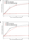
HA–Rae1 expression restores mitotic checkpoint activation in both Rae1 and Bub3 haplo-insufficient cells. (A) Mitotic index of nocodazole-treated Rae1+/−/empty vector (n = 2) and Rae1+/−/HA–Rae1 (n = 2) cell lines. (B) Mitotic index of nocodazole-treated Bub3+/−/empty vector (n = 2) and Bub3+/−/HA–Rae1 (n = 2) cell lines. Note that HA–Rae1-expressing cultures accumulate mitotic cells over time, illustrating that their mitotic checkpoint activity is restored.
Figure 8.
Figure 8.
Rae1 synergizes with Bub3 in the development of aneuploidy. (A) Growth curves of MEFs derived from wild-type, Rae1+/−, Bub3+/−, and Rae1+/−/Bub3+/− 13.5-d-old embryos (all harvested from Rae1+/− x Bub3+/− intercrosses). Data shown are means and standard deviations derived from three independent MEF lines per genotype. (B) Chromosome analysis of MEFs (at passage 5) from 13.5-d-old embryos collected from Rae1+/− females that were fertilized by Bub3+/− males. Data shown are means and standard deviations derived from three independent MEF lines per genotype. We counted the chromosomes of 50 metaphase spreads per independent MEF line. 200 mitotic figures per MEF line were screened for premature sister chromatid separation (PMSCS). (C) Metaphase spreads showing sister chromatid cohesion in a wild-type MEF (left) and PMSCS in a double heterozygote MEF (right). (D) Comparison of chromosome number distributions between wild-type, single heterozygous, and double heterozygous MEF lines (combined values of each genotype). (E) Aneuploidy and PMSCS in primary splenocytes from 5-mo-old mice of various genotypes. Data shown are means and standard deviations derived from three mice per genotype. We counted the chromosomes of 50 metaphase spreads per mouse. 100 spreads per mouse were screened for PMSCS.
Figure 9.
Figure 9.
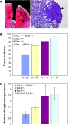
DMBA-induced tumor formation in checkpoint-defective mice. (A) Gross photograph of lung tumors of a DMBA-treated Rae1+/−/Bub3+/− mouse (left), and a section of a Rae1+/−/Bub3+/− lung tumor (right; 10×, hematoxylin + eosin). Arrows point out the tumors. (B) The occurrence of lung tumors in 5-mo-old mice plotted as percentage incidence. The asterisk indicates P < 0.05 compared with wild-type mice by Chi-squared test. We note that the tumor incidence of the checkpoint-defective group of mice as a whole is significantly increased relative to wild-type mice (P < 0.05 by Chi-squared test). (C) The average number of lung adenomas per mouse (±SEM). The asterisks indicate P < 0.05 compared with wild-type mice by Wilcoxon rank sum test.
Figure 10.
Figure 10.
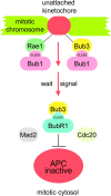
Model for Rae1 function in mitosis. We propose that Rae1 targets to unattached kinetochores at the onset of mitosis together with Bub1, just like Bub3. There, Rae1–Bub1 and Bub3–Bub1 proteins act to activate the mitotic checkpoint, potentially by producing anaphase wait signals. Unlike Rae1, Bub3 also interacts with BubR1 and localizes to the cytosolic component of the mitotic checkpoint complex that acts to inhibit the APC activator Cdc20 when anaphase wait signals are present. For simplicity, only part of the components of the checkpoint is shown in the model.

Similar articles

Cited by

References

    1. Bachi, A., I.C. Braun, J.P. Rodrigues, N. Pante, K. Ribbeck, C. Von Kobbe, U. Kutay, M. Wilm, D. Gorlich, M. Carmo-Fonseca, and E. Izaurralde. 2000. The C-terminal domain of TAP interacts with the nuclear pore complex and promotes export of specific CTE-bearing RNA substrates. RNA. 6:136–158. - PMC - PubMed
    1. Bailer, S.M., S. Siniossoglou, A. Podtelejnikov, A. Hellwig, M. Mann, and E. Hurt. 1998. Nup116p and nup100p are interchangeable through a conserved motif which constitutes a docking site for the mRNA transport factor gle2p. EMBO J. 17:1107–1119. - PMC - PubMed
    1. Basu, J., H. Bousbaa, E. Logarinho, Z. Li, B.C. Williams, C. Lopes, C.E. Sunkel, and M.L. Goldberg. 1999. Mutations in the essential spindle checkpoint gene bub1 cause chromosome missegregation and fail to block apoptosis in Drosophila. J. Cell Biol. 146:13–28. - PMC - PubMed
    1. Blajeski, A.L., V.A. Phan, T.J. Kottke, and S.H. Kaufmann. 2002. G(1) and G(2) cell-cycle arrest following microtubule depolymerization in human breast cancer cells. J. Clin. Invest. 110:91–99. - PMC - PubMed
    1. Braun, I.C., E. Rohrbach, C. Schmitt, and E. Izaurralde. 1999. TAP binds to the constitutive transport element (CTE) through a novel RNA-binding motif that is sufficient to promote CTE-dependent RNA export from the nucleus. EMBO J. 18:1953–1965. - PMC - PubMed

Publication types

MeSH terms

Substances