. 2003 Jan 15;17(2):201-13.
doi: 10.1101/gad.1050003.

Senescence, aging, and malignant transformation mediated by p53 in mice lacking the Brca1 full-length isoform

Affiliations

Senescence, aging, and malignant transformation mediated by p53 in mice lacking the Brca1 full-length isoform

Liu Cao et al. Genes Dev. .

Abstract

Senescence may function as a two-edged sword that brings unexpected consequences to organisms. Here we provide evidence to support this theory by showing that the absence of the Brca1 full-length isoform causes senescence in mutant embryos and cultured cells as well as aging and tumorigenesis in adult mice. Haploid loss of p53 overcame embryonic senescence but failed to prevent the adult mutant mice from prematurely aging, which included decreased life span, reduced body fat deposition, osteoporosis, skin atrophy, and decreased wound healing. We further demonstrate that mutant cells that escaped senescence had undergone clonal selection for faster proliferation and extensive genetic/molecular alterations, including overexpression of cyclin D1 and cyclin A and loss of p53. These observations provide the first in vivo evidence that links cell senescence to aging due to impaired function of Brca1 at the expense of tumorigenesis.


Figures
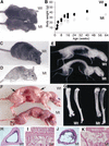
Figure 1
Figure 1
Aging-related phenotypes in Brca1Δ11/Δ11 p53+/− mice. (A) Photograph of 3-month-old p53+/− (all p53+/− mice are wild type for Brca1 and are simply referred to as Wt) and Brca1Δ11/Δ11 p53+/− (Mt) male mice. (B) Body weights of p53+/− and Brca1Δ11/Δ11 p53+/− mice (including both male and female, n = 65). Photograph of 8-month-old p53+/− (C) and Brca1Δ11/Δ11 p53+/− (D) female mice. (E) X-Ray radiograph of an 8-month-old Brca1Δ11/Δ11 p53+/− male mouse showing kyphosis (5 out of 18 mice examined exhibited this phenotype). (F) Photograph of 8-month-old female mouse, showing that all examined mutant mice (n > 10) have less fat (arrows). (G) Long-bone radiograph of 9-month-old p53+/− and Brca1Δ11/Δ11 p53+/− mice; 18 out of 25 mutant mice examined showed decreased bone density. (HK) Haematoxylin-eosin staining section of 10-month-old p53+/− (H,I) and Brca1Δ11/Δ11 p53+/− (J,K) female mice. The mutant mice exhibited decreased bone density (G), thinner cortical bone (J), and reduced trabecular bones (K). Most mice were tumor-free at the time when they were analyzed.
Figure 2
Figure 2
Skin-aging phenotypes in Brca1Δ11/Δ11 p53+/− mice. (A,B) Haematoxylin-eosin-stained section of 8-month-old p53+/− (A) and Brca1Δ11/Δ11 p53+/− (B) mice. The thickness of the dermal layer (d) and subcutaneous fat (f) is reduced in mutant mice. (C,D) Quantitative measurement of thickness of dermal layer (C) and fat layer (D; n = 14). (E,F) Haematoxylin-eosin-stained section at day 4 postwounding in 8-month-old p53+/− (E) and Brca1Δ11/Δ11 p53+/− (F) female mice. Epithelialization is visible in control mice, but this process is delayed in mutant mice (arrow in F‘). (G) Comparison of wound healing in 11-month-old p53+/− and Brca1Δ11/Δ11 p53+/− male mice (data were summarized from 12 wounds in 4 mutant mice and 12 wounds in 4 control mice). (H) Western blot analysis of p53 expression in 1-month-old (1M) and 6-month-old (6M) p53+/− and Brca1Δ11/Δ11 p53+/− mice. Protein extracts (50 μg/lane) from spleen were used for the assay. Three mutant and three control mice are shown at each time point.
Figure 3
Figure 3
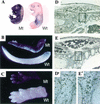
Senescence phenotypes in Brca1Δ11/Δ11 embryos. (AC) Photograph of acidic β-galactosidase staining of Brca1Δ11/Δ11 (Mt) and Brca1+/+ wild-type (Wt) E18 embryos. Strong staining was found in the tail (B) and limbs (C) of all tested mutant embryos (n = 6) that were E16.5 and older. (D,E) BrdU incorporation of Brca1Δ11/Δ11 (D) and wild-type (E) E16 embryos in thoracic region. Mutant embryos contained fewer BrdU-positive cells than wild-type embryos. The boxed areas in D and E are shown in D‘ and E‘. To provide a quantitative comparison, BrdU+ cells in comparable areas equivalent to the size shown in D‘ and E‘ in wild-type (n = 4) and mutant (n = 4) embryos were counted and subjected to the T-test. The average number of BrdU+ cells was 118 ± 11.9 cells/area and 88.5 ± 12.2 cells/area in wild-type and mutant, respectively (p ≤ 0.017).
Figure 4
Figure 4
Senescence phenotypes in Brca1Δ11/Δ11 MEF cells. Morphology (A,B) and X-gal staining (C,D) of passage 4 MEF cells. The mutant MEF cells show flattened and enlarged morphology (B) and strong staining of X-gal (D). (E) Percentage of senescent cells at each passages in Brca1+/+ MEF cells (closed squares) and Brca1Δ11/Δ11 MEF cells (open squares). (F) Proliferation analysis of wild-type MEF cells (closed squares) and Brca1Δ11/Δ11 MEF cells (open squares). (G) Expression analysis of p53, p21, p16, and p19, at passage 2 (P2), passage 3 (P3), and passage 5 (P5) cells.
Figure 5
Figure 5
Effects of p53 and p21 on senescence of Brca1Δ11/Δ11 embryos and MEF cells. (A,B) Morphology and X-gal staining of passage 4 Brca1Δ11/Δ11 MEF cells (A) and Brca1Δ11/Δ11 p53−/− MEF cells (B). The Brca1Δ11/Δ11 MEF cells showed a senescence-like morphology, and strong X-gal staining. (C,D) Photograph of X-gal staining of E18 embryos (C), and postnatal (P) day 1 pups (D). All Brca1Δ11/Δ11 p21−/− pups died within 24 h after birth. (E) Percentages of senescent MEF cells at passage 1 (P1), passage 3 (P3), and passage 5 (P5). (F) Cell proliferation of MEF cells at passages 1–5.
Figure 6
Figure 6
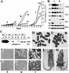
Transformation and tumorigenesis of immortalized Brca1Δ11/Δ11 MEF cells. (A) Cell proliferation analysis of immortalized Brca1+/+ (Wt) and Brca1Δ11/Δ11 (Mt) MEF cells at early (E, 5–10 passages), mediate (M, ∼50 passages), and later (L, >150 passages) passages after immortalization. (B) Expression of cyclin A (CyA), cyclin D1 (CyD1), and p53 in MEF cells at early and late passages. (C) p53 activity as reflected by p21 induction upon γ-irradiation of immortalized Brca1Δ11/Δ11 MEF cells at early passages. (D,E) Chromosome spreads of immortalized Wt control (D) and Brca1Δ11/Δ11 (E) MEF cells. Note that only parts of spreads are shown in order to highlight the short chromosomes (arrows) found in mutant cells. (FK) Soft agar colony formation assay of immortalized Brca1Δ11/Δ11 (F,H,J) and control (G,I,K) MEF cells at early (F,G), mediate (H,I), and late (J,K) passages, respectively. (L,M) Tumorigenicity assay of MEF cells at mediate passages in nude mice. Nude mice were tumor-free even 3 mo after injection of wild-type MEF cells (L), whereas tumors (arrows) were observed 3 wk after injection of Brca1Δ11/Δ11 MEF cells (M).
Figure 7
Figure 7
A working model summarizing how the Brca1 deficiency causes cell senescence, premature aging, and tumorigenesis.

Similar articles

Cited by

References

    1. Alberg AJ, Helzlsouer KJ. Epidemiology, prevention, and early detection of breast cancer. Curr Opin Oncol. 1997;9:505–511. - PubMed
    1. Alcorta DA, Xiong Y, Phelps D, Hannon G, Beach D, Barrett JC. Involvement of the cyclin-dependent kinase inhibitor p16INK4a in replicative senescence of normal human fibroblasts. Proc Natl Acad Sci. 1996;93:13742–13747. - PMC - PubMed
    1. Allsopp RC, Vaziri H, Patterson C, Goldstein S, Younglai EV, Futcher AB, Greider CW, Harley CB. Telomere length predicts replicative capacity of human fibroblasts. Proc Natl Acad Sci. 1992;89:10114–10118. - PMC - PubMed
    1. Atadja P, Wong H, Garkavtsev I, Veillette C, Riabowol K. Increased activity of p53 in senescing fibroblasts. Proc Natl Acad Sci. 1995;92:8348–8352. - PMC - PubMed
    1. Bachelier, R., Xu, X., Wang, X., Li, W., Naramura, M., Gu, H., and Deng, C.X. 2003. Normal lymphocyte development and thymic lymphoma formation in Brca1 exon 11-deficient mice. Oncogene (In Press). - PubMed