. 2003 Jan;14(1):129-41.
doi: 10.1091/mbc.e02-08-0483.

Piecemeal microautophagy of nucleus in Saccharomyces cerevisiae

Affiliations

Piecemeal microautophagy of nucleus in Saccharomyces cerevisiae

Paul Roberts et al. Mol Biol Cell. 2003 Jan.

Abstract

Nucleus-vacuole (NV) junctions in Saccharomyces cerevisiae are formed through specific interactions between Vac8p on the vacuole membrane and Nvj1p in the nuclear envelope. Herein, we report that NV junctions in yeast promote piecemeal microautophagy of the nucleus (PMN). During PMN, teardrop-like blebs are pinched from the nucleus, released into the vacuole lumen, and degraded by soluble hydrolases. PMN occurs in rapidly dividing cells but is induced to higher levels by carbon and nitrogen starvation and is under the control of the Tor kinase nutrient-sensing pathway. Confocal and biochemical assays demonstrate that Nvj1p is degraded in a PMN-dependent manner. PMN occurs normally in apg7-delta cells and is, therefore, not dependent on macroautophagy. Transmission electron microscopy reveals that portions of the granular nucleolus are often sequestered into PMN structures. These results introduce a novel mode of selective microautophagy that targets nonessential components of the yeast nucleus for degradation and recycling in the vacuole.


Figures

Figure 1
Figure 1
Frequencies of NV junctions increase during growth in rich medium. The percentage of cNVJ1::EYFP YEF473a cells containing cNvj1p-EYFP–stained NV junctions (filled circles, dashed gray line) as a function of growth in YPD (open circles, black line). At least 100 cells were scored for junctions at each point.
Figure 2
Figure 2
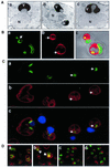
Vacuole-associated nuclear bulges, blebs, and intravacuolar vesicles. (A) Electron micrographs of a vacuole-associated nuclear envelope bulge (a) and blebs (b and c) (V, vacuole; N, nucleus). YEF473a cells were processed for TEM as described in MATERIAL AND METHODS. (B) Confocal images of extreme nuclear exvaginations in two starved pep4-Δ cells expressing PCUP1-NVJ1-EYFP and stained with FM4-64 (see MATERIALS AND METHODS). Nvj1p-EYFP (a), FM4-64 (b), and differential interference contrast overlay (c). Arrow in a points to a thin tether connecting the intravacuolar structure to the nucleus. Arrows in b point to FM4-64–stained intravacuolar membrane vesicles. (C) Nuclear envelope blebs and vesicles in cNvj1p-EYFP–expressing cells. Images show cNvj1p-EYFP (a), FM4-64 (b), and overlay (c) of a and b with Hoechst and differential interference contrast. Arrows point to intravacuolar vesicles. (D) Nuclear envelope blebbing is dependent on Vac8p. Nvj1p-EYFP–labeled blebs and vesicles were monitored in VAC8 (a and b) and vac8-Δ cells (c and d) in rich (YPD) (a and c) and starvation (SD-N) (b and d) media. Cells were grown in ScGlu, PCUP1-NVJ1:EYFP expression induced for 1 h with 0.1 mM Cu2+, stained with FM4-64, and starved for 3 h in SD-N, or incubated in YPD for the same time period, as described in MATERIALS AND METHODS. Arrows indicate PMN structures.
Figure 3
Figure 3
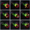
Movement of intravacuolar nuclear envelope vesicles. Images taken at 90-s intervals of an Nvj1-EYFP–expressing wt cell containing a number of blebs and vesicles. At least two Nvj1p-EYFP/FM4-64–stained vesicles moved freely in the vacuole (white arrowheads). An immobile Nvj1p-EYFP/FM4-64–stained bleb/vesicle located away from the NV junction but adjacent the vacuole membrane (pink arrowhead). In addition to blebs associated with the NV junction, this cell contains a mobile vesicle stained only with FM4-64 (purple arrowhead). Cells were grown in YPD, stained with FM4-64, starved for 3 h in SD-N, and stained with Hoechst as described in MATERIALS AND METHODS.
Figure 4
Figure 4
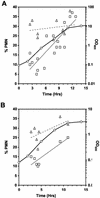
Physiological control of nuclear envelope blebbing. Frequencies of cNvj1p-EYFP–labeled blebs and vesicles as a function of growth, starvation, and rapamycin treatment in cNVJ1::EYFP YEF473a cells. (A) During growth in YPD (open circles, black line), cells at different OD600 were scored for vacuole-associated nuclear envelope blebs and vesicles (squares, gray regression line). Cells in YPD were transferred to SD-N medium at different times during the growth curve, and after 3 h, were scored for blebs and vesicles (triangles, dashed regression line). (B) Cells grown in SCGlu were scored for blebs and vesicles (squares, gray regression line) as a function of the growth curve (circles, black line) and after treatment with rapamycin (gray triangles, dashed regression line) as described in MATERIALS AND METHODS. At least 100 cells were scored at each point.
Figure 5
Figure 5
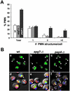
Degradation of nuclear envelope blebs is PEP4-dependent and APG7-independent. (A) Frequencies of Nvj1p-EYFP/FM4-64–stained nuclear envelope blebs and vesicles in wt (white bars), apg7-Δ (gray bars) and pep4-Δ (black bars) BY4741 cells. Each bar represents the mean of three independent counts of at least 100 cells. (B) Nvj1p-EYFP–labeled blebs and/or vesicles in wt, apg7-Δ, and pep4-Δ cells (arrowheads). (a–c) Cells stained with Nvj1p-EYFP (green). (d–f) Overlays of a, b, and c with Hoechst-stained chromatin (blue), FM4-64 for the vacuole membrane (red), and differential interference contrast. PCUP1-NVJ1::EYFP expression in the indicated strains was induced for 1 h with 0.1 mM Cu2+, shifted to SD-N medium for 3 h, and stained with Hoechst as described in MATERIALS AND METHODS.
Figure 6
Figure 6
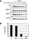
The degradation of Nvj1p-EYFP is dependent on Pep4p and Vac8p but not Apg7p. (A) wt, pep4-Δ, vac8-Δ, and apg7-Δ cells harboring PGAL1-NVJ1-EYFP were induced to express Nvj1p-EYFP for 3 h in 2% galactose and then shifted to glucose-containing starvation media (SD-N). Two OD units of culture were collected at the indicated times postinduction. Protein extracts were prepared and Nvj1p-EYFP levels (see arrows) were analyzed by immunoblot as described in MATERIALS AND METHODS. Uninduced control extracts are denoted as C. (B) Degradation levels of Nvj1p-EYFP after 20 h in SD-N. Nvj1p-EYFP protein levels were quantified at times 0 and 20 h (see arrows) and normalized against a nonspecific band (see asterisk) and adjusted for cell growth as described in MATERIALS AND METHODS.
Figure 7
Figure 7
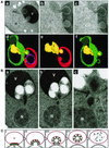
Partitioning of the granular nucleolus into nuclear envelope blebs and a model for PMN. (A) Thin sections of a cell containing two nuclear envelope blebs (a–c). Nucleus (N), nucleolus (Nu), vacuole (V). Three-dimensional reconstruction (d–f) from serial thin sections of the cell shown in panels a-c. Nuclear envelope (green), nuclear pore complexes (blue spheres), nucleolus (yellow), vacuole membrane (red), and an intravacuolar vesicle of unknown origin (magenta). (B) a–c, PMN blebs containing highly condensed material, which may or may not have a nucleolar origin, within the neck and bulb of an elongated bleb that protrudes into a section of the vacuole containing other intravacuolar vesicles. myo1-Δ cells were grown to early log phase in SCGlu and transferred to SD-N for 3 h before cryofixation and TEM (see MATERIALS AND METHODS). (C) Model for PMN based on observed structures (stage I). NV junctions are formed by the interaction and clustering of Nvj1p (green circles) in the nuclear envelope with Vac8p (red squares) in the vacuolar membrane (N, nucleus; V, vacuole). Nuclear bulges (stage II) develop into tethered blebs (stage III). Bulging and blebbing could be considered to be early and late steps of the same stage. Intravacuolar vesicles are formed by scission of the vacuolar and two nuclear envelope membranes of the blebs and the release of intravacuolar vesicles into the vacuole lumen (stage IV). Finally, the PMN vesicle and its contents are degraded by vacuolar hydrolases (stage V).

Similar articles

Cited by

References

    1. Abeliovich H, Darsow T, Emr SD. Cytoplasm to vacuole trafficking of aminopeptidase I requires a t-SNARE-Sec1p complex composed of Tlg2p and Vps45p. EMBO J. 1999;18:6005–6016. - PMC - PubMed
    1. Abeliovich H, Dunn WA, Jr, Kim J, Klionsky DJ. Dissection of autophagosome biogenesis into distinct nucleation and expansion steps. J Cell Biol. 2000;151:1025–1034. - PMC - PubMed
    1. Achleitner G, Gaigg B, Krasser A, Kainersdorfer E, Kohlwein SD, Perktold A, Zellnig G, Daum G. Association between the endoplasmic reticulum and mitochondria of yeast facilitates interorganelle transport of phospholipids through membrane contact. Eur J Biochem. 1999;264:545–553. - PubMed
    1. Baba M, Takeshige K, Baba N, Ohsumi Y. Ultrastructural analysis of the autophagic process in yeast: detection of autophagosomes and their characterization. J Cell Biol. 1994;124:903–913. - PMC - PubMed
    1. Darsow T, Rieder SE, Emr SD. A multispecificity syntaxin homologue, Vam3p, essential for autophagic and biosynthetic protein transport to the vacuole. J Cell Biol. 1997;138:517–529. - PMC - PubMed

Publication types

MeSH terms