. 2003 Jan;14(1):40-53.
doi: 10.1091/mbc.e02-08-0454.

STAT5a activation mediates the epithelial to mesenchymal transition induced by oncogenic RhoA

Affiliations

STAT5a activation mediates the epithelial to mesenchymal transition induced by oncogenic RhoA

Salvador Aznar Benitah et al. Mol Biol Cell. 2003 Jan.

Abstract

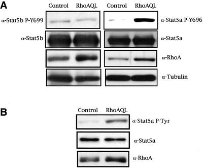
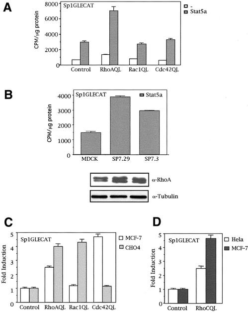
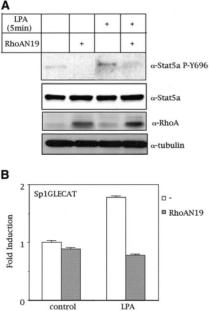
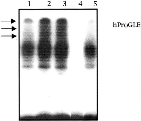
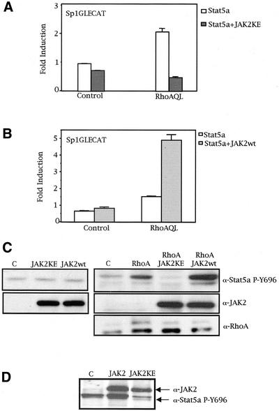
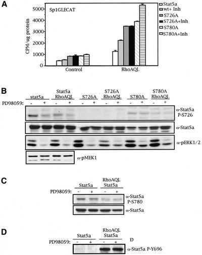
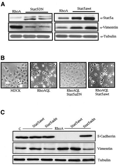
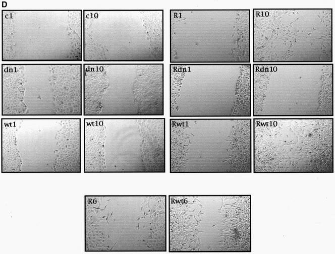
The involvement of Rho GTPases in signal transduction pathways leading to transcription activation is one of the major roles of this family of GTPases. Thus, the identification of transcription factors regulated by Rho GTPases and the understanding of the mechanisms of their activation and its biological outcome are of great interest. Here, we provide evidence that Rho GTPases modulate Stat5a, a transcription factor of the family of signal transducers and activators of transcription. RhoA triggers tyrosine phosphorylation (Y696) of Stat5a via a JAK2-dependent mechanism and promotes DNA-binding activity of Stat5a. Tyrosine phosphorylation of Stat5a is also stimulated physiologically by lysophosphatidic acid (LPA) in a Rho-dependent manner. Simultaneously, RhoA reduces serine phosphorylation of Stat5a at both serine residues S726 and S780, resulting in a further increase of activity as defined by mutagenesis experiments. Furthermore, serine dephosphorylation of Stat5a by RhoA does not take place by down-modulation of either JNK1, MEK1, or p38 MAP kinases, as determined by transfection experiments or chemical inhibition of both MEK1, p38, and JNK serine kinases. Thus, RhoA regulates Stat5a via tyrosine phosphorylation and via a yet to be determined novel down-modulating pathway that involves serine dephosphorylation. Finally, we provide evidence for a role of Stat5a in RhoA-induced epithelial-to-mesenchymal transition with concomitant increase in vimentin expression, E-cadherin down-regulation, and cell motility.


Figures

Figure 1
Figure 1
Oncogenic RhoA induces tyrosine phosphorylation (Y694) of Stat5a but not Stat5b. (A) MDCK cells transfected with either 2.0 μg of pcDNAIIIB or RhoA (QL), together with 2.0 μg of Stat5a, and harvested 48 h after transfection. Incubation of whole-cell extracts with a phosphospecific Stat5a-Y696 antibody revealed that RhoA induces tyrosine phosphorylation of Stat5a (right). Equal Stat5a transfection was verified with anti-Stat5a antibody. Expression of RhoA was detected with anti-RhoA antibody, and equal loading was verified by determining the levels of tubulin. The same type of experiment was performed with respect to Stat5b, and no tyrosine phosphorylation of residue 699 was observed under RhoA signaling (left). (B) RhoAQL induces tyrosine phosphorylation of endogenous Stat5a. RhoAQL or control vector was transfected in MDCK cells as in A, and tyrosine phosphorylation of Stat5a was determined by Western analysis. A and B show representative blots obtained in three independent experiments with similar results.
Figure 2
Figure 2
Rho GTPases differentially induce Stat5-dependent transcription of the Sp1GLECAT reporter in different cell lines. (A) RhoA but not Rac1 or Cdc42 (QL) activates Stat5a-dependent transcription of the Sp1GLECAT reporter via the endogenous and ectopically expressed Stat5a in MDCK cells. Cells transfected with Stat5a (2.0 μg) or not transfected, together with control vector or RhoA (QL), along with 0.5 μg of Sp1GLECAT reporter, were assayed for CAT activity 48 h after transfection. (B) RhoA (QL) activates Stat5a transactivation in two stable clones that constitutively express oncogenic RhoA (QL). MDCK or either clone was transfected with 0.5 μg of Sp1GLECAT reporter and 2.0 μg of Stat5a, and CAT activity was measured 48 h after transfection. The levels of expression of RhoA (QL) were determined with an anti-RhoA polyclonal antibody. Equal loading was verified with anti-tubulin antibody. (C) RhoA, Cdc42, and Rac1 differentially activate Stat5-dependent transcription in MCF-7 and CHO cells. Transfection of 1.0 μg of RhoA, Rac1, or Cdc42 (QL) along with the Sp1GLECAT reporter was performed in MCF-7 or CHO cells, and CAT activity was measured 24 h after transfection. (D) RhoC (QL) promotes Stat5-dependent transcription in HeLa and MCF-7 cells. Same experiment as in C was performed in both cell lines, and CAT activity was measured 24 h after transfection. All CAT experiments shown in this figure represent the mean of a single experiment performed in triplicate ±SD and are representative of at least three independent experiments with similar results.
Figure 3
Figure 3
LPA induces tyrosine phosphorylation and transcriptional activation of Stat5a via RhoA. (A) RhoA mediates LPA-induced Stat5a tyrosine phosphorylation. MDCK cells transfected with 2.0 μg of Stat5a and RhoAN19 expression vectors (where indicated) were treated with 50 μM LPA for 5 min, and tyrosine phosphorylation of Stat5a was determined by Western blot analysis. (B) LPA-induced Stat5a transcriptional activation is mediated by RhoA: 2.0 μg of control pcDNAIIIB or RhoAN19 (where indicated) expression vectors, along with the Sp1GLECAT reporter, were transfected in MDCK cells. Forty-eight hours after transfection, cells were treated with 50 μM LPA for 12 h, and CAT activity was determined. CAT experiment shown in this figure represents the mean of a single experiment performed in triplicate ±SD and is representative of at least three independent experiments with similar results. A shows a representative result obtained in three independent experiments.
Figure 4
Figure 4
RhoA (QL) induces Stat5a DNA binding to the GAS sequence of the human prolactin promoter. Two micrograms of MDCK-Stat5a or MDCK-RhoAQL/Stat5a nuclear extracts was subjected to EMSA. Lanes 1 and 2 represent control cells and RhoAQL-transfected cells, respectively. Lanes 3 and 4 correspond to unspecific competition and specific competition with excess hProGLE probe, respectively. Lane 5 corresponds to competition with α-Stat5a antibody. Results shown are representative of four independent experiments.
Figure 5
Figure 5
JAK2 mediates RhoA-induced Stat5a tyrosine phosphorylation. (A) Dominant negative JAK2 inhibits Stat5a transcriptional activation by RhoA (QL). One microgram of control pcDNAIIIB or RhoAQL was cotransfected with 1 μg of Stat5a, with either 3 μg of pRK-JAK2-KE or pRK control vector, along with 0.25 μg of Sp1GLECAT reporter. CAT activity was measured 48 h after transfection (0.5% FBS). (B) Wild-type JAK2 synergizes with RhoA (QL) to promote Stat5a-dependent transcription. Same amounts of control, RhoAQL, and Sp1GLECAT vectors were transfected as in A. Three micrograms of wild-type JAK2 was transfected where indicated. Data shown in A and B represent a single experiment performed in triplicate ±SD and are representative of three independent experiments. (C) JAK2 acts downstream of RhoA to promote Stat5a tyrosine phosphorylation, with no effect on basal tyrosine phosphorylation of Stat5a in control (C) cells. Thirty micrograms of the same extracts as in A and B were used for Western immunoblotting. Levels of JAK2 (KE and wt) and RhoA (QL) were verified with specific antibodies. (D) Same effect as in C is observed using a stable RhoA (QL) expressing MDCK cell line (clone SP7.29). Three micrograms of pRK-JAK2-KE or JAK2wt was transfected, and 48 h after transfection, protein expression was verified by Western blot. JAK2 (KE; wt) and Stat5a PY-694 were detected with specific antibodies. Immunoblot shown is representative of two independent experiments.
Figure 6
Figure 6
RhoAQL reduces the level of Stat5a serine phosphorylation of serine residues 726 and 780, which leads to an increase in Stat5a activity. (A) RhoA inhibits Stat5a serine phosphorylation of serines 726 and 780. Cells were transfected with 2.0 μg of control vector, RhoA (QL), and wild-type Stat5a or serine mutants S726A and S780A as indicated. Forty-eight hours after transfection, cells were harvested and lysed. Western immunoblotting was performed with phospho-Stat5a S726 and phospho-Stat5a S780 antibodies where indicated. Equal expression of Stat5a RhoAQL was verified with anti-Stat5a and anti-RhoA antibodies, respectively. Equal loading was confirmed with anti-tubulin. The blots shown are representative of five independent experiments. (B) Stat5a serine phosphorylation of both serines 726 and 780 is inhibitory for Stat5a-dependent Sp1GLE transcription. Control vector or RhoAQL was cotransfected with the indicated Stat5a expression vectors, along with the Sp1GLECAT promoter, and CAT activity was measured. (C) Same extracts were used for Western immunoblotting, and the levels of Stat5a serine phosphorylation on serine 726 were confirmed to be lower in RhoA transfectants. Levels of Stat5a and RhoA were confirmed to be equal by anti-Stat5a and anti-RhoA antibodies. Experiments shown in B and C are representative of three independent results.
Figure 7
Figure 7
MEK1 and RhoA (QL) modulate Stat5a serine phosphorylation of serine 726 by two independent mechanisms. (A) Treatment with MEK1 inhibitor PD98059 results in an increase of transcriptional activity of Stat5a and S780A mutant but not S726 mutant. MDCK cells transfected with 2.0 μg of the expression vectors indicated, together with 0.5 μg of Sp1GLECAT reporter, were maintained in 10% FBS for 24 h after transfection, and CAT activity was measured. (B) MEK-1 phosphorylates Stat5a serine 726 by a mechanism independent of RhoA. Same extracts as in A were used to verify the level of Stat5a serine phosphorylation on MEK-1 inhibition. As a control of inhibition, the amount of phospho-ERK1/2 was shown to decrease significantly on PD98059 treatment. The levels of transfected Stat5a or serine mutants were verified with specific anti-Stat5a antibody. (C) Treatment with PD98059 does not affect phosphorylation of serine 780. Same extracts as in A and B were used to determine the levels of Stat5a serine phosphorylation on serine 780. (D) PD98059 does not affect Stat5a tyrosine phosphorylation. Same extracts as in A were used to perform this experiment. Results shown here are representative of three independent experiments.
Figure 8
Figure 8
Stat5a is necessary for RhoA-induced EMT, vimentin expression, and loss of E-cadherin and motility in MDCK cells. (A) Stable expression of Stat5aDN or Stat5wt inhibits and enhances expression of vimentin induced by oncogenic RhoA, respectively. Stat5 stable cells lines were generated as described in MATERIALS AND METHODS and were verified for Stat5DN (left) and Stat5awt (right) stable expression with anti-Stat5a antibody and expression of endogenous vimentin with anti-vimentin. (B) Pictures of MDCK, MDCK-RhoAQL, RhoAQL/Stat5DN, and RhoAQL/Stat5awt stable cell lines with representative morphological changes. (C) Stat5a is necessary for loss of E-cadherin and vimentin expression accompanying RhoA-induced EM transition. Equal loading was verified with anti-tubulin. (D) Stat5a modulates cell motility induced by oncogenic RhoA. Cell motility was monitored at 1-h intervals after scratching, and representative pictures at 1 and 10 h are shown for each clone. An intermediate time point (6 h) is shown for RhoAQL- and RhoAQL/Stat5awt-expressing clones (c, MDCK-control vector; dn, MDCK-Stat5aDN; wt, MDCK-Stat5awt; R, MDCK-RhoAQL; Rwt, MDCK-RhoAQL/Stat5awt; Rdn, MDCK-RhoAQL/Stat5dn).
Figure 8
Figure 8
Stat5a is necessary for RhoA-induced EMT, vimentin expression, and loss of E-cadherin and motility in MDCK cells. (A) Stable expression of Stat5aDN or Stat5wt inhibits and enhances expression of vimentin induced by oncogenic RhoA, respectively. Stat5 stable cells lines were generated as described in MATERIALS AND METHODS and were verified for Stat5DN (left) and Stat5awt (right) stable expression with anti-Stat5a antibody and expression of endogenous vimentin with anti-vimentin. (B) Pictures of MDCK, MDCK-RhoAQL, RhoAQL/Stat5DN, and RhoAQL/Stat5awt stable cell lines with representative morphological changes. (C) Stat5a is necessary for loss of E-cadherin and vimentin expression accompanying RhoA-induced EM transition. Equal loading was verified with anti-tubulin. (D) Stat5a modulates cell motility induced by oncogenic RhoA. Cell motility was monitored at 1-h intervals after scratching, and representative pictures at 1 and 10 h are shown for each clone. An intermediate time point (6 h) is shown for RhoAQL- and RhoAQL/Stat5awt-expressing clones (c, MDCK-control vector; dn, MDCK-Stat5aDN; wt, MDCK-Stat5awt; R, MDCK-RhoAQL; Rwt, MDCK-RhoAQL/Stat5awt; Rdn, MDCK-RhoAQL/Stat5dn).

Similar articles

Cited by

References

    1. Abraham MT, Kuriakose MA, Sacks PG, Yee H, Chiriboga L, Bearer EL, Delacure MD. Motility-related proteins as markers for head and neck squamous cell cancer. Laryngoscope. 2001;111:1285–1289. - PMC - PubMed
    1. Alberts AS, Geneste O, Treisman R. Activation of SRF-regulated chromosomal templates by Rho-family GTPases requires a signal that also induces H4 hyperacetylation. Cell. 1998;92:475–487. - PubMed
    1. Al-Shami A, Mahanna W, Naccache PH. Granulocyte-macrophage colony-stimulating factor-activated signaling pathways in human neutrophils: selective activation of Jak2, Stat3, and Stat5b. J Biol Chem. 1998;273:1058–1063. - PubMed
    1. Aznar S, Lacal JC. Searching new targets for anticancer drug design: the families of Ras and RhoGTPases and their effectors. Prog Nucleic Acid Res Mol Biol. 2001a;67:193–234. - PubMed
    1. Aznar S, Lacal JC. Rho signals to cell growth and apoptosis. Cancer Lett. 2001b;165:1–10. - PubMed