. 2003 Feb;77(3):2063-70.
doi: 10.1128/jvi.77.3.2063-2070.2003.

Depletion of Wee-1 kinase is necessary for both human immunodeficiency virus type 1 Vpr- and gamma irradiation-induced apoptosis

Affiliations

Depletion of Wee-1 kinase is necessary for both human immunodeficiency virus type 1 Vpr- and gamma irradiation-induced apoptosis

Huidong Yuan et al. J Virol. 2003 Feb.

Abstract

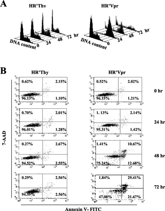
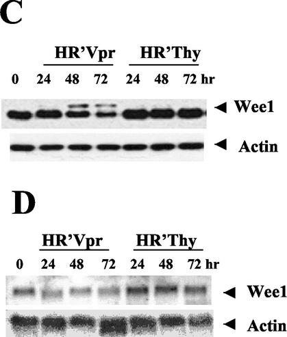
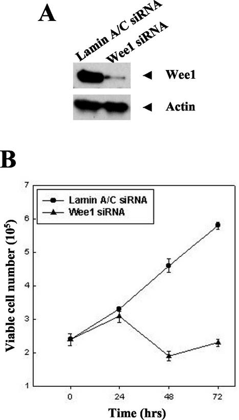
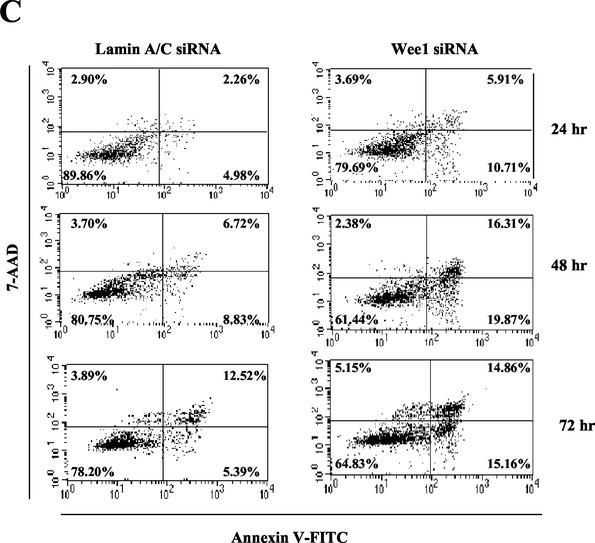
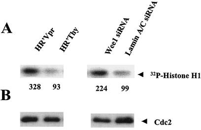
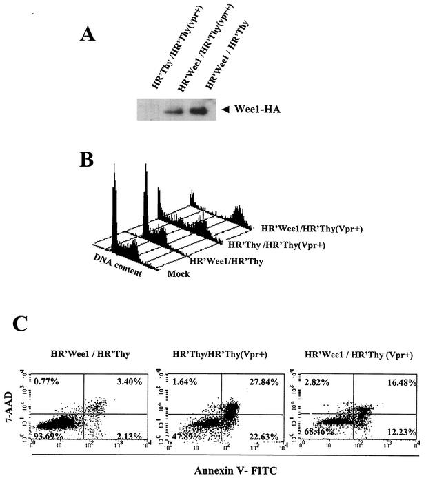
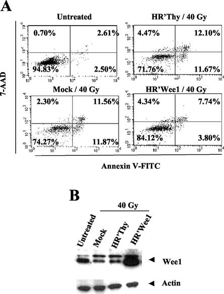
Human immunodeficiency virus (HIV) protein R (Vpr) induces G2 arrest, and prolonged G2 arrest leads to apoptosis. We find that in HeLa cells the cell cycle regulatory kinase, Wee-1, is depleted following prolonged G2 arrest induced by Vpr. Of note, small interfering RNAs directed to Wee-1 triggered apoptosis, suggesting a direct role for Wee-1 in apoptosis. In support of this hypothesis, overexpression of Wee-1 suppressed Vpr-mediated apoptosis. Importantly, similar results were observed with cells induced to undergo apoptosis gamma irradiation. Thus, Wee-1 may serve as a key regulator of both HIV type 1 Vpr- and gamma irradiation-mediated apoptosis and possibly serve as a general regulator linking the cell cycle to some pathways of apoptosis.


Figures

FIG. 1.
FIG. 1.
Wee-1 levels decrease in Vpr-induced apoptotic cells. HeLa cells were infected and analyzed for both cell cycle and apoptosis as previously described (9, 12). All stained cells were analyzed on a FACScan II (Becton Dickinson) and acquired by the Cell Quest software package. (A) Cell cycle profile of HeLa cells in response to HR′Vpr infection. Virus stock was produced and titrated to ensure that more than 95% of HeLa cells were arrested in G2 phase at 24 h postinfection (the equivalent of 1.2 μg of viral p24 was used for 2 × 10⁵ HeLa cells per well of a six-well plate [Falcon]). Equivalent amounts of HR′Thy virus were used as negative control. Cells were stained with PI, and DNA profiles of 5,000 cells were analyzed at the indicated times. (B) Kinetics of apoptosis after virus infection. HeLa cells were stained with Annexin V-FITC and 7-AAD at the indicated times. Percentages of positive cells within a quadrant are indicated. The lower right quadrant represents apoptotic cells. The upper right quadrant represents dead cells. (C) Protein immunoblot of Wee-1. Twenty micrograms of cell lysate was loaded in each lane and separated by SDS-10% PAGE, followed by immunoblotting with antibodies to Wee-1 or β-actin. (D) Northern blot analysis of Wee-1 RNA. Ten micrograms of total RNA was transferred to a nitrocellulose membrane and hybridized with human Wee-1 (nt 648 to 1938) and β-actin cDNAs in ExpressHyb hybridization solution.
FIG. 1.
FIG. 1.
Wee-1 levels decrease in Vpr-induced apoptotic cells. HeLa cells were infected and analyzed for both cell cycle and apoptosis as previously described (9, 12). All stained cells were analyzed on a FACScan II (Becton Dickinson) and acquired by the Cell Quest software package. (A) Cell cycle profile of HeLa cells in response to HR′Vpr infection. Virus stock was produced and titrated to ensure that more than 95% of HeLa cells were arrested in G2 phase at 24 h postinfection (the equivalent of 1.2 μg of viral p24 was used for 2 × 10⁵ HeLa cells per well of a six-well plate [Falcon]). Equivalent amounts of HR′Thy virus were used as negative control. Cells were stained with PI, and DNA profiles of 5,000 cells were analyzed at the indicated times. (B) Kinetics of apoptosis after virus infection. HeLa cells were stained with Annexin V-FITC and 7-AAD at the indicated times. Percentages of positive cells within a quadrant are indicated. The lower right quadrant represents apoptotic cells. The upper right quadrant represents dead cells. (C) Protein immunoblot of Wee-1. Twenty micrograms of cell lysate was loaded in each lane and separated by SDS-10% PAGE, followed by immunoblotting with antibodies to Wee-1 or β-actin. (D) Northern blot analysis of Wee-1 RNA. Ten micrograms of total RNA was transferred to a nitrocellulose membrane and hybridized with human Wee-1 (nt 648 to 1938) and β-actin cDNAs in ExpressHyb hybridization solution.
FIG. 2.
FIG. 2.
Depletion of Wee-1 by siRNA transfection leads to apoptosis. (A) Immunoblot of Wee-1. HeLa cells (1.5 × 10⁵ cells per well of a six-well plate) were transfected with 21-nt siRNAs targeting Wee-1 or Lamin A/C as described in the text. Immunoblotting of cell lysates prepared at 24 h after transfection was performed with antibodies against Wee-1 or β-actin. Twenty micrograms of cell lysate was loaded in each lane and separated by SDS-10% PAGE, followed by immunoblotting with antibodies to Wee-1 or β-actin. (B) Cell proliferation after siRNA transfection. Cells were stained with 0.4% trypan blue and were counted under a microscope at different time points. (C) Apoptosis occurred following Wee-1 siRNA transfection. Apoptosis was measured by flow cytometric analysis of Annexin V-FITC- and 7-AAD-stained cells.
FIG. 2.
FIG. 2.
Depletion of Wee-1 by siRNA transfection leads to apoptosis. (A) Immunoblot of Wee-1. HeLa cells (1.5 × 10⁵ cells per well of a six-well plate) were transfected with 21-nt siRNAs targeting Wee-1 or Lamin A/C as described in the text. Immunoblotting of cell lysates prepared at 24 h after transfection was performed with antibodies against Wee-1 or β-actin. Twenty micrograms of cell lysate was loaded in each lane and separated by SDS-10% PAGE, followed by immunoblotting with antibodies to Wee-1 or β-actin. (B) Cell proliferation after siRNA transfection. Cells were stained with 0.4% trypan blue and were counted under a microscope at different time points. (C) Apoptosis occurred following Wee-1 siRNA transfection. Apoptosis was measured by flow cytometric analysis of Annexin V-FITC- and 7-AAD-stained cells.
FIG.3.
FIG.3.
Cdc2 kinase activity is increased in HR′Vpr and Wee-1 siRNA-transfected HeLa cells. Cells were infected as described in the legend for Fig. 1 and transfected in parallel with those shown in Fig. 2. Cells were collected at 72 h post-virus infection or 24 h post-siRNA transfection. (A) Kinase assay of Cdc2. The assay was performed as described in Materials and Methods. The results were visualized and quantified using a PhosphorImager (Molecular Dynamics), and the numbers represent the relative density of the 32P-labeled histone H1. (B) Cdc2 protein level the kinase assay shown in panel A. Five micrograms of cell lysates was fractionated by SDS-12% PAGE and immunoblotted with Cdc2 antibody as described in the legend for Fig. 1. Relative Cdc2 activities were determined after normalization with the Cdc2 protein amount.
FIG. 4.
FIG. 4.
Ectopic expression of Wee-1 inhibits Vpr-induced apoptosis. Wee-1 with a C-terminal HA tag was expressed with a lentiviral vector derived from pHR′CMV-EGFP (1). HR′Wee1 and HR′Thy (Vpr+) viruses were made as described in the text. HR′Thy (Vpr+) packages Vpr but does not express Vpr de novo (30). HR′Thy (Vpr+) was titrated as described in the legend for Fig. 1. A virus inoculum equivalent to 2.5 μg of p24 was needed to achieve more than 95% G2 arrest in 2 × 10⁵ HeLa cells. Cells were infected by HR′Wee-1 or HR′Thy (control) 1 day before HR′Thy (Vpr+) or HR′Thy (control) infection. Viruses with an equivalent amount of p24 were used in each infection. Cells were stained either with Annexin V and 7-AAD or with PI or were lysed for Western blotting 72 h postinfection. (A) Expression of HA-tagged Wee-1 in the coinfected HeLa cells. Cell lysates were fractionated by SDS-10% PAGE and immunoblotted with HA antibody (Convence) as described in the legend for Fig. 1. (B) Cell cycle analysis of coinfected HeLa cells. (C) Apoptosis of HeLa cells coinfected with viruses as indicated.
FIG. 5.
FIG. 5.
Expression of exogenous Wee-1 suppresses gamma irradiation-induced apoptosis. A total of 2 × 10⁵ HeLa cells were infected with HR′Wee-1 or HR′Thy 1 day before exposure to 40 Gy of gamma irradiation. Cells were harvested and divided equally for assessment of apoptosis and Western blotting at 50 h after irradiation, as described in the legend for Fig. 1. (A) HR′Wee-1 infection alleviated gamma irradiation-induced apoptosis. (B) Wee-1 protein levels in the samples shown in panel A.

Similar articles

Cited by

References

    1. An, D. S., R. P. Wersto, B. Agricola, M. Metzger, S. Lu, R. G. Amado, I. S. Y. Chen, and R. E. Donahue. 2000. Marking and gene expression by a lentiviral vector in transplanted human and nonhuman primate CD34+ cells. J. Virol. 74:1286-1295. - PMC - PubMed
    1. Ayyavoo, V., A. Mahboubi, S. Mahalingam, R. Ramalingam, S. Kudchodkar, W. V. Williams, D. R. Green, and D. B. Weiner. 1997. HIV-1 Vpr suppresses immune activation and apoptosis through regulation of nuclear factor kappa B. Nat. Med. 3:1117-1123. - PubMed
    1. Balotta, C., P. Lusso, R. Crowley, R. C. Gallo, and G. Franchini. 1993. Antisense phosphorothioate oligodeoxynucleotides targeted to the vpr gene inhibit human immunodeficiency virus type 1 replication in primary human macrophages. J. Virol. 67:4409-4414. - PMC - PubMed
    1. de Noronha, C. M., M. P. Sherman, H. W. Lin, M. V. Cavrois, R. D. Moir, R. D. Goldman, and W. C. Greene. 2001. Dynamic disruptions in nuclear envelope architecture and integrity induced by HIV-1 Vpr. Science 294:1105-1108. - PubMed
    1. Elbashir, S. M., J. Harborth, W. Lendeckel, A. Yalcin, K. Weber, and T. Tuschl. 2001. Duplexes of 21-nucleotide RNAs mediate RNA interference in cultured mammalian cells. Nature 411:494-498. - PubMed

Publication types

LinkOut - more resources