

Distinct functional roles of the metabotropic glutamate receptors 1 and 5 in the rat globus pallidus
- PMID: 12514208
- PMCID: PMC6742153
- DOI: 10.1523/JNEUROSCI.23-01-00122.2003
Distinct functional roles of the metabotropic glutamate receptors 1 and 5 in the rat globus pallidus
Abstract
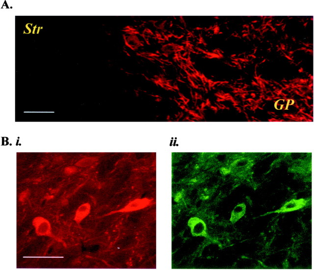
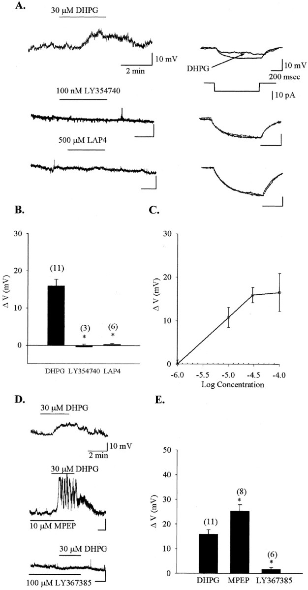
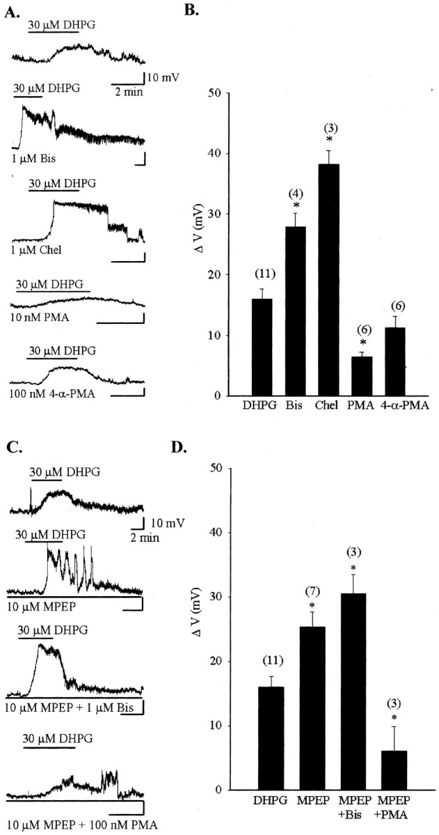
Group I metabotropic glutamate receptors (mGluRs) 1 and 5 frequently colocalize in the same neurons throughout the CNS. Because both receptors can couple to the same effector systems, the purpose of their cellular coexpression remains unclear. Here, we report that group I mGluR1 and mGluR5 have distinct functional roles in type II neurons of the rat globus pallidus (GP). Type II GP neurons form a large population of GABAergic projection neurons that are characterized by the presence of inwardly rectifying current I(h), low-threshold voltage-activated calcium current I(t), and activity at rest. Although immunocytochemical analysis reveals a high degree of neuronal colocalization of the two group I mGluRs in the GP, activation of mGluR1 only directly depolarizes type II GP neurons. Interestingly, blockade of mGluR5 by a highly selective antagonist, methylphenylethynylpyridine, leads to the potentiation of the mGluR1-mediated depolarization in this neuronal subpopulation. Metabotropic GluR1 desensitizes during repeated activation with the agonist in type II GP neurons, and blocking mGluR5 prevents the desensitization of the mGluR1-mediated depolarization. Elimination of the activity of protein kinase C (PKC) by an application of 1 microm bisendolylmaleimide or 1 microm chelerythrine, both protein kinase C inhibitors, potentiates the mGluR1-mediated response and prevents the desensitization of mGluR1 in type II GP neurons, suggesting that the effect of mGluR5 on mGluR1 signaling may involve PKC. Together, these data illustrate a novel mechanism by which mGluR1 and mGluR5, members of the same family of G-protein-coupled receptors, can interact to modulate neuronal activity in the rat GP.
Figures


Similar articles
-
Molecular mechanisms of group I metabotropic glutamate receptor mediated LTP and LTD in basolateral amygdala in vitro.Psychopharmacology (Berl). 2017 Feb;234(4):681-694. doi: 10.1007/s00213-016-4503-7. Epub 2016 Dec 28. Psychopharmacology (Berl). 2017. PMID: 28028604
-
Group I metabotropic glutamate receptor-mediated activation of PKC gamma in the nucleus accumbens core promotes the reinstatement of cocaine seeking.Addict Biol. 2015 Mar;20(2):285-96. doi: 10.1111/adb.12122. Epub 2014 Feb 9. Addict Biol. 2015. PMID: 24506432 Free PMC article.
-
Metabotropic glutamate receptor 5 mediates the potentiation of N-methyl-D-aspartate responses in medium spiny striatal neurons.Neuroscience. 2001;106(3):579-87. doi: 10.1016/s0306-4522(01)00297-4. Neuroscience. 2001. PMID: 11591458
-
Group I metabotropic glutamate receptors: implications for brain diseases.Prog Neurobiol. 1999 Sep;59(1):55-79. doi: 10.1016/s0301-0082(98)00095-1. Prog Neurobiol. 1999. PMID: 10416961 Review.
-
Glutamate receptors: brain function and signal transduction.Brain Res Brain Res Rev. 1998 May;26(2-3):230-5. doi: 10.1016/s0165-0173(97)00033-7. Brain Res Brain Res Rev. 1998. PMID: 9651535 Review.
Cited by
-
Therapeutic potential of targeting metabotropic glutamate receptors for Parkinson's disease.Neurodegener Dis Manag. 2012 Apr 1;2(2):221-232. doi: 10.2217/nmt.12.6. Neurodegener Dis Manag. 2012. PMID: 23526920 Free PMC article.
-
Opposite Effects of mGluR1a and mGluR5 Activation on Nucleus Accumbens Medium Spiny Neuron Dendritic Spine Density.PLoS One. 2016 Sep 12;11(9):e0162755. doi: 10.1371/journal.pone.0162755. eCollection 2016. PLoS One. 2016. PMID: 27618534 Free PMC article.
-
Metaplasticity at the addicted tetrapartite synapse: A common denominator of drug induced adaptations and potential treatment target for addiction.Neurobiol Learn Mem. 2018 Oct;154:97-111. doi: 10.1016/j.nlm.2018.02.007. Epub 2018 Feb 9. Neurobiol Learn Mem. 2018. PMID: 29428364 Free PMC article. Review.
-
Differential localization and function of GABA transporters, GAT-1 and GAT-3, in the rat globus pallidus.Eur J Neurosci. 2011 Apr;33(8):1504-18. doi: 10.1111/j.1460-9568.2011.07636.x. Epub 2011 Mar 17. Eur J Neurosci. 2011. PMID: 21410779 Free PMC article.
-
Quantification of D1 and D5 dopamine receptor localization in layers I, III, and V of Macaca mulatta prefrontal cortical area 9: coexpression in dendritic spines and axon terminals.J Comp Neurol. 2008 Jun 20;508(6):893-905. doi: 10.1002/cne.21710. J Comp Neurol. 2008. PMID: 18399540 Free PMC article.
References
-
- Alagarsamy S, Marino MJ, Rouse ST, Geraeau IVRW, Heinemann SF, Conn PJ. Activation of NMDA receptors reverses desensitization of mGluR5 in native and recombinant systems. Nat Neurosci. 1999;2:234–240. - PubMed
-
- Alagarsamy S, Sorensen SD, Conn PJ. Coordinate regulation of metabotropic glutamate receptors. Curr Opin Neurobiol. 2001;11:357–362. - PubMed
-
- Anwyl R. Metabotropic glutamate receptors: electrophysiological properties and role in plasticity. Brain Res Brain Res Rev. 1999;29:83–120. - PubMed
Publication types
MeSH terms
Substances
LinkOut - more resources
Full Text Sources
Other Literature Sources
Molecular Biology Databases