. 2003 Jan;77(2):1469-80.
doi: 10.1128/jvi.77.2.1469-1480.2003.

Human immunodeficiency virus type 1 nucleocapsid zn(2+) fingers are required for efficient reverse transcription, initial integration processes, and protection of newly synthesized viral DNA

Affiliations

Human immunodeficiency virus type 1 nucleocapsid zn(2+) fingers are required for efficient reverse transcription, initial integration processes, and protection of newly synthesized viral DNA

James S Buckman et al. J Virol. 2003 Jan.

Abstract

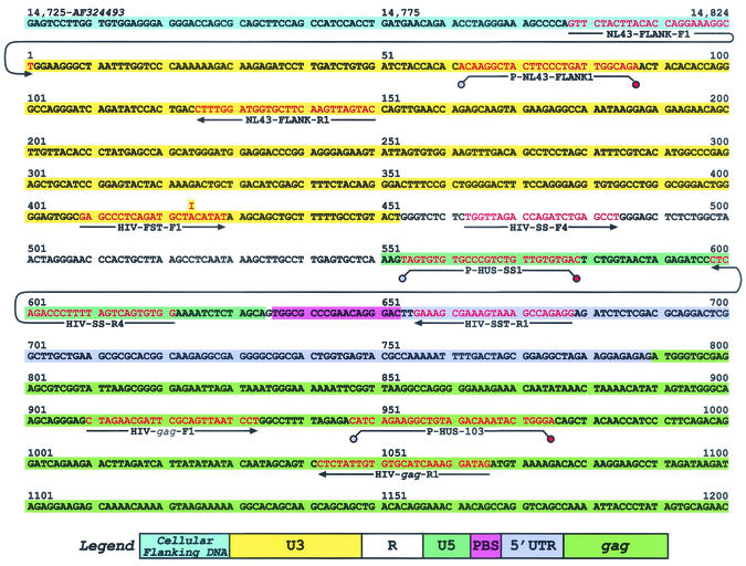
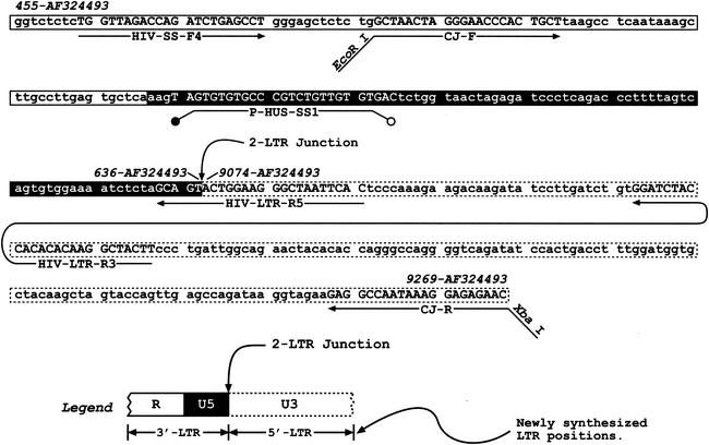
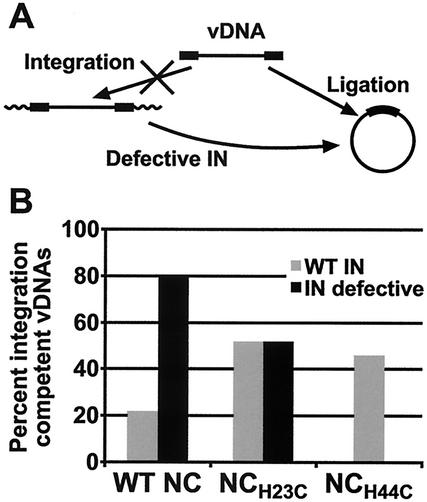
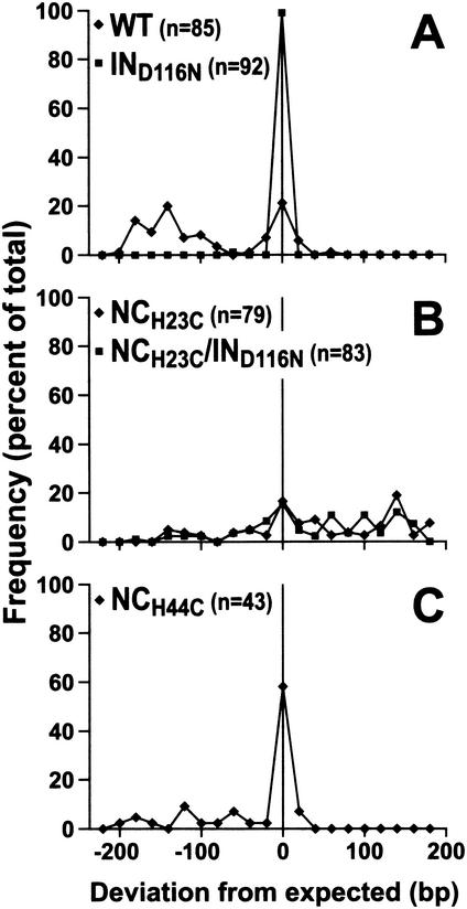
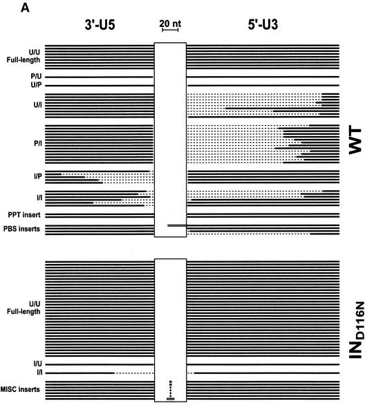
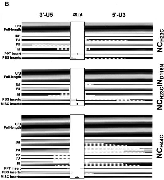
Human immunodeficiency virus type 1 (HIV-1) containing mutations in the nucleocapsid (NC) Zn(2+) finger domains have greatly reduced infectivity, even though genome packaging is largely unaffected in certain cases. To examine replication defects, viral DNA (vDNA) was isolated from cells infected with viruses containing His-to-Cys changes in their Zn(2+) fingers (NC(H23C) and NC(H44C)), an integrase mutant (IN(D116N)), a double mutant (NC(H23C)/IN(D116N)), or wild-type HIV-1. In vitro assays have established potential roles for NC in reverse transcription and integration. In vivo results for these processes were obtained by quantitative PCR, cloning of PCR products, and comparison of the quantity and composition of vDNA generated at discrete points during reverse transcription. Quantitative analysis of the reverse transcription intermediates for these species strongly suggests decreased stability of the DNA produced. Both Zn(2+) finger mutants appear to be defective in DNA synthesis, with the minus- and plus-strand transfer processes being affected while interior portions of the vDNA remain more intact. Sequences obtained from PCR amplification and cloning of 2-LTR circle junction fragments revealed that the NC mutants had a phenotype similar to the IN mutant; removal of the terminal CA dinucleotides necessary for integration of the vDNA is disabled by the NC mutations. Thus, the loss of infectivity in these NC mutants in vivo appears to result from defective reverse transcription and integration processes stemming from decreased protection of the full-length vDNA. Finally, these results indicate that the chaperone activity of NC extends from the management of viral RNA through to the full-length vDNA.


Figures

FIG. 1.
FIG. 1.
Sequence used for real-time PCR detection of reverse transcription intermediates. A portion of the sequence contained in pRB1008 that is used as a standard template for real-time PCR is presented. The sequence and positions in the pNL4-3 sequence of the 5′ cellular flanking region, the 5′ LTR, and a portion of the gag region are shown. The location in the pNL4-3 sequence is denoted by the numbers at the top of the sequence. Positions of primers and probes used are shown, with the sequences depicted in red text (probes are designated with a P- prefix). Primers and probes used for the detection of cellular flanking sequences are NL43-FLANK-F1, NL43-FLANK- R1, and P-NL43-FLANK1, respectively. The R-U5 region, representative of minus-strand strong-stop vDNA, is detected using HIV-SS-F4, HIV-SS-R4, and P-HUS-SS1. Detection of the U3-U5 region generated after minus-strand transfer is performed using HIV-FST-F1, HIV-SS-R4, and P-HUS-SS1. HIV-FST-F1 has an inosine at position 16 (from the 5′ end) instead of an adenosine. The inosine forms a base pair with thymidine in the standard template pRB1008 (the U3 region was derived from the 5′ LTR of pNL4-3), and in the vDNA target the inosine forms a base pair with cytosine since the source of the U3 region is derived from the 3′ end of the genome (i.e., the 3′ LTR of pNL4-3). The inosine-thymidine and inosine-cytosine interactions involve the same number of hydrogen bonds, and thus the HIV-FST-F1 will have roughly the same Tm with either target. Sequences in gag are quantitated using HIV-gag-F1, HIV-gag-R1, and P-HUS-103, and this region represents sequences generated after late-minus-strand synthesis. R-5′UTR targets, representative of sequences generated after plus-strand transfer, are detected using HIV-SS-F4, HIV-SST-R1, and P-HUS-SS1.
FIG. 2.
FIG. 2.
HIV-1 2-LTR circle junction fragment sequence, with PCR and cloning primers, and the probe. A portion of the full-length 2-LTR junction sequence is presented, along with the locations of various primers and the probe. Nucleotide positions in the pNL4-3 sequence are denoted at the top in italics. The position of the 2-LTR junction is also shown. This sequence region contained in the pJB1041 plasmid was used as a standard template for real-time PCR. The legend depicting the various regions (R, U5, and U3) is shown at the bottom of the figure. The primers and probe used to quantify full-junction 2-LTR circles are HIV-SS-F4, HIV-LTR-R5, and P-HUS, SS1, respectively. Total 2-LTR circles are detected using HIV-SS-F4, HIV-LTR-R3, and P-HUS-SS1. The CJ-F and CJ-R primers have been described as the upstream and downstream primers, respectively, by Julias et al. (37).
FIG. 3.
FIG. 3.
Fates of vDNA with and without a competent IN protein. (A) Schematic representation of the fate of integrated vDNA when the IN protein is knocked out. Viral DNA that could have been integrated now becomes ligated when IN is defective. The vDNA is represented by the black boxes (LTR regions) and the black line, and the cellular flanking DNA into which the provirus in inserted is represented by a thick wavy line. (B) Graph showing the percentage of integration-competent vDNAs isolated from cells infected with mutant and wild-type viruses. Values were calculated by assuming that the four types of linear vDNA sequences in Fig. 5 (U/U, U/P, P/U, and P/P) would have been integration competent if not for some defect other than vDNA end truncations or insertions. Values are reported as the percentage of integration-competent sequences of the total sequences obtained in Fig. 5.
FIG. 4.
FIG. 4.
Frequency distribution of the sizes of cloned 2-LTR circular vDNA PCR products from cells incubated with mutant and wild-type viruses. Inserts of the PCR product clones were analyzed for their sizes by either agarose gel electrophoresis or sequence analysis. Results are reported as the deviation from the expected size of 346 bp (Fig. 2). The frequencies were compiled in 20-nt windows from the expected size. Frequencies of the variance of insert sizes are reported as a percentage of the total number of samples examined. Distributions for wild-type (⧫) and IND116N (▪) (A), NCH23C (⧫) and NCH23C/IND116N (▪) (B), and NCH44C (⧫) (C) virus infections are shown.
FIG. 5.
FIG. 5.
Schematic of PCR product insert alignments. The cloned inserts of products from the PCR of vDNA isolated from cells incubated with mutant and wild-type viruses were sequenced. The virus examined is indicated on the right of each collection, and the classification of each sequence type is indicated on the left as follows: U, unprocessed end (full length); P, processed end (the CA dinucleotides were removed from the end, presumably by the viral IN protein); I, incomplete end (truncated compared to the full-length sequence). The letter to the left of the slash represents the state of the 3′-U5 end, and the letter to the right represents the state of the 5′-U3 end of the vDNA prior to ligation. The thick black lines in each panel represent nucleotides that are present in each of the clones. The dashed regions depict the nucleotides that are missing from the vDNA product insert. The grey regions depict DNA insertions of viral origin (PPT or PBS), and the horizontal boxes represent insertions of nonviral origin. The vertical boxes are spacer regions to accommodate the insertions. Black lines that are butted against the vertical boxes are full-length 2-LTR viral sequences. The lengths of lines and horizontal boxes are proportional to the length of the DNA sequence, with a 20-nt reference shown at the top of the figure. The PPT insert in the NCH23C collection of alignments, denoted by *, is 229 bp and is contiguous with the 5′-U3 end. A summary of the types of sequences for each virus is presented in Table 3. (A) Alignment of sequences from cloned PCR products of vDNA from infections with virus containing wild-type NC protein. (B) Alignment of sequences from cloned PCR products of vDNA from infections with virus containing mutant NC proteins.
FIG. 5.
FIG. 5.
Schematic of PCR product insert alignments. The cloned inserts of products from the PCR of vDNA isolated from cells incubated with mutant and wild-type viruses were sequenced. The virus examined is indicated on the right of each collection, and the classification of each sequence type is indicated on the left as follows: U, unprocessed end (full length); P, processed end (the CA dinucleotides were removed from the end, presumably by the viral IN protein); I, incomplete end (truncated compared to the full-length sequence). The letter to the left of the slash represents the state of the 3′-U5 end, and the letter to the right represents the state of the 5′-U3 end of the vDNA prior to ligation. The thick black lines in each panel represent nucleotides that are present in each of the clones. The dashed regions depict the nucleotides that are missing from the vDNA product insert. The grey regions depict DNA insertions of viral origin (PPT or PBS), and the horizontal boxes represent insertions of nonviral origin. The vertical boxes are spacer regions to accommodate the insertions. Black lines that are butted against the vertical boxes are full-length 2-LTR viral sequences. The lengths of lines and horizontal boxes are proportional to the length of the DNA sequence, with a 20-nt reference shown at the top of the figure. The PPT insert in the NCH23C collection of alignments, denoted by *, is 229 bp and is contiguous with the 5′-U3 end. A summary of the types of sequences for each virus is presented in Table 3. (A) Alignment of sequences from cloned PCR products of vDNA from infections with virus containing wild-type NC protein. (B) Alignment of sequences from cloned PCR products of vDNA from infections with virus containing mutant NC proteins.

Similar articles

Cited by

References

    1. Adachi, A., H. E. Gendelman, S. Koenig, T. Folks, R. Willey, A. Rabson, and M. A. Martin. 1986. Production of acquired immunodeficiency syndrome- associated retrovirus in human and nonhuman cells transfected with an infectious molecular clone. J. Virol. 59:284-291. - PMC - PubMed
    1. Aiken, C. 1997. Pseudotyping human immunodeficiency virus type 1 (HIV-1) by the glycoprotein of vesicular stomatitis virus targets HIV-1 entry to an endocytic pathway and suppresses both the requirement for Nef and the sensitivity to cyclosporin A. J. Virol. 71:5871-5877. - PMC - PubMed
    1. Allain, B., M. Lapadat-Tapolsky, C. Berlioz, and J.-L. Darlix. 1994. Transactivation of the minus-strand DNA transfer by nucleocapsid protein during reverse transcription of the retroviral genome. EMBO J. 13:973-981. - PMC - PubMed
    1. Berg, J. M. 1986. Potential metal-binding domains in nucleic acid binding proteins. Science 232:485-487. - PubMed
    1. Berthoux, L., C. Pechoux, M. Ottmann, G. Morel, and J. L. Darlix. 1997. Mutations in the N-terminal domain of human immunodeficiency virus type 1 nucleocapsid protein affect virion core structure and proviral DNA synthesis. J. Virol. 71:6973-6981. - PMC - PubMed

Publication types

LinkOut - more resources