Comparative Study
. 2002 Dec 9;159(5):881-91.
doi: 10.1083/jcb.200206043. Epub 2002 Dec 9.

Recruitment of the Arp2/3 complex to vinculin: coupling membrane protrusion to matrix adhesion

Affiliations
Comparative Study

Recruitment of the Arp2/3 complex to vinculin: coupling membrane protrusion to matrix adhesion

Kris A DeMali et al. J Cell Biol. .

Abstract

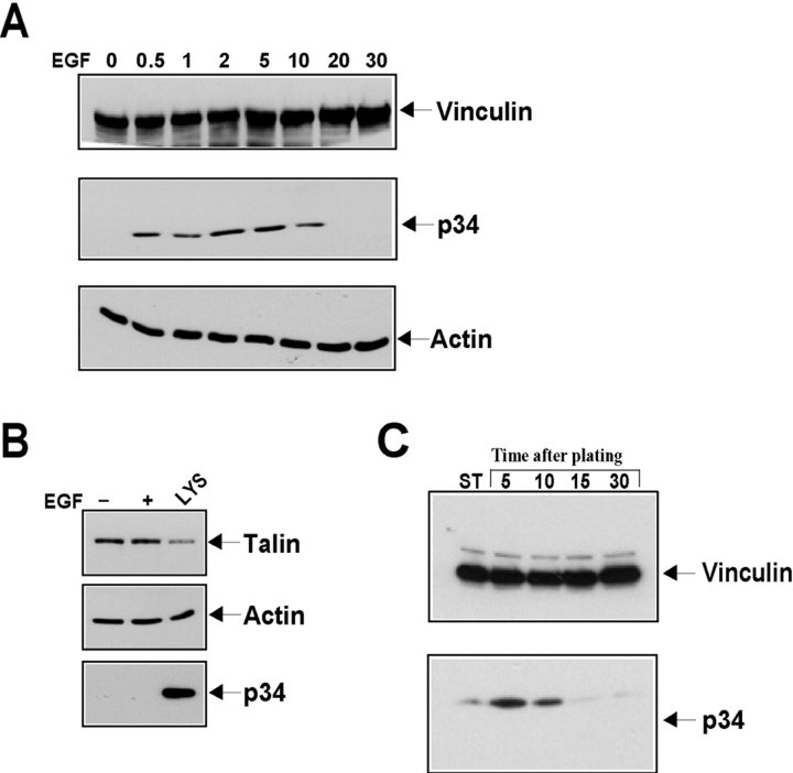
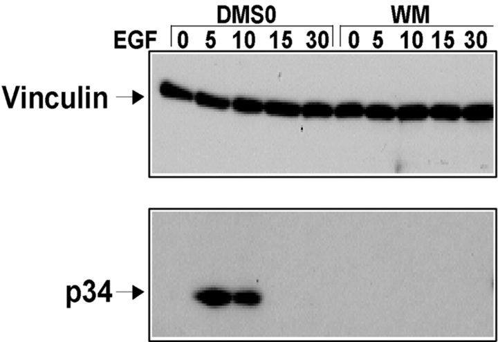
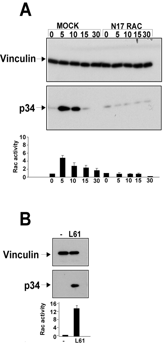
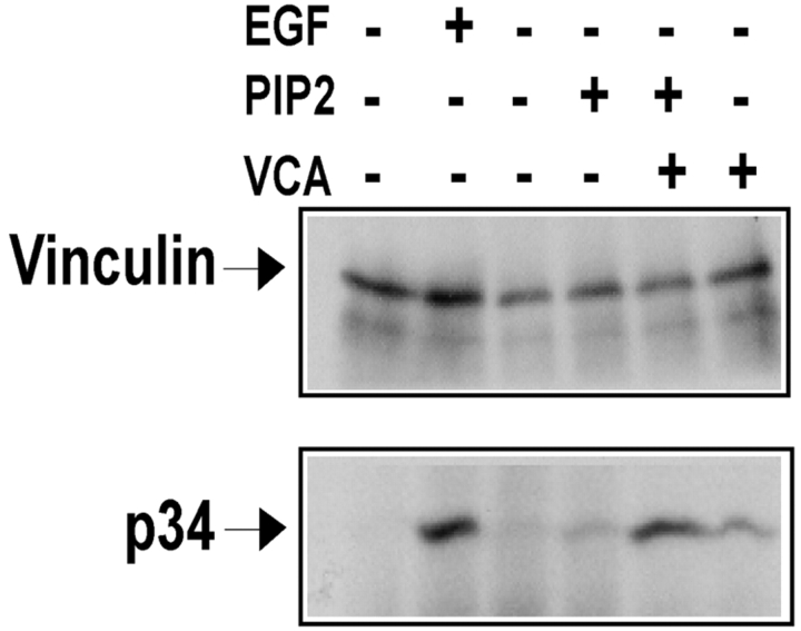
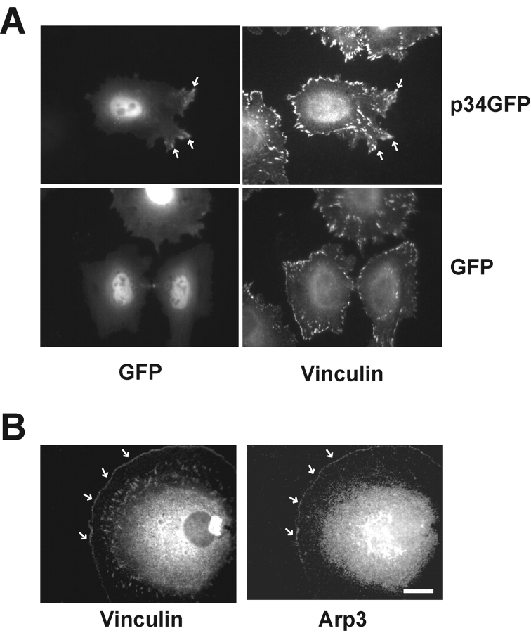
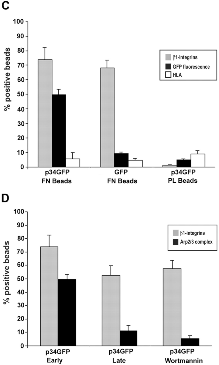
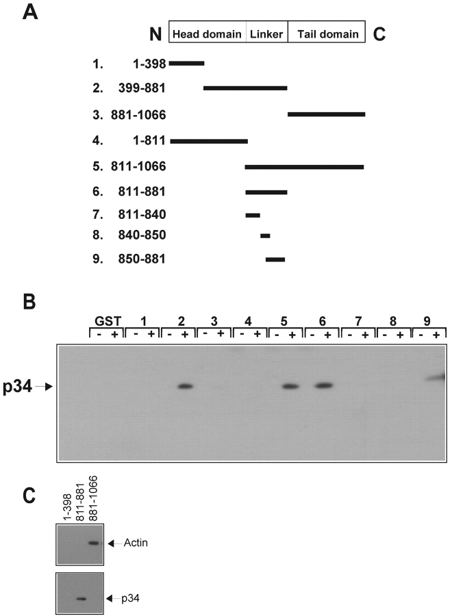
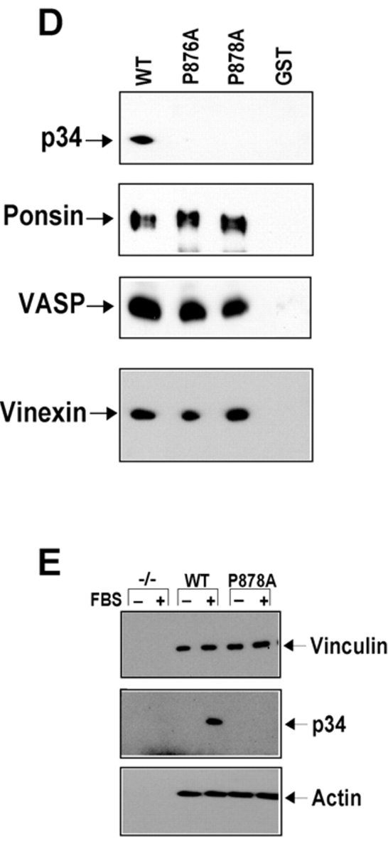
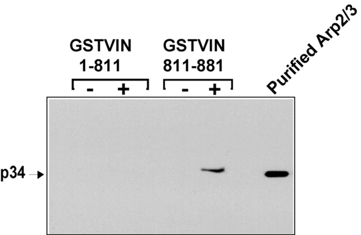
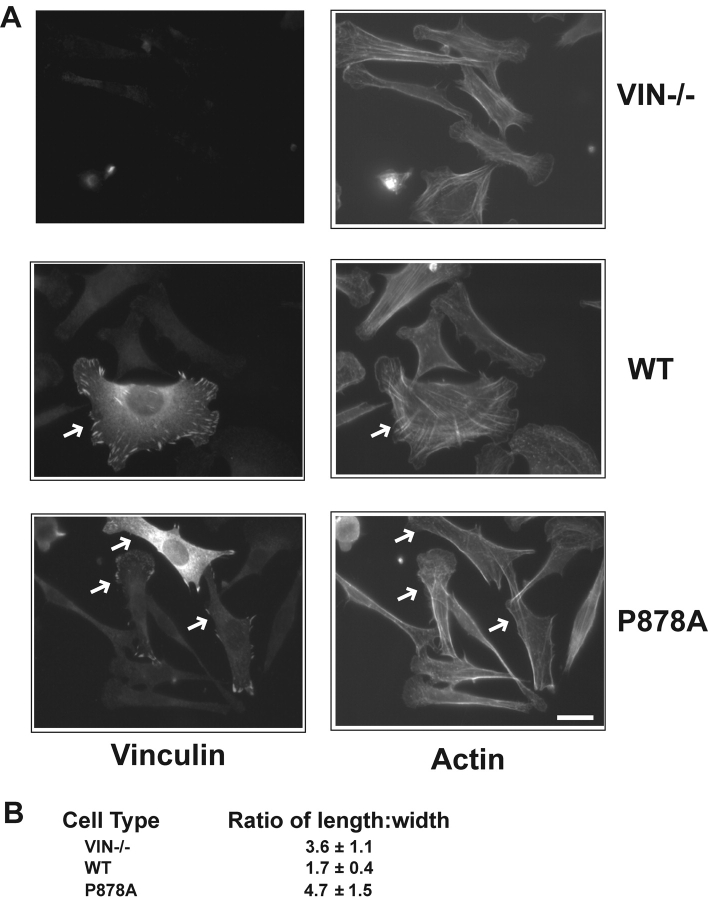
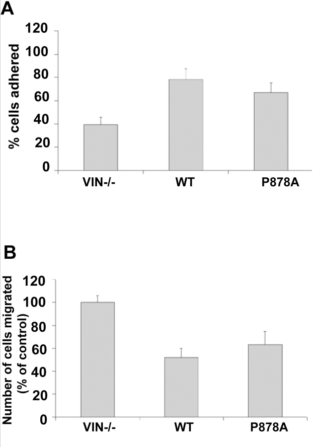
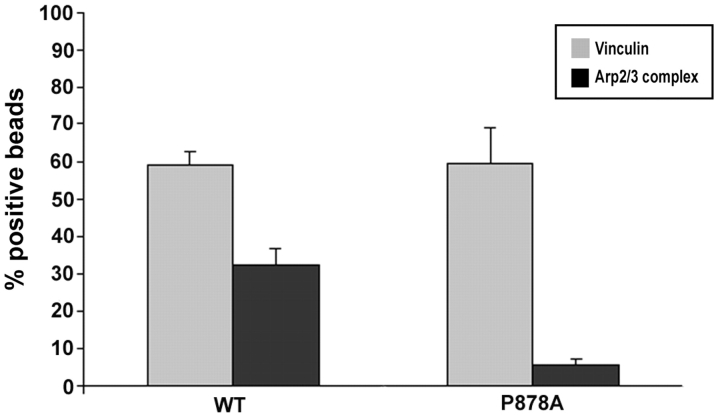
Cell migration involves many steps, including membrane protrusion and the development of new adhesions. Here we have investigated whether there is a link between actin polymerization and integrin engagement. In response to signals that trigger membrane protrusion, the actin-related protein (Arp)2/3 complex transiently binds to vinculin, an integrin-associated protein. The interaction is regulated, requiring phosphatidylinositol-4,5-bisphosphate and Rac1 activation, and is sufficient to recruit the Arp2/3 complex to new sites of integrin aggregation. Binding of the Arp2/3 complex to vinculin is direct and does not depend on the ability of vinculin to associate with actin. We have mapped the binding site for the Arp2/3 complex to the hinge region of vinculin, and a point mutation in this region selectively blocks binding to the Arp2/3 complex. Compared with WT vinculin, expression of this mutant in vinculin-null cells results in diminished lamellipodial protrusion and spreading on fibronectin. The recruitment of the Arp2/3 complex to vinculin may be one mechanism through which actin polymerization and membrane protrusion are coupled to integrin-mediated adhesion.


Figures

Figure 1.
Figure 1.
Factors that trigger membrane protrusion induce the association of the Arp2/3 complex with vinculin. (A and B) Growth factor treatment of cells. Serum-starved A431 cells were stimulated with buffer (0) or human EGF (100 ng/ml) for the times indicated (min). The cells were lysed and vinculin or talin was immunoprecipitated. 20% of resulting immunoprecipitates were subjected to Western blot analysis with antibodies against vinculin (A) or talin (B), 10% was used for the actin blots (A and B, bottom), and 70% was used for the blots of p34 subunit of the Arp2/3 complex (p34, bottom). LYS denotes a sample of whole cell lysate. (C) Cells spreading on FN. Hs68, human foreskin fibroblasts, were plated into each well of a FN-coated 6-well plate in serum-free DME. The cells were centrifuged onto the dish and allowed to recover at 37°C for the times indicated. The cells were lysed, vinculin was immunoprecipitated, and cells were blotted as described in A and B. ST, stationary cells that had been plated for 4 h on a similar concentration of FN.
Figure 2.
Figure 2.
PI3K is required for recruitment of the Arp2/3 complex to vinculin. Effect of inhibition of PI3K by wortmannin (A) on recruitment of the Arp2/3 complex to vinculin. Serum-starved A431 cells were pretreated with DMSO or 100 nM wortmannin (WM) for 30 min and then left resting (0) or stimulated with EGF for the times indicated (min). Vinculin was immunoprecipitated and blotted for vinculin or p34-Arc as described in Fig. 1.
Figure 3.
Figure 3.
Rac1 is necessary and sufficient for recruitment of the Arp2/3 complex to vinculin. (A) Effect of dominant negative Rac1 on the recruitment of proteins to vinculin. A431 cells were transiently transfected with vector alone (MOCK) or myc-tagged N17Rac1 (N17Rac1), allowed to recover for 12 h, and then serum starved for an additional 18–24 h in DME. Cells were stimulated with 100 ng/ml of EGF as in Fig. 1 for the times given (min), and vinculin immunoprecipitated. (B) Effect of constitutively active Rac1 on the recruitment of proteins to vinculin. A431 cells were transiently transfected with AU-tagged L61Rac1 (L61), allowed to recover for 12h, and then serum starved for an additional 18–24 h in DME. Vinculin immunoprecipitates were blotted for p34. The graphs beneath each panel indicate the levels of GTP-bound Rac1. Each number represents the mean fold activation ± SEM in Rac1 activity over unstimulated cells from three independent experiments.
Figure 4.
Figure 4.
PIP 2 and GST-VCA induce binding of the Arp2/3 complex to vinculin. Serum-starved A431 cells were stimulated with buffer alone or with 100 ng/ml EGF for 5 min. The cells were lysed in 500 μl vol as described in the text, and the lysates were incubated in the presence of no exogenous proteins or 30 μg of GST, 10 mM PIP2, 10 mM PIP2 +30 μg GST–VCA, or 30 μg of GST–VCA for 20 min at 4°C. Vinculin immunoprecipitates were prepared and analyzed as described above. In three independent experiments, compared with the level of Arp2/3 binding to vinculin in response to 5 min EGF stimulation (100%), the level of Arp2/3 complex recruitment in response to the various stimuli was 13% ± 7% (PIP2), 27% ± 15% (GST–VCA), and 94% ± 11% (PIP2 + GST–VCA).
Figure 5.
Figure 5.
Recruitment of the Arp2/3 complex to sites of integrin aggregation. (A) REF52 cells expressing GFP or p34GFP were plated on FN-coated coverslips for 1.5 h at 37°C. (B) Serum-starved Swiss3T3 cells were plated on FN-coated coverslips, and microinjected with 61LRac and C3. The cells were examined by immunofluorescence using antibodies against vinculin and the Arp3 subunit of the Arp2/3 complex. Sites of colocalization of the Arp2/3 complex with vinculin (focal complexes) are indicated with arrows in A. Bar, 20 μm. (C and D) Hela cells expressing GFP or p34GFP were plated on collagen and presented with beads coated with fibronectin (FN) or poly-lysine (PL beads) for 20 min (C and D marked early) or 35 min (D, late). Cells were stained for β1 integrins or HLA. The percentage of beads able to recruit the β1 integrins (gray bars), the GFP fusion proteins (black bars), and/or HLA (white bars) was scored using immunofluorescence. In some experiments, cells were preincubated with 100 nM wortmannin (D, wortmannin) for 30 min before incubation with the beads.
Figure 5.
Figure 5.
Recruitment of the Arp2/3 complex to sites of integrin aggregation. (A) REF52 cells expressing GFP or p34GFP were plated on FN-coated coverslips for 1.5 h at 37°C. (B) Serum-starved Swiss3T3 cells were plated on FN-coated coverslips, and microinjected with 61LRac and C3. The cells were examined by immunofluorescence using antibodies against vinculin and the Arp3 subunit of the Arp2/3 complex. Sites of colocalization of the Arp2/3 complex with vinculin (focal complexes) are indicated with arrows in A. Bar, 20 μm. (C and D) Hela cells expressing GFP or p34GFP were plated on collagen and presented with beads coated with fibronectin (FN) or poly-lysine (PL beads) for 20 min (C and D marked early) or 35 min (D, late). Cells were stained for β1 integrins or HLA. The percentage of beads able to recruit the β1 integrins (gray bars), the GFP fusion proteins (black bars), and/or HLA (white bars) was scored using immunofluorescence. In some experiments, cells were preincubated with 100 nM wortmannin (D, wortmannin) for 30 min before incubation with the beads.
Figure 6.
Figure 6.
Mapping the Arp2/3 complex binding site of vinculin. (A) Linear schematic is shown of vinculin and the fragments of vinculin as fusion proteins of GST (GST–VIN). (B and C) Association of the Arp2/3 complex with GST–VIN proteins. Thirty micrograms of fusion protein attached to beads were incubated for 1.5 h in the absence (−) or presence (+) of 10 μl of a crude platelet fraction. The beads were sedimented, washed, and subjected to Western blot analysis as described. Note that in C, the binding of the Arp2/3 complex to vinculin fragments does not involve binding to actin. (D) Substitution of proline 878 or 876 with alanine prevents binding of the Arp2/3 complex to vinculin in vitro. 30 μg of GST or GST fusions of VIN 811–881, VIN 811–881 with a proline to alanine mutation at 876 (P876A) or at 878 (P878A) were examined for their ability to retrieve the Arp2/3 complex, ponsin, vinexin, or VASP from cell lysates obtained from platelets, MDCK, C2C12, or A431 cells, respectively. Different cell types were used for preparing the lysates because of failure to detect the antigens in some cell types. (E) Full-length vinculin harboring a proline to alanine mutation at 878 prevents recruitment of the Arp2/3 complex, but has no effect on actin binding. Serum-starved, Vin−/− MEFs expressing an empty vector (−/−), full-length vinculin (WT), or full-length vinculin with a P878A substitution (P878A) were left resting (−) or stimulated (+) with 10% FBS for 5 min, vinculin immunoprecipitates were obtained and analyzed.
Figure 6.
Figure 6.
Mapping the Arp2/3 complex binding site of vinculin. (A) Linear schematic is shown of vinculin and the fragments of vinculin as fusion proteins of GST (GST–VIN). (B and C) Association of the Arp2/3 complex with GST–VIN proteins. Thirty micrograms of fusion protein attached to beads were incubated for 1.5 h in the absence (−) or presence (+) of 10 μl of a crude platelet fraction. The beads were sedimented, washed, and subjected to Western blot analysis as described. Note that in C, the binding of the Arp2/3 complex to vinculin fragments does not involve binding to actin. (D) Substitution of proline 878 or 876 with alanine prevents binding of the Arp2/3 complex to vinculin in vitro. 30 μg of GST or GST fusions of VIN 811–881, VIN 811–881 with a proline to alanine mutation at 876 (P876A) or at 878 (P878A) were examined for their ability to retrieve the Arp2/3 complex, ponsin, vinexin, or VASP from cell lysates obtained from platelets, MDCK, C2C12, or A431 cells, respectively. Different cell types were used for preparing the lysates because of failure to detect the antigens in some cell types. (E) Full-length vinculin harboring a proline to alanine mutation at 878 prevents recruitment of the Arp2/3 complex, but has no effect on actin binding. Serum-starved, Vin−/− MEFs expressing an empty vector (−/−), full-length vinculin (WT), or full-length vinculin with a P878A substitution (P878A) were left resting (−) or stimulated (+) with 10% FBS for 5 min, vinculin immunoprecipitates were obtained and analyzed.
Figure 7.
Figure 7.
Binding of the Arp2/3 complex to vinculin is direct. Vinculin fusion proteins attached to beads were incubated with 2 μg of purified, human platelet Arp2/3 complex for 1.5 h at 4°C. The beads were washed and the pellets were examined for the ability of the fusion proteins to bind the Arp2/3 complex. Purified Arp2/3 denotes a sample of Arp2/3 complex purified from platelets.
Figure 8.
Figure 8.
A vinculin mutant unable to associate with the Arp2/3 complex is impaired in lamellipodial extension and spreading on fibronectin. Vinculin null cells reexpressing vector alone (Vin−/−), WT or mutant vinculin (P878A) were allowed to spread onto 50 μg/ml of FN for 120 min (A) and then examined by immunofluorescence with antibodies against vinculin or using Alexa-conjugated phalloidin. The white arrows indicate the transfected cells. Bars, 20 μm. (B) the extent of spreading was quantified by expressing the ratio ± SD of the length/width of 50 cells at the 120-min time point. The WT reexpressing cells are significantly more spread than either the null or mutant reexpressers (P < 0.001).
Figure 9.
Figure 9.
Recruitment of the Arp2/3 complex to vinculin is not required for cell adhesion or cell migration. Vinculin-null cells reexpressing vector alone (Vin−/−), WT vinculin, or mutant vinculin (P878A) were examined for the ability to adhere to 50 μg/ml FN (A) or migrate through Transwell filters (B). The mean number of cells expressing vector alone that migrated through the filter was set to 100%. The relative number of cells/field ± SEM was calculated from the means of three independent experiments.
Figure 10.
Figure 10.
Mutation of the proline 878 ablates recruitment of the Arp2/3 complex to FN-coated beads. Vinculin-null cells reexpressing WT vinculin or vinculin with a mutated Arp2/3 complex binding site (P878A) were presented with beads coated with FN (50 μg/ml) for 15 min. The percentage of beads able to recruit vinculin (light gray) and p34GFP (dark gray) was scored by immunofluorescence microscopy.

Similar articles

Cited by

References

    1. Aspenstrom, P., U. Lindberg, and A. Hall. 1996. Two GTPases, Cdc42 and Rac, bind directly to a protein implicated in the immunodeficiency disorder Wiskott-Aldrich syndrome. Curr. Biol. 6:70–75. - PubMed
    1. Bagrodia, S., S.J. Taylor, K.A. Jordon, L. Van Aelst, and R.A. Cerione. 1998. A novel regulator of p21-activated kinases. J. Biol. Chem. 273:23633–23636. - PubMed
    1. Bailly, M., I. Ichetovkin, W. Grant, N. Zebda, L.M. Machesky, J.E. Segall, and J. Condeelis. 2001. The F-actin side binding activity of the Arp2/3 complex is essential for actin nucleation and lamellipod extension. Curr. Biol. 11:620–625. - PubMed
    1. Blanchoin, L., K.J. Amann, H.N. Higgs, J.B. Marchand, D.A. Kaiser, and T.D. Pollard. 2000. Direct observation of dendritic actin filament networks nucleated by Arp2/3 complex and WASP/Scar proteins. Nature. 404:1007–1011. - PubMed
    1. Borisy, G.G., and T.M. Svitkina. 2000. Actin machinery: pushing the envelope. Curr. Opin. Cell Biol. 12:104–112. - PubMed

Publication types

MeSH terms