. 2002 Nov;13(11):3915-29.
doi: 10.1091/mbc.e02-05-0291.

Modulation of fibroblast morphology and adhesion during collagen matrix remodeling

Affiliations

Modulation of fibroblast morphology and adhesion during collagen matrix remodeling

Elisa Tamariz et al. Mol Biol Cell. 2002 Nov.

Abstract

When fibroblasts are placed within a three-dimensional collagen matrix, cell locomotion results in translocation of the flexible collagen fibrils of the matrix, a remodeling process that has been implicated in matrix morphogenesis during development and wound repair. In the current experiments, we studied formation and maturation of cell-matrix interactions under conditions in which we could distinguish local from global matrix remodeling. Local remodeling was measured by the movement of collagen-embedded beads towards the cells. Global remodeling was measured by matrix contraction. Our observations show that no direct relationship occurs between protrusion and retraction of cell extensions and collagen matrix remodeling. As fibroblasts globally remodel the collagen matrix, however, their overall morphology changes from dendritic to stellate/bipolar, and cell-matrix interactions mature from punctate to focal adhesion organization. The less well organized sites of cell-matrix interaction are sufficient for translocating collagen fibrils, and focal adhesions only form after a high degree of global remodeling occurs in the presence of growth factors. Rho kinase activity is required for maturation of fibroblast morphology and formation of focal adhesions but not for translocation of collagen fibrils.


Figures
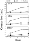
Figure 1
Figure 1
Global remodeling of collagen matrices. HD matrices (□), LD matrices (▪), or matrices without cells (×) were incubated for the times shown in basal medium (BSA) or medium containing LPA or PDGF. At the end of the incubations, contraction was determined by measuring reduction in cell height. In the presence of LPA or PDGF, matrix contraction was ∼50% by 1 h and 80–90% by 6 h (starting height ∼1.8–1.9 mm). In basal medium, the rate of contraction was slower with little contraction observed after 1 h and only 60–70% by 6 h. Studies were carried out in triplicate. SDs were smaller than the size of the points.
Figure 2
Figure 2
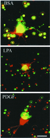
Local remodeling of collagen matrices. Images of cells from LD matrices at 1 h (green) and 4 h (red) in basal medium (BSA) or medium containing LPA or PDGF as indicated. Overlay shows displacement of beads during the incubations. Little bead displacement occurred in LD matrices in basal medium, although cell extensions were prominent and undergoing protrusion and withdrawal. Centripetal displacement of beads occurred in matrices in medium containing LPA or PDGF. Data shown are from representative films; two to four films were made for each condition. Bar, 20 μM.
Figure 3
Figure 3
Time course of local remodeling of collagen matrices in LPA-containing medium. Images from LD matrices at the times shown. Overlaid images show displacement of beads during selected intervals. An initial period of centripetal bead displacement (9′ 14′) was followed by reversal (19′ 32′). During this time cells had retracted their extensions and started to bleb. After blebbing ceased, displacement of beads began again (63′ to 101′), which became more pronounced as the cells began to protrude new extensions (163′ to 207′). Data shown are from a representative film; three films were made. Bar, 14 μM.
Figure 4
Figure 4
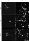
(A) Morphology of fibroblasts in LD and HD matrices after 1 h. LD matrices and HD matrices were incubated for 1 h in basal medium (BSA) or medium containing LPA or PDGF. At the end of the incubations, samples were fixed and stained for actin. Cells in matrices in basal medium or PDGF-containing medium formed a dendritic network of cell extensions. In LPA-containing medium these extensions were retracted completely in LD matrices and partly in HD matrices. In HD matrices and PDGF or LPA medium, actin stress fibers were evident. Bar, 17 μm. (B) Morphology of fibroblasts in LD and HD matrices after 4 h. LD matrices and HD matrices were incubated for 4 h in basal medium or medium containing LPA or PDGF. At the end of the incubations, samples were fixed and stained for actin. In LD matrices, cells in basal medium or PDGF-containing medium extended further the dendritic network of cell extensions. In LPA-containing medium, small extensions reappeared and tended to be organized in a bipolar manner in contrast to the circumferential distribution of extensions around cells in basal or PDGF medium. In HD matrices, cells became stellate or bipolar and contained stressed fibers in PDGF- and LPA-containing medium. Bar, 17 μm.
Figure 4
Figure 4
(A) Morphology of fibroblasts in LD and HD matrices after 1 h. LD matrices and HD matrices were incubated for 1 h in basal medium (BSA) or medium containing LPA or PDGF. At the end of the incubations, samples were fixed and stained for actin. Cells in matrices in basal medium or PDGF-containing medium formed a dendritic network of cell extensions. In LPA-containing medium these extensions were retracted completely in LD matrices and partly in HD matrices. In HD matrices and PDGF or LPA medium, actin stress fibers were evident. Bar, 17 μm. (B) Morphology of fibroblasts in LD and HD matrices after 4 h. LD matrices and HD matrices were incubated for 4 h in basal medium or medium containing LPA or PDGF. At the end of the incubations, samples were fixed and stained for actin. In LD matrices, cells in basal medium or PDGF-containing medium extended further the dendritic network of cell extensions. In LPA-containing medium, small extensions reappeared and tended to be organized in a bipolar manner in contrast to the circumferential distribution of extensions around cells in basal or PDGF medium. In HD matrices, cells became stellate or bipolar and contained stressed fibers in PDGF- and LPA-containing medium. Bar, 17 μm.
Figure 5
Figure 5
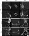
(A) Codistribution of vinculin and actin in fibroblasts in LD and HD matrices after 1 h. LD matrices and HD matrices were incubated for 1 h in basal medium (BSA) or medium containing LPA or PDGF. At the end of the incubations, samples were fixed and stained for actin and vinculin. Fibroblasts in LD matrices had diffuse vinculin staining. The large punctate spots of vinculin in cells stimulated by LPA might have been related to cell-blebbing activity. Actin stress fibers were undetectable. Cells in HD matrices had streaks of vinculin and short actin stress fibers at the tips of cell extensions in medium containing LPA or PDGF. Bar, 7 μm. (B) Codistribution of vinculin and actin in fibroblasts in LD and HD matrices after 4 h. LD matrices and HD matrices were incubated for 4 h in basal medium containing LPA or PDGF. At the end of the incubations, samples were fixed and stained for actin and vinculin. In LD matrices after 1 h, vinculin staining was diffuse. In HD matrices and PDGF or LPA medium, vinculin streaks were prominent and widely distributed over the extensions, and actin stress fibers seemed to insert into the focal adhesions. Bar, 7 μm.
Figure 5
Figure 5
(A) Codistribution of vinculin and actin in fibroblasts in LD and HD matrices after 1 h. LD matrices and HD matrices were incubated for 1 h in basal medium (BSA) or medium containing LPA or PDGF. At the end of the incubations, samples were fixed and stained for actin and vinculin. Fibroblasts in LD matrices had diffuse vinculin staining. The large punctate spots of vinculin in cells stimulated by LPA might have been related to cell-blebbing activity. Actin stress fibers were undetectable. Cells in HD matrices had streaks of vinculin and short actin stress fibers at the tips of cell extensions in medium containing LPA or PDGF. Bar, 7 μm. (B) Codistribution of vinculin and actin in fibroblasts in LD and HD matrices after 4 h. LD matrices and HD matrices were incubated for 4 h in basal medium containing LPA or PDGF. At the end of the incubations, samples were fixed and stained for actin and vinculin. In LD matrices after 1 h, vinculin staining was diffuse. In HD matrices and PDGF or LPA medium, vinculin streaks were prominent and widely distributed over the extensions, and actin stress fibers seemed to insert into the focal adhesions. Bar, 7 μm.
Figure 6
Figure 6
(A) Distribution of β1 integrin in fibroblasts in LD and HD matrices after 1 h. LD matrices and HD matrices were incubated for 1 h in basal medium (BSA) or medium containing LPA or PDGF. At the end of the incubations, samples were fixed and stained for β1 integrin. Ligand-occupied β1 integrin could be detected in a punctate distribution on the cell body and extensions of fibroblasts in LD and HD matrices with or without growth factors. Bar, 10 μm. (B) Distribution of β1 integrin in fibroblasts in HD and LD matrices after 4 h. HD matrices and LD matrices were incubated for 4 h in basal medium or medium containing LPA or PDGF. At the end of the incubations, samples were fixed and stained for β1 integrin. In LD matrices, some linear arrays of β1 integrin were evident by 4 h. In HD matrices, linear arrays and streaks resembling focal adhesions occurred by 4 h in LPA- or PDGF-containing medium. Bar, 10 μm.
Figure 6
Figure 6
(A) Distribution of β1 integrin in fibroblasts in LD and HD matrices after 1 h. LD matrices and HD matrices were incubated for 1 h in basal medium (BSA) or medium containing LPA or PDGF. At the end of the incubations, samples were fixed and stained for β1 integrin. Ligand-occupied β1 integrin could be detected in a punctate distribution on the cell body and extensions of fibroblasts in LD and HD matrices with or without growth factors. Bar, 10 μm. (B) Distribution of β1 integrin in fibroblasts in HD and LD matrices after 4 h. HD matrices and LD matrices were incubated for 4 h in basal medium or medium containing LPA or PDGF. At the end of the incubations, samples were fixed and stained for β1 integrin. In LD matrices, some linear arrays of β1 integrin were evident by 4 h. In HD matrices, linear arrays and streaks resembling focal adhesions occurred by 4 h in LPA- or PDGF-containing medium. Bar, 10 μm.
Figure 7
Figure 7
Distribution of α-actinin in fibroblasts in LD and HD matrices after 1 or 4 h. HD matrices and LD matrices were incubated for 1 or 4 h in basal medium (BSA) or medium containing LPA or PDGF. At the end of the incubations, samples were fixed and stained for α-actinin. α-Actinin staining accumulated at the tips of cell extensions in both LD and HD matrices, but staining was more evident in LPA or PDGF than basal medium. In HD matrices, the α-actinin staining pattern of the extensions became organized into linear streaks after 4 h. Bar, 7 μm.
Figure 8
Figure 8
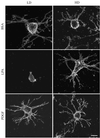
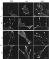
Effect of Rho kinase inhibitor on codistribution of vinculin and actin in fibroblasts in HD matrices after 4 h. HD matrices were incubated for 4 h in basal medium (BSA) or medium containing LPA or PDGF in the presence of Rho kinase inhibitor Y27632 (10 μM). At the end of the incubations, samples were fixed and stained for actin and vinculin. Maturation of cells from the dendritic network of extensions to stellate/bipolar appearance was completely blocked and actin stress fibers and streaks of vinculin staining were absent. Bar, 7 μm.
Figure 9
Figure 9
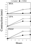
Effect of Rho kinase inhibitor on global remodeling of collagen matrices in the presence of Rho kinase inhibitor. HD matrices were incubated for the times shown in basal medium (BSA) or medium containing LPA or PDGF (□) or plus Rho kinase inhibitor Y27632 (10 μM) (▪). At the end of the incubations, contraction was determined by measuring cell height. Addition of Y27632 inhibited global remodeling in basal medium almost completely but only partly reduced remodeling in LPA and PDGF medium. Studies were carried out in triplicate. Data show averages and SDs (most of which were smaller than the size of the points).

Similar articles

Cited by

References

    1. Amano M, Chihara K, Kimura K, Fukata Y, Nakamura N, Matsuura Y, Kaibuchi K. Formation of actin stress fibers and focal adhesions enhanced by Rho-kinase. Science. 1997;275:1308–1311. - PubMed
    1. Bell E, Ivarsson B, Merrill C. Production of a tissue-like structure by contraction of collagen lattices by human fibroblasts of different proliferative potential in vitro. Proc Natl Acad Sci USA. 1979;76:1274–1278. - PMC - PubMed
    1. Beningo KA, Dembo M, Kaverina I, Small JV, Wang YL. Nascent focal adhesions are responsible for the generation of strong propulsive forces in migrating fibroblasts. J Cell Biol. 2001;153:881–888. - PMC - PubMed
    1. Brown RA, Prajapati R, McGrouther DA, Yannas IV, Eastwood M. Tensional homeostasis in dermal fibroblasts: mechanical responses to mechanical loading in three-dimensional substrates. J Cell Physiol. 1998;175:323–332. - PubMed
    1. Burridge K, Chrzanowska-Wodnicka M. Focal adhesions, contractility, and signaling. Annu Rev Cell Dev Biol. 1996;12:463–518. - PubMed

Publication types