

Heme deficiency may be a factor in the mitochondrial and neuronal decay of aging
- PMID: 12417755
- PMCID: PMC137500
- DOI: 10.1073/pnas.192585799
Heme deficiency may be a factor in the mitochondrial and neuronal decay of aging
Abstract
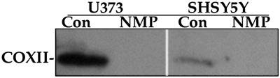
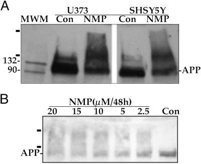
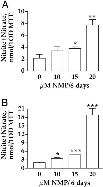
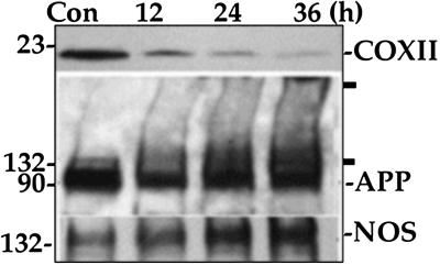
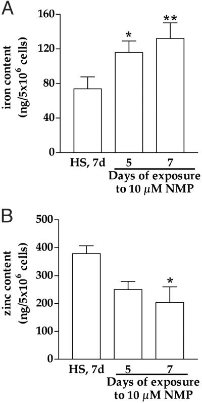
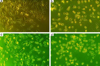
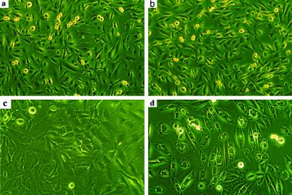
Heme, a major functional form of iron in the cell, is synthesized in the mitochondria by ferrochelatase inserting ferrous iron into protoporphyrin IX. Heme deficiency was induced with N-methylprotoporphyrin IX, a selective inhibitor of ferrochelatase, in two human brain cell lines, SHSY5Y (neuroblastoma) and U373 (astrocytoma), as well as in rat primary hippocampal neurons. Heme deficiency in brain cells decreases mitochondrial complex IV, activates nitric oxide synthase, alters amyloid precursor protein, and corrupts iron and zinc homeostasis. The metabolic consequences resulting from heme deficiency seem similar to dysfunctional neurons in patients with Alzheimer's disease. Heme-deficient SHSY5Y or U373 cells die when induced to differentiate or to proliferate, respectively. The role of heme in these observations could result from its interaction with heme regulatory motifs in specific proteins or secondary to the compromised mitochondria. Common causes of heme deficiency include aging, deficiency of iron and vitamin B6, and exposure to toxic metals such as aluminum. Iron and B6 deficiencies are especially important because they are widespread, but they are also preventable with supplementation. Thus, heme deficiency or dysregulation may be an important and preventable component of the neurodegenerative process.
Figures
Similar articles
-
Heme, iron, and the mitochondrial decay of ageing.Ageing Res Rev. 2004 Jul;3(3):303-18. doi: 10.1016/j.arr.2004.02.002. Ageing Res Rev. 2004. PMID: 15231238 Review.
-
Mineral and vitamin deficiencies can accelerate the mitochondrial decay of aging.Mol Aspects Med. 2005 Aug-Oct;26(4-5):363-78. doi: 10.1016/j.mam.2005.07.007. Mol Aspects Med. 2005. PMID: 16102804 Review.
-
Frataxin deficiency alters heme pathway transcripts and decreases mitochondrial heme metabolites in mammalian cells.Hum Mol Genet. 2005 Dec 15;14(24):3787-99. doi: 10.1093/hmg/ddi393. Epub 2005 Oct 20. Hum Mol Genet. 2005. PMID: 16239244
-
Enzymatic formation of zinc-protoporphyrin by rat liver and its potential effect on hepatic heme metabolism.Gastroenterology. 1983 Sep;85(3):663-8. Gastroenterology. 1983. PMID: 6873612
-
Heme deficiency is associated with senescence and causes suppression of N-methyl-D-aspartate receptor subunits expression in primary cortical neurons.Mol Pharmacol. 2006 Mar;69(3):697-705. doi: 10.1124/mol.105.016675. Epub 2005 Nov 23. Mol Pharmacol. 2006. PMID: 16306232
Cited by
-
Transition between acute and chronic hepatotoxicity in mice is associated with impaired energy metabolism and induction of mitochondrial heme oxygenase-1.PLoS One. 2013 Jun 6;8(6):e66094. doi: 10.1371/journal.pone.0066094. Print 2013. PLoS One. 2013. PMID: 23762471 Free PMC article.
-
Iron accumulation during cellular senescence in human fibroblasts in vitro.Antioxid Redox Signal. 2003 Oct;5(5):507-16. doi: 10.1089/152308603770310158. Antioxid Redox Signal. 2003. PMID: 14580305 Free PMC article.
-
The mechanism for heme to prevent Aβ(1-40) aggregation and its cytotoxicity.J Biol Inorg Chem. 2011 Jun;16(5):809-16. doi: 10.1007/s00775-011-0783-x. Epub 2011 Apr 27. J Biol Inorg Chem. 2011. PMID: 21523435
-
Biomarkers in autism.Front Psychiatry. 2014 Aug 12;5:100. doi: 10.3389/fpsyt.2014.00100. eCollection 2014. Front Psychiatry. 2014. PMID: 25161627 Free PMC article. Review.
-
Recent Development in the Understanding of Molecular and Cellular Mechanisms Underlying the Etiopathogenesis of Alzheimer's Disease.Int J Mol Sci. 2023 Apr 14;24(8):7258. doi: 10.3390/ijms24087258. Int J Mol Sci. 2023. PMID: 37108421 Free PMC article. Review.
References
-
- Parker W. D., Parks, J., Filley, C. M. & Kleinschmidt-DeMasters, B. K. (1994) Neurology 44, 1090-1096. - PubMed
-
- Albers D. S. & Beal, M. F. (2000) J. Neural Transm. Suppl. 59, 133-154. - PubMed
-
- Beal M. F. (2000) Trends Neurosci. 23, 298-304. - PubMed
-
- Smith M. A., Nunomura, A., Zhu, X., Takeda, A. & Perry, G. (2000) Antioxid. Redox Signal. 2, 413-420. - PubMed
Publication types
MeSH terms
Substances
Grants and funding
LinkOut - more resources
Full Text Sources
Other Literature Sources
