. 2002 Dec;76(23):11889-903.
doi: 10.1128/jvi.76.23.11889-11903.2002.

Mapping the encapsidation determinants of feline immunodeficiency virus

Affiliations

Mapping the encapsidation determinants of feline immunodeficiency virus

Iris Kemler et al. J Virol. 2002 Dec.

Abstract

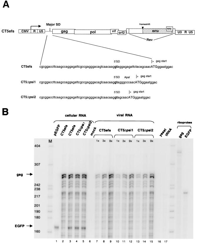
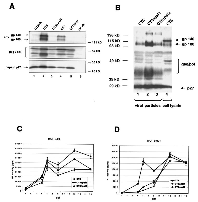
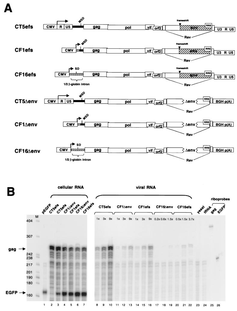
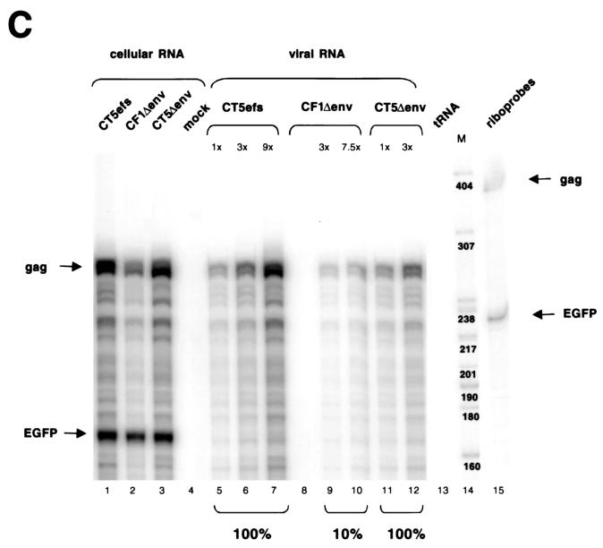
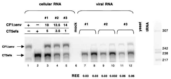
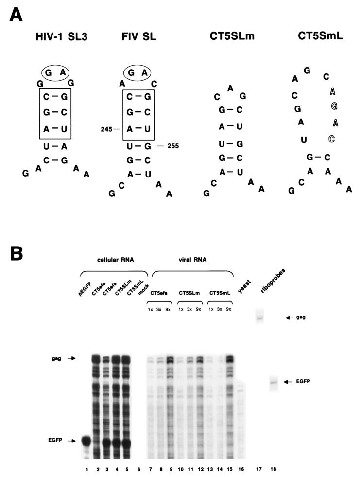
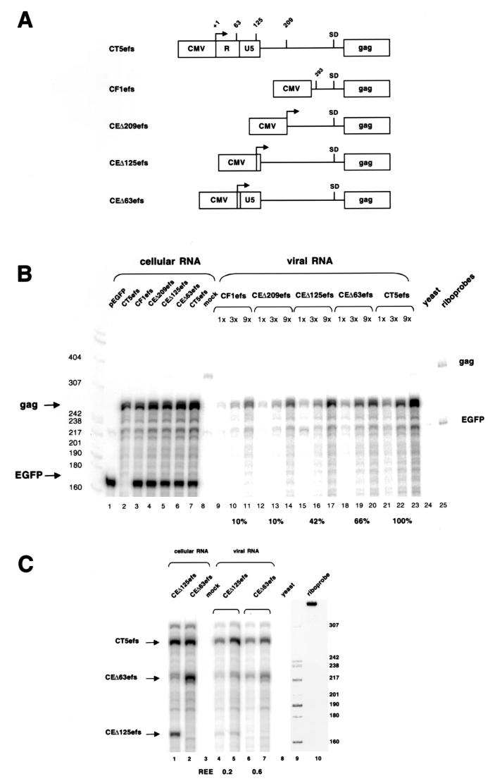
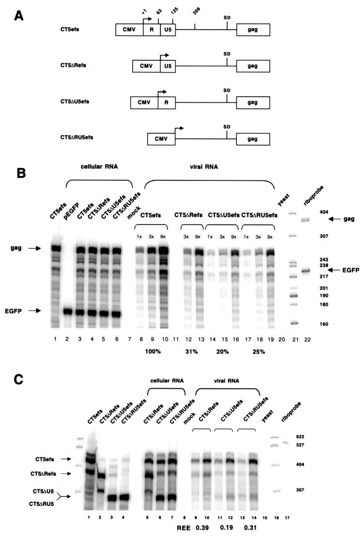
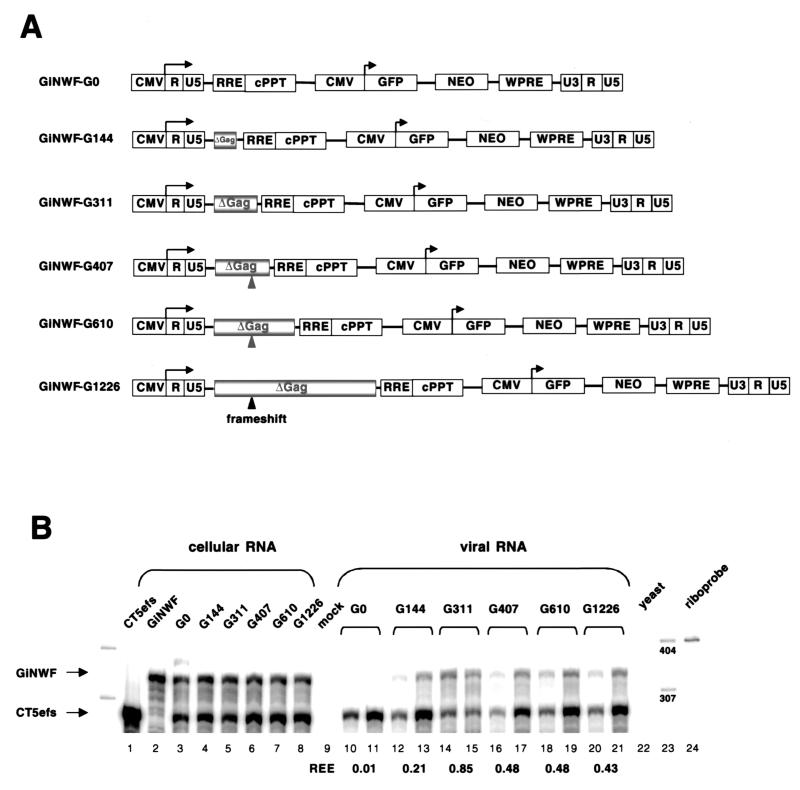
Encapsidation of retroviral RNA involves specific interactions between viral proteins and cis-acting genomic RNA sequences. Human immunodeficiency virus type 1 (HIV-1) RNA encapsidation determinants appear to be more complex and dispersed than those of murine retroviruses. Feline lentiviral (feline immunodeficiency virus [FIV]) encapsidation has not been studied. To gain comparative insight into lentiviral encapsidation and to optimize FIV-based vectors, we used RNase protection assays of cellular and virion RNAs to determine packaging efficiencies of FIV deletion mutants, and we studied replicative phenotypes of mutant viruses. Unlike the case for other mammalian retroviruses, the sequences between the major splice donor (MSD) and the start codon of gag contribute negligibly to FIV encapsidation. Moreover, molecular clones having deletions in this region were replication competent. In contrast, sequences upstream of the MSD were important for encapsidation, and deletion of the U5 element markedly reduced genomic RNA packaging. The contribution of gag sequences to packaging was systematically investigated with subgenomic FIV vectors containing variable portions of the gag open reading frame, with all virion proteins supplied in trans. When no gag sequence was present, packaging was abolished and marker gene transduction was absent. Inclusion of the first 144 nucleotides (nt) of gag increased vector encapsidation to detectable levels, while inclusion of the first 311 nt increased it to nearly wild-type levels and resulted in high-titer FIV vectors. However, the identified proximal gag sequence is necessary but not sufficient, since viral mRNAs that contain all coding regions, with or without as much as 119 nt of adjacent upstream 5' leader, were excluded from encapsidation. The results identify a mechanism whereby FIV can encapsidate its genomic mRNA in preference to subgenomic mRNAs.


Figures

FIG. 1.
FIG. 1.
Sequences between the MSD and the start codon of gag contribute negligibly to encapsidation. (A) Schematic representation of CT5efs, a provirus with a frameshifting (29-nt) oligonucleotide insertion in the SU domain of env. In CT5Δpsi1 and CT5Δpsi2,sequences between the MSD and the gag start codon are deleted. (B) 293T cells were transiently transfected with CT5efs, CT5Δpsi1, or CT5Δpsi2 together with pEGFP-N1. Cellular RNA and viral RNA were subjected to an RPA with antisense riboprobes pSPT-CT5PB and pSPT-EGFP. Protected fragments are 279 and 163 nt long, respectively. RNAs from cells transfected with either pEGFP or CT5efs alone are shown in lanes 1 and 2, respectively. Viral RNAs were analyzed in three different amounts (1×, 3×, and 9×).
FIG. 2.
FIG. 2.
Deletions in CT5Δpsi1 and CT5Δpsi2 have no effect on FIV protein expression and viral replication. (A) Western blot with cell lysates from 293T cells transfected with the indicated plasmids. (B) Western blot with lysates from viral particles CT5, CT5Δpsi1, and CT5Δpsi2 (lanes 1 to 3), harvested from CrFK.CXCR4 cells at 12 days postinfection, and a cell lysate from CT5-transfected 293T cells (lane 4). Both membranes were probed with Petaluma serum, a FIV-infected-cat serum. The secondary antibody was a peroxidase-conjugated goat anti-feline immunoglobulin G. (C and D) CrFK.CXCR4 cells were infected with CT5, CT5Δpsi1, or CT5Δpsi2 viral preparations at an MOI of 0.01 (C) or 0.001 (D). At different days postinfection (dpi), supernatant was collected and virus growth was measured by an RT assay. Error bars indicate standard deviations.
FIG. 3.
FIG. 3.
The 5′ LTR and 154 nt of the leader are required for FIV encapsidation. (A) Schematic representation of plasmids analyzed. CT5efs, CF1efs, and CF16efs have a frameshifting mutation in the env gene. In CT5Δenv, CF1Δenv, and CF16Δenv, 875 nt is deleted from the env gene and the 3′ LTR is replaced by the bovine growth hormone (BGH) polyadenylation signal. The FIV leader is represented by a black bar; non-FIV sequences are stippled. In CF1efs and CF1Δenv, the 5′ LTR and 154 nt of the leader are replaced by the CMV promoter. In CF16efs and CF16Δenv, all viral sequences between the CMV promoter and the ATG gag codon are replaced by β-globin intron sequences. (B) RPA of a noncompetitive encapsidation experiment with CT5efs, CF1Δenv, CF1efs, CF16Δenv, and CF16efs transfected with pEGFP-N1 into 293T cells. Antisense riboprobes pSPT-CT5PB and pSPT-EGFP were used to detect viral gag mRNA and eGFP mRNA, respectively. (C) Noncompetitive encapsidation assay with CT5efs, CF1Δenv, and CT5Δenv. The same riboprobes as for panel B were used.
FIG. 3.
FIG. 3.
The 5′ LTR and 154 nt of the leader are required for FIV encapsidation. (A) Schematic representation of plasmids analyzed. CT5efs, CF1efs, and CF16efs have a frameshifting mutation in the env gene. In CT5Δenv, CF1Δenv, and CF16Δenv, 875 nt is deleted from the env gene and the 3′ LTR is replaced by the bovine growth hormone (BGH) polyadenylation signal. The FIV leader is represented by a black bar; non-FIV sequences are stippled. In CF1efs and CF1Δenv, the 5′ LTR and 154 nt of the leader are replaced by the CMV promoter. In CF16efs and CF16Δenv, all viral sequences between the CMV promoter and the ATG gag codon are replaced by β-globin intron sequences. (B) RPA of a noncompetitive encapsidation experiment with CT5efs, CF1Δenv, CF1efs, CF16Δenv, and CF16efs transfected with pEGFP-N1 into 293T cells. Antisense riboprobes pSPT-CT5PB and pSPT-EGFP were used to detect viral gag mRNA and eGFP mRNA, respectively. (C) Noncompetitive encapsidation assay with CT5efs, CF1Δenv, and CT5Δenv. The same riboprobes as for panel B were used.
FIG. 4.
FIG. 4.
RPA of a competitive encapsidation experiment. CF1Δenv (10, 12.5, or 14 μg) and CT5efs (5, 2.5, or 1 μg) were cotransfected into 293T cells. Antisense riboprobe pSPT-CF1MM was used to detect 5 μg of cellular RNA (lanes 1 to 5) and 5 μl (lanes 7, 9, and 11) or 15 μl (lanes 8, 10, and 12) of viral RNA. Protected RNA species are 281 nt for CF1Δenv and 233 nt for CT5efs. The REE was determined by calculating the ratio of mutant RNA to wild-type RNA in the virion, relative to the ratio of the two RNAs in the cytoplasm (28).
FIG. 5.
FIG. 5.
A putative FIV stem-loop does not contribute to FIV mRNA encapsidation. (A) Schematic representation of HIV-1 SL3 and a putative stem-loop structure in the FIV leader (FIV SL). In CT5SLm the loop was mutated, and in CT5SmL the stem was disrupted. (B) RPA of a noncompetitive encapsidation experiment with CT5efs, CT5SLm, and CT5SmL transfected with pEGFP-N1 into 293T cells. Antisense riboprobes pSPT-CT5PB and pSPT-EGFP were used to detect viral gag mRNA and eGFP mRNA, respectively.
FIG. 6.
FIG. 6.
(A) Schematic representation of CT5efs, CF1efs, CEΔ209efs, CEΔ125efs, and CEΔ63efs (the numbers refer to the amount of left-hand genomic mRNA sequences missing). (B) RPA of a noncompetitive encapsidation experiment. The plasmids depicted in panel A were cotransfected with pEGFP-N1. Antisense riboprobes were the same as in Fig. 5B. (C) RPA of a competitive encapsidation experiment. Wild-type CT5efs was cotransfected with either CEΔ125efs or CEΔ63efs. Antisense riboprobe pSPT-CT5SR was used. Protected RNA species are 293 nt for CT5efs, 230 nt for CEΔ63efs, and 168 nt for CEΔ125efs.
FIG. 7.
FIG. 7.
Encapsidation determinants are located in R and U5. (A) Schematic representation of CT5efs, CT5ΔRefs, CT5ΔU5efs, and CT5ΔRU5efs. (B) RPA of a noncompetitive encapsidation experiment with CT5efs, CT5ΔRefs, CT5ΔU5efs, and CT5ΔRU5efs transfected with pEGFP-N1. Antisense riboprobes pSPT-CT5PB and pSPT-EGFP were used. (C) RPA of a competitive encapsidation experiment. Wild-type CT5efs was cotransfected with either CT5ΔRefs, CT5ΔU5efs, or CT5ΔRU5efs. Antisense riboprobe pSPT-CT5MM was used. Protected RNA species are 447 nt for CT5efs, 373 nt for CT5ΔRefs, and 307 nt for CT5ΔU5efs and CT5ΔRU5efs.
FIG. 8.
FIG. 8.
The proximal 311 nt of gag is necessary for efficient FIV RNA encapsidation. (A) Schematic representation of GiNWF-G clones, subgenomic FIV transfer vectors containing increasing amounts of gag sequences. (B) RPA of a competitive encapsidation experiment. Wild-type CT5efs was cotransfected with GiNWF-G0, -G144, -G311, -G407, -G610, or -G1226. Antisense riboprobe pSPT-FLAP was used. Protected RNA species are 358 nt for all GiNWF-G clones and 296 nt for CT5efs.

Similar articles

Cited by

References

    1. Adam, M. A., and A. D. Miller. 1988. Identification of a signal in a murine retrovirus that is sufficient for packaging of nonretroviral RNA into virions. J. Virol. 62:3802-3806. - PMC - PubMed
    1. Aldovini, A., and R. A. Young. 1990. Mutations of RNA and protein sequences involved in human immunodeficiency virus type 1 packaging result in production of noninfectious virus. J. Virol. 64:1920-1926. - PMC - PubMed
    1. Banks, J. D., and M. L. Linial. 2000. Secondary structure analysis of a minimal avian leukosis-sarcoma virus packaging signal. J. Virol. 74:456-464. - PMC - PubMed
    1. Banks, J. D., A. Yeo, K. Green, F. Cepeda, and M. L. Linial. 1998. A minimal avian retroviral packaging sequence has a complex structure. J. Virol. 72:6190-6194. - PMC - PubMed
    1. Bender, M. A., T. D. Palmer, R. E. Gelinas, and A. D. Miller. 1987. Evidence that the packaging signal of Moloney murine leukemia virus extends into the Gag region. J. Virol. 61:1639-1646. - PMC - PubMed

Publication types

MeSH terms