. 2002 Oct;13(10):3730-46.
doi: 10.1091/mbc.e02-04-0213.

Continual production of phosphatidic acid by phospholipase D is essential for antigen-stimulated membrane ruffling in cultured mast cells

Affiliations

Continual production of phosphatidic acid by phospholipase D is essential for antigen-stimulated membrane ruffling in cultured mast cells

Niamh O'Luanaigh et al. Mol Biol Cell. 2002 Oct.

Abstract

Phospholipase Ds (PLDs) are regulated enzymes that generate phosphatidic acid (PA), a putative second messenger implicated in the regulation of vesicular trafficking and cytoskeletal reorganization. Mast cells, when stimulated with antigen, show a dramatic alteration in their cytoskeleton and also release their secretory granules by exocytosis. Butan-1-ol, which diverts the production of PA generated by PLD to the corresponding phosphatidylalcohol, was found to inhibit membrane ruffling when added together with antigen or when added after antigen. Inhibition by butan-1-ol was completely reversible because removal of butan-1-ol restored membrane ruffling. Measurements of PLD activation by antigen indicate a requirement for continual PA production during membrane ruffling, which was maintained for at least 30 min. PLD1 and PLD2 are both expressed in mast cells and green fluorescent protein-tagged proteins were used to identify PLD2 localizing to membrane ruffles of antigen-stimulated mast cells together with endogenous ADP ribosylation factor 6 (ARF6). In contrast, green fluorescent protein-PLD1 localized to intracellular vesicles and remained in this location after stimulation with antigen. Membrane ruffling was independent of exocytosis of secretory granules because phorbol 12-myristate 13-acetate increased membrane ruffling in the absence of exocytosis. Antigen or phorbol 12-myristate 13-acetate stimulation increased both PLD1 and PLD2 activity when expressed individually in RBL-2H3 cells. Although basal activity of PLD2-overexpressing cells is very high, membrane ruffling was still dependent on antigen stimulation. In permeabilized cells, antigen-stimulated phosphatidylinositol(4,5)bisphosphate synthesis was dependent on both ARF6 and PA generated from PLD. We conclude that both activation of ARF6 by antigen and a continual PLD2 activity are essential for local phosphatidylinositol(4,5)bisphosphate generation that regulates dynamic actin cytoskeletal rearrangements.


Figures

Figure 1
Figure 1
Inhibition of antigen-induced membrane ruffling by butan-1-ol. (A) Confocal slices of RBL mast cells fixed and stained with TRITC-phalloidin before and after stimulation with antigen. Control cells (a–d), stimulated with antigen for 10 min (e–h), or stimulated with antigen for 10 min in the presence of 0.5% butan-1-ol (i–l). (B) Cells were treated with antigen, antigen plus 0.5% butan-1-ol, and antigen plus 0.5% butan-2-ol. For each condition, >250 cells were examined for their morphology. The results are from three separate experiments and are expressed as a percentage of total cells.
Figure 1
Figure 1
Inhibition of antigen-induced membrane ruffling by butan-1-ol. (A) Confocal slices of RBL mast cells fixed and stained with TRITC-phalloidin before and after stimulation with antigen. Control cells (a–d), stimulated with antigen for 10 min (e–h), or stimulated with antigen for 10 min in the presence of 0.5% butan-1-ol (i–l). (B) Cells were treated with antigen, antigen plus 0.5% butan-1-ol, and antigen plus 0.5% butan-2-ol. For each condition, >250 cells were examined for their morphology. The results are from three separate experiments and are expressed as a percentage of total cells.
Figure 2
Figure 2
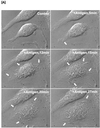
Time-dependent stimulation of membrane ruffling compared with activation of PLD activity by antigen. (A) Phase contrast, time-lapse recording of antigen-stimulated RBL mast cells, demonstrating that membrane ruffling is continuous for at least 30 min. White arrows indicate sites of ruffling. (B) Time course of PLD activation in antigen-stimulated RBL mast cells as monitored by PA formation. (C) Time course of PLD activation in antigen-stimulated RBL mast cells as monitored by PBut formation in the presence of 0.5% butan-1-ol. (D) Determination of ongoing PLD activity: RBL mast cells were incubated with butan-1-ol for 10 or 20 min with and without antigen (c, control and s, stimulated). c20* and s20*, cells incubated with buffer or antigen for 20 min with butan-1-ol present during the last 10 min.
Figure 2
Figure 2
Time-dependent stimulation of membrane ruffling compared with activation of PLD activity by antigen. (A) Phase contrast, time-lapse recording of antigen-stimulated RBL mast cells, demonstrating that membrane ruffling is continuous for at least 30 min. White arrows indicate sites of ruffling. (B) Time course of PLD activation in antigen-stimulated RBL mast cells as monitored by PA formation. (C) Time course of PLD activation in antigen-stimulated RBL mast cells as monitored by PBut formation in the presence of 0.5% butan-1-ol. (D) Determination of ongoing PLD activity: RBL mast cells were incubated with butan-1-ol for 10 or 20 min with and without antigen (c, control and s, stimulated). c20* and s20*, cells incubated with buffer or antigen for 20 min with butan-1-ol present during the last 10 min.
Figure 3
Figure 3
Butano-1-ol but not butan-2-ol inhibits membrane ruffling when added after membrane ruffling is initiated. Inhibition by butan-1-ol is reversible. (A) RBL mast cells were stimulated with antigen and bright field images of the cells were acquired over a 30-min period by using Nomarski phase contrast optics. Control cells (a), same cells imaged 5 min after antigen addition (b); at 5 min after antigen addition, butan-1-ol was added to halt membrane ruffling and the cells imaged at 10 min (c); the same cells imaged at 15 min to illustrate that membrane ruffling is still inhibited in the presence of butan-1-ol (d). At this point the cells were washed free of butan-1-ol and antigen added. Membrane ruffling 5 min after washout of butan-1-ol (e) and membrane ruffling still ongoing 10 min after washout of butan-1-ol (f). (B) Butan-2-ol does not inhibit membrane ruffling. Control cells (a); same field of cells after 5 min with antigen (b); 10 min with antigen and 5 min with butan-2-ol (c); 15 min with antigen and 10 min with butan-2-ol (d).
Figure 4
Figure 4
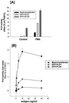
Antigen or PMA can regulate the activity of PLD1 and PLD2 in RBL mast cells. (A) Basal and PMA-stimulated PLD activity measured by formation of PBut is enhanced in GFP-PLD1– and GFP-PLD2–transfected cells. PMA, 100 nM for 20 min. (B) Antigen stimulates PLD activity as measured by formation of PBut in a concentration-dependent manner. The cells were stimulated for 20 min with antigen. (C) PLD1-transfected RBL mast cells: Top, control cells and bottom, same field of cells after stimulation with antigen for 10 min. The Nomarski image (a) of the field of cells is shown together with three confocal slices, showing the adhesion plane (b), the midplane (c), and the top of the cell (d). PLD1 localizes mainly to intracellular vesicles in unstimulated cells, and this pattern of staining is unchanged after 10 min of stimulation. No change in staining was observed at 5 min or 20 min poststimulation either. (D) PLD2-transfected RBL mast cells: Top, control cells and bottom, same field of cells after stimulation with antigen for 10 min. The Nomarski image (a) of the field of cells is shown together with three confocal slices, showing the adhesion plane (b), the midplane (c), and the top of the cell (d). PLD2 localizes to the plasma membrane and upon antigen addition, PLD2 is found in membrane ruffles and in pinocytic vesicles (indicated by arrow) that accompany membrane ruffling.
Figure 4
Figure 4
Antigen or PMA can regulate the activity of PLD1 and PLD2 in RBL mast cells. (A) Basal and PMA-stimulated PLD activity measured by formation of PBut is enhanced in GFP-PLD1– and GFP-PLD2–transfected cells. PMA, 100 nM for 20 min. (B) Antigen stimulates PLD activity as measured by formation of PBut in a concentration-dependent manner. The cells were stimulated for 20 min with antigen. (C) PLD1-transfected RBL mast cells: Top, control cells and bottom, same field of cells after stimulation with antigen for 10 min. The Nomarski image (a) of the field of cells is shown together with three confocal slices, showing the adhesion plane (b), the midplane (c), and the top of the cell (d). PLD1 localizes mainly to intracellular vesicles in unstimulated cells, and this pattern of staining is unchanged after 10 min of stimulation. No change in staining was observed at 5 min or 20 min poststimulation either. (D) PLD2-transfected RBL mast cells: Top, control cells and bottom, same field of cells after stimulation with antigen for 10 min. The Nomarski image (a) of the field of cells is shown together with three confocal slices, showing the adhesion plane (b), the midplane (c), and the top of the cell (d). PLD2 localizes to the plasma membrane and upon antigen addition, PLD2 is found in membrane ruffles and in pinocytic vesicles (indicated by arrow) that accompany membrane ruffling.
Figure 5
Figure 5
Membrane ruffling in PLD2-transfected cells stimulated with antigen. GFP-PLD2–transfected RBL mast cells were imaged live at 37°C. Still images from time-lapse recording are shown. Arrows point to PLD2-containing circular structures forming over time.
Figure 6
Figure 6
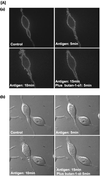
Membrane ruffling in PLD2-transfected cells is sensitive to inhibition by butan-1-ol but not butan-2-ol. (A, a) PLD2-transfected cell. The cells were stimulated with antigen for 10 min (images at 5 and 10 min postantigen are shown) to illustrate the pattern of membrane ruffling. Butan-1-ol (0.5%) was added at 10 min and the cells were imaged for another 5 min. Membrane ruffling was inhibited upon addition of butan-1-ol. (b) Same field of cells as observed by Normaski optics. (B, c) PLD2-transfected cell. The cells were stimulated with antigen for 10 min (images at 5 and 10 min postantigen are shown) to illustrate the pattern of membrane ruffling. Butan-2-ol (0.5%) was added at 10 min, and the cells were imaged for another 10 min. Membrane ruffling was unaffected by the presence of 0.5% butan-2-ol. (d) Same field of cells as observed by Normaski optics. Note that membrane ruffling of the transfected cells is similar to the membrane ruffling observed in the nontransfected counterparts.
Figure 6
Figure 6
Membrane ruffling in PLD2-transfected cells is sensitive to inhibition by butan-1-ol but not butan-2-ol. (A, a) PLD2-transfected cell. The cells were stimulated with antigen for 10 min (images at 5 and 10 min postantigen are shown) to illustrate the pattern of membrane ruffling. Butan-1-ol (0.5%) was added at 10 min and the cells were imaged for another 5 min. Membrane ruffling was inhibited upon addition of butan-1-ol. (b) Same field of cells as observed by Normaski optics. (B, c) PLD2-transfected cell. The cells were stimulated with antigen for 10 min (images at 5 and 10 min postantigen are shown) to illustrate the pattern of membrane ruffling. Butan-2-ol (0.5%) was added at 10 min, and the cells were imaged for another 10 min. Membrane ruffling was unaffected by the presence of 0.5% butan-2-ol. (d) Same field of cells as observed by Normaski optics. Note that membrane ruffling of the transfected cells is similar to the membrane ruffling observed in the nontransfected counterparts.
Figure 7
Figure 7
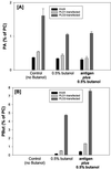
Measurements of PA and PBut in PLD1- and PLD2-overexpressing RBL mast cells before and after stimulation with antigen. Cells transfected with GFP-PLD2 were stimulated with antigen for 20 min in the presence and absence of butan-1-ol. The samples were analyzed by two-dimensional TLC to separate PA (A) and PBut (B). In the absence of butan-1-ol, PA levels are enhanced fourfold in PLD2-transfected cells. Presence of butan-1-ol decreases the PA levels, and this is accompanied by a pronounced increase in PBut (note the scale for PA and PBut is different), illustrating that PLD activity is high but does not result in accumulation of PA. In the presence of antigen and butano-1-ol, levels of PA are not elevated further, although an increase in PBut is clearly observed.
Figure 8
Figure 8
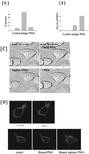
PMA stimulates membrane ruffling but not exocytosis in RBL mast cells. (A) Antigen but not PMA stimulates release of hexosaminidase. (B) Antigen and PMA both stimulate PLD activity. (C) Butan-1-ol inhibits PMA-induced membrane ruffling. Cells were incubated with 0.5% butan-1-ol. PMA (10 nM) was subsequently added to the cells to stimulate membrane ruffling. No membrane ruffling was observed as long as butan-1-ol was present. After removal of butan-1-ol, membrane ruffling commenced. (D) Membrane ruffling stimulated in PLD2-transfected cells with PMA (top). Membrane ruffling is inhibited by butan-1-ol and can be restored upon washout of butan-1-ol (bottom). Butan-1-ol was washed out at 10 min after PMA addition.
Figure 9
Figure 9
ARF6 and PA are both required for the stimulation of PIP2 synthesis in antigen-stimulated RBL mast cells. (A) In streptolysin-O–permeabilized RBL mast cells, ARF1 but not ARF6 leaks out of cells. (B) Synthesis of PI(4,5)P2 by antigen is dependent on the addition of ARF6 proteins to permeabilized RBL mast cells. (C) ARF6 plus antigen-stimulated PIP2 synthesis requires PLD-generated PA production. Butan-1-ol but not butan-2-ol (0.5%) blocks PIP2 synthesis stimulated with antigen plus ARF6.

Similar articles

Cited by

References

    1. Apgar JR. Activation of protein kinase C in rat basophilic leukemia cells stimulates increased production of phosphatidylinositol 4-phosphate and phosphatidylinositol 4,5-bisphosphate: correlation with actin polymerization. Mol Biol Cell. 1995;6:97–108. - PMC - PubMed
    1. Azuma T, Witke W, Stossel TP, Hartwig JH, Kwiatkowski DJ. Gelsolin is a downstream effector of rac for fibroblast motility. EMBO J. 2000;17:1362–1370. - PMC - PubMed
    1. Barker SA, Caldwell KK, Hall A, Martinez AM, Pfeiffer JR, Oliver JM, Wilson BS. Wortmannin blocks lipid and protein kinase activities associated with PI 3-kinase and inhibits a subset of responses induced by FcεR1 cross linking. Mol Biol Cell. 1995;6:1145–1158. - PMC - PubMed
    1. Brown MT, Andrade J, Radhakrishna H, Donaldson J, Cooper JA, Randazzo P. ASAP1, a phospholipid-dependent Arf GTPase-activating protein that associates with and is phosphorylated by Src. Mol Cell Biol. 1998b;18:7038–7051. - PMC - PubMed
    1. Brown FD, Thompson N, Saqib KM, Clark JM, Powner D, Thompson NT, Solari R, Wakelam MJO. Phospholipase D1 localizes to secretory granules and lysosomes and is plasma-membrane located on cellular stimulation. Curr Biol. 1998a;8:835–838. - PubMed

Publication types

MeSH terms

LinkOut - more resources