Identification of the orthopoxvirus p4c gene, which encodes a structural protein that directs intracellular mature virus particles into A-type inclusions
- PMID: 12388681
- PMCID: PMC136765
- DOI: 10.1128/jvi.76.22.11216-11225.2002
Identification of the orthopoxvirus p4c gene, which encodes a structural protein that directs intracellular mature virus particles into A-type inclusions
Abstract
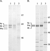
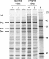
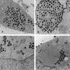
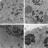
The orthopoxvirus gene p4c has been identified in the genome of the vaccinia virus strain Western Reserve. This gene encodes the 58-kDa structural protein P4c present on the surfaces of the intracellular mature virus (IMV) particles. The gene is disrupted in the genome of cowpox virus Brighton Red (BR), demonstrating that although the P4c protein may be advantageous for virus replication in vivo, it is not essential for virus replication in vitro. Complementation and recombination analyses with the p4c gene have shown that the P4c protein is required to direct the IMV into the A-type inclusions (ATIs) produced by cowpox virus BR. The p4c gene is highly conserved among most members of the orthopoxvirus genus, including viruses that produce ATIs, such as cowpox, ectromelia, and raccoonpox viruses, as well as those such as variola, monkeypox, vaccinia, and camelpox viruses, which do not. The conservation of the p4c gene among the orthopoxviruses, irrespective of their capacities to produce ATIs, suggests that the P4c protein provides functions in addition to that of directing IMV into ATIs. These findings, and the presence of the P4c protein in IMV but not extracellular enveloped virus (D. Ulaeto, D. Grosenbach, and D. E. Hruby, J. Virol. 70:3372-3377, 1996), suggest a model in which the P4c protein may play a role in the retrograde movement of IMV particles, thereby contributing to the retention of IMV particles within the cytoplasm and within ATIs when they are present. In this way, the P4c protein may affect both viral morphogenesis and processes of virus dissemination.
Figures


Similar articles
-
Comparative sequence analysis of A-type inclusion (ATI) and P4c proteins of orthopoxviruses that produce typical and atypical ATI phenotypes.Virus Genes. 2009 Oct;39(2):200-9. doi: 10.1007/s11262-009-0376-8. Virus Genes. 2009. PMID: 19533319
-
Vaccinia virus gene A36R encodes a M(r) 43-50 K protein on the surface of extracellular enveloped virus.Virology. 1994 Oct;204(1):376-90. doi: 10.1006/viro.1994.1542. Virology. 1994. PMID: 8091668
-
Elimination of A-type inclusion formation enhances cowpox virus replication in mice: implications for orthopoxvirus evolution.Virology. 2014 Mar;452-453:59-66. doi: 10.1016/j.virol.2013.12.030. Epub 2014 Jan 29. Virology. 2014. PMID: 24606683 Free PMC article.
-
Vaccinia virus morphogenesis and dissemination.Trends Microbiol. 2008 Oct;16(10):472-9. doi: 10.1016/j.tim.2008.07.009. Epub 2008 Sep 12. Trends Microbiol. 2008. PMID: 18789694 Review.
-
[Research progress in the structure and fuction of Orthopoxvirus host range genes].Bing Du Xue Bao. 2013 Jun;29(4):437-41. Bing Du Xue Bao. 2013. PMID: 23895011 Review. Chinese.
Cited by
-
Orthopoxvirus species and strain differences in cell entry.Virology. 2012 Nov 25;433(2):506-12. doi: 10.1016/j.virol.2012.08.044. Epub 2012 Sep 20. Virology. 2012. PMID: 22999097 Free PMC article.
-
Host-derived pathogenicity islands in poxviruses.Virol J. 2005 Apr 11;2:30. doi: 10.1186/1743-422X-2-30. Virol J. 2005. PMID: 15823205 Free PMC article.
-
Crocodilepox Virus Evolutionary Genomics Supports Observed Poxvirus Infection Dynamics on Saltwater Crocodile (Crocodylus porosus).Viruses. 2019 Dec 2;11(12):1116. doi: 10.3390/v11121116. Viruses. 2019. PMID: 31810339 Free PMC article.
-
In vitro host range, multiplication and virion forms of recombinant viruses obtained from co-infection in vitro with a vaccinia-vectored influenza vaccine and a naturally occurring cowpox virus isolate.Virol J. 2009 May 12;6:55. doi: 10.1186/1743-422X-6-55. Virol J. 2009. PMID: 19435511 Free PMC article.
-
Modern uses of electron microscopy for detection of viruses.Clin Microbiol Rev. 2009 Oct;22(4):552-63. doi: 10.1128/CMR.00027-09. Clin Microbiol Rev. 2009. PMID: 19822888 Free PMC article. Review.
References
-
- Amegadzie, B. Y., J. R. Sisler, and B. Moss. 1992. Frame-shift mutations within the vaccinia virus A-type inclusion protein gene. Virology 186:777-782. - PubMed
-
- Antoine, G., F. Scheiflinger, F. Dorner, and F. G. Falkner. 1998. The complete genomic sequence of the modified vaccinia Ankara strain: comparison with other orthopoxviruses. Virology 244:365-396. - PubMed
-
- Cudmore, S., P. Cossart, G. Griffiths, and M. Way. 1995. Actin-based motility of vaccinia virus. Nature 378:636-638. - PubMed
Publication types
MeSH terms
Substances
Associated data
- Actions
- Actions
Grants and funding
LinkOut - more resources
Full Text Sources
Other Literature Sources

