. 2002 Aug 19;158(4):647-57.
doi: 10.1083/jcb.200205057. Epub 2002 Aug 19.

The human silent information regulator (Sir)2 homologue hSIRT3 is a mitochondrial nicotinamide adenine dinucleotide-dependent deacetylase

Affiliations

The human silent information regulator (Sir)2 homologue hSIRT3 is a mitochondrial nicotinamide adenine dinucleotide-dependent deacetylase

Bjorn Schwer et al. J Cell Biol. .

Abstract

The yeast silent information regulator (Sir)2 protein links cellular metabolism and transcriptional silencing through its nicotinamide adenine dinucleotide (NAD)-dependent histone deacetylase activity. We report that mitochondria from mammalian cells contain intrinsic NAD-dependent deacetylase activity. This activity is inhibited by the NAD hydrolysis product nicotinamide, but not by trichostatin A, consistent with a class III deacetylase. We identify this deacetylase as the nuclear-encoded human Sir2 homologue hSIRT3, and show that hSIRT3 is located within the mitochondrial matrix. Mitochondrial import of hSIRT3 is dependent on an NH2-terminal amphipathic alpha-helix rich in basic residues. hSIRT3 is proteolytically processed in the mitochondrial matrix to a 28-kD product. This processing can be reconstituted in vitro with recombinant mitochondrial matrix processing peptidase (MPP) and is inhibited by mutation of arginines 99 and 100. The unprocessed form of hSIRT3 is enzymatically inactive and becomes fully activated in vitro after cleavage by MPP. These observations demonstrate the existence of a latent class III deacetylase that becomes catalytically activated upon import into the human mitochondria.


Figures
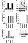
Figure 1.
Figure 1.
Mitochondria contain Sir2-like deacetylase activity. (A) Mitochondrial lysates were prepared from HEK293T cells and assayed for deacetylase activity on a histone H4 peptide in the presence or absence of NAD (1 mM) or in combination with nicotinamide (5 mM) or TSA (400 nM) for 2 h at 25°C. Released acetyl was quantitated as described in Materials and methods. A representative experiment is shown. (B) Western blot analysis of anti-FLAG immunoprecipitates obtained from whole-cell lysates (top) or purified mitochondria (bottom) after transfection of hSIRT3. Because different amounts of cellular and mitochondrial lysates were used in the immunoprecipitation, the top and bottom panels cannot be compared quantitatively. (C) hSIRT proteins were immunoprecipitated from whole-cell lysates of transfected HEK293T cells with anti-FLAG antibodies and assayed in the presence or absence of NAD (1 mM). (D) Purified mitochondria from HEK293T cells transfected with hSIRT proteins were lysed and FLAG-tagged proteins were immunoprecipitated and analyzed for in vitro deacetylase activity. (E) Mitochondria were isolated from HEK293T cells transfected with hSIRT3–FLAG (WT), hSIRT3N229A–FLAG, hSIRT3H248Y–FLAG, or control vector (pFLAG). Lysates were prepared and tested for deacetylase activity. (F) Mitochondria were analyzed for wild-type and mutant hSIRT3 by Western blotting. Two hSIRT3–FLAG specific forms (asterisks) were detected.
Figure 2.
Figure 2.
Subcellular localization of hSIRT3 in mitochondria. (A) hSIRT3–GFP was transfected into HeLa cells grown on coverslips. Cells were stained with the mitochondrial marker MitoTracker, embedded, and analyzed by confocal laser scanning microscopy. (Left) Fluorescence from the hSIRT3–GFP fusion protein. (Middle) Fluorescence from the MitoTracker–stained mitochondria in the same focal plane. (Right) Merged image showing complete overlap of the two staining patterns. (B) HEK293T cells transfected with hSIRT3–FLAG were homogenized and fractionated by differential centrifugation. Equal amounts (30 μg) of heavy membranes (HM), light membranes (LM) and cytosolic proteins (S-100) fraction were analyzed by immunoblotting. hSIRT3–FLAG was revealed by detection with monoclonal M2 anti-FLAG antibodies. Two hSIRT3–FLAG specific forms (asterisks) were detected. Nitrocellulose membranes were stripped and reprobed with antibodies against cytochrome c (cyt c) and Hsp90α. (C) Mitochondria were prepared from HEK293T cells and lysates were analyzed by Western blotting with a polyclonal rabbit hSIRT3 antiserum or preimmune serum obtained from the same rabbit. (D) hSIRT3 was immunoprecipitated from HEK293T cells with hSIRT3 antiserum (0.35 mg/ml), preimmune serum (0.35 mg/ml), or protein G Sepharose. Equal amounts of immunoprecipitate were analyzed for in vitro deacetylase activity in the absence (−) or presence (+) of NAD.
Figure 3.
Figure 3.
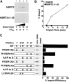
The NH 2 -terminal region of hSIRT3 is required for mitochondrial import. (A) Schematic diagram of hSIRT3. The orange box illustrates the region involved in mitochondrial targeting (left). Parts of the NH2-terminal region show a high probability of forming an amphiphatic α-helix (middle). A helical wheel plot of residues 4–21 reveals a cluster of basic amino acids (black) on one side of the putative helix (right). (B) HeLa cells grown on coverslips were transfected with hSIRT3Δ1–25–GFP for 36 h, stained with MitoTracker, and analyzed by confocal laser scanning microscopy. (left) GFP fluorescence emitted by the fusion protein (green). (Middle) MitoTracker signal (red). (Right) Merged image.
Figure 4.
Figure 4.
Mitochondrial import of hSIRT3 in vitro.(A) [35S]-labeled hSIRT3–FLAG or hSIRT3Δ1–25–FLAG synthesized in rabbit reticulocyte lysate was imported into isolated mammalian mitochondria at 30°C. Import in the absence of Δψm (lane 4), was arranged by adding valinomycin (1 μM), antimycin (8 μM), and oligomycin (20 μM) to mitochondria 5 min before the addition of proteins. Import was stopped after 2, 5, or 15 min by dissipating Δψm (addition of 1 μM valinomycin) and incubation at 0°C. Samples were treated with proteinase K to remove nonimported proteins. Imported proteins were visualized by autoradiography after reisolation of mitochondria and SDS-PAGE. (B) Quantitation of data from panel A by phosphorimaging. (C, left) Mutants were generated to assess the role of the amphipathic helix of hSIRT3 in mitochondrial import. [35S]-labeled hSIRT3 wild-type or mutants were imported into isolated mitochondria for 20 min at 30°C. Import was stopped as described above and nonimported proteins were removed by proteinase K treatment. Reisolated and washed mitochondria were lysed in SDS sample buffer and analyzed by SDS-PAGE. Standards representing 50% of the input used in the individual import reactions were loaded adjacent to each import sample. (Right) Import efficiency of individual hSIRT3 mutants was quantitated in relation to their standards by phosphorimaging. The import efficiency of hSIRT3 (WT) was set to 100%.
Figure 5.
Figure 5.
hSIRT3 is localized in the mitochondrial matrix. (A) Mitochondria were isolated from HEK293T cells transfected with hSIRT3–FLAG and treated with proteinase K to remove proteins bound to the outer mitochondrial surface. Mitochondrial preparations were divided, and one half was diluted with hypotonic buffer to create mitoplasts (MP), while the other half was maintained under isotonic conditions (M). After incubation (20 min at 0°C), mitochondria and mitoplasts were treated again with proteinase K and reisolated by centrifugation followed by Western blotting. Rupture of the outer mitochondrial membrane was confirmed by detection of endogenous intermembrane space protein cytochrome c (cyt c). Integrity of the inner mitochondrial membrane was determined with the matrix protein Hsp60 as a marker. hSIRT3-FLAG was detected using anti-FLAG M2 antibodies. (B) Mitochondria were isolated from HEK293T cells transfected with hSIRT3–FLAG and treated with proteinase K. The preparation was divided, and one half was resuspended in SDS sample buffer (Total, left lane). The other half of the preparation was resuspended in sodium carbonate (Na2CO3) buffer. The extract was centrifuged at 100,000 g at 4°C, and the mitochondrial membranes (Pellet, middle lane) were resuspended in SDS sample buffer. The supernatant containing the soluble and peripheral membrane proteins (Soluble, right lane ) was precipitated with TCA. Samples were analyzed by Western blotting. hSIRT3 was detected with anti–FLAG antibodies. Alkaline extraction was controlled by detection of the marker proteins COXIV and mtHsp70.
Figure 6.
Figure 6.
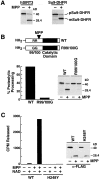
Proteolytic processing of hSIRT3 by MPP leads to enzymatic activation. (A) [35S]-labeled hSIRT3–FLAG (left) or pSu9–DHFR (right) was incubated with purified recombinant yeast MPP for 45 min at 27°C. Samples were analyzed by SDS-PAGE and autoradiography. m, mature form of pSu9–DHFR; p, precursor form. (B) [35S]-labeled hSIRT3–FLAG (WT) and hSIRT3R99/100G–FLAG (R99/1006) were incubated with MPP and analyzed as in A. (Left) Efficiency of proteolytic processing by recombinant yeast MPP was quantitated by phosphorimaging. (Right) Autoradiography of the same experiment. (C) Unlabeled hSIRT3–FLAG (WT) or hSIRT3H248Y–FLAG (H2484) synthesized in vitro in rabbit reticulocyte lysates was incubated with recombinant yeast MPP for 45 min at 27°C. FLAG-tagged proteins were immunoprecipitated with anti-FLAG M2-agarose beads and analyzed for deacetylase activity in vitro with the H4 histone peptide assay in the presence or absence of NAD (1 mM; left). Western blot analysis of immunoprecipitates used in the deacetylase assay (right).

Similar articles

Cited by

References

    1. Abe, Y., T. Shodai, T. Muto, K. Mihara, H. Torii, S. Nishikawa, T. Endo, and D. Kohda. 2000. Structural basis of presequence recognition by the mitochondrial protein import receptor Tom20. Cell. 100:551–560. - PubMed
    1. Afshar, G., and J.P. Murnane. 1999. Characterization of a human gene with sequence homology to Saccharomyces cerevisiae SIR2. Gene. 234:161–168. - PubMed
    1. Arretz, M., H. Schneider, U. Wienhues, and W. Neupert. 1991. Processing of mitochondrial precursor proteins. Biomed. Biochim. Acta. 50:403–412. - PubMed
    1. Bernardi, P. 1999. Mitochondrial transport of cations: channels, exchangers, and permeability transition. Physiol. Rev. 79:1127–1155. - PubMed
    1. Bernardi, P., L. Scorrano, R. Colonna, V. Petronilli, and F. Di Lisa. 1999. Mitochondria and cell death. Mechanistic aspects and methodological issues. Eur. J. Biochem. 264:687–701. - PubMed

Publication types

MeSH terms