

Activation of signal transducer and activator of transcription (STAT) 1 in human chronic inflammatory bowel disease
- PMID: 12171960
- PMCID: PMC1773344
- DOI: 10.1136/gut.51.3.379
Activation of signal transducer and activator of transcription (STAT) 1 in human chronic inflammatory bowel disease
Abstract
Background: Increased expression of proinflammatory cytokines, including tumour necrosis factor alpha, interleukin 6, and interferon gamma, as well as activation of proinflammatory signalling molecules such as nuclear factor kappa B, is characteristic of inflammatory bowel disease (IBD).
Aims: To investigate expression and activation of signal transducer and activator of transcription (STAT) 1 in patients with IBD.
Patients: Patients with active IBD (n=42), disease specificity controls (n=8), and normal controls (n=12) were investigated.
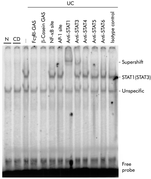
Methods: Expression and activation of STAT1 were assessed by western blotting and electrophoretic mobility shift assays in extracts of endoscopic colonic biopsies. Cellular localisation was determined by immunohistochemistry.
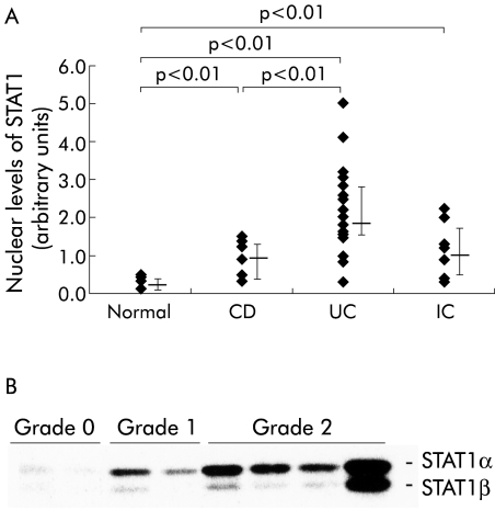
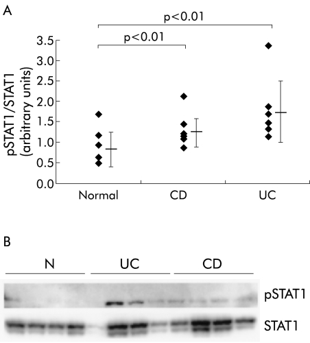
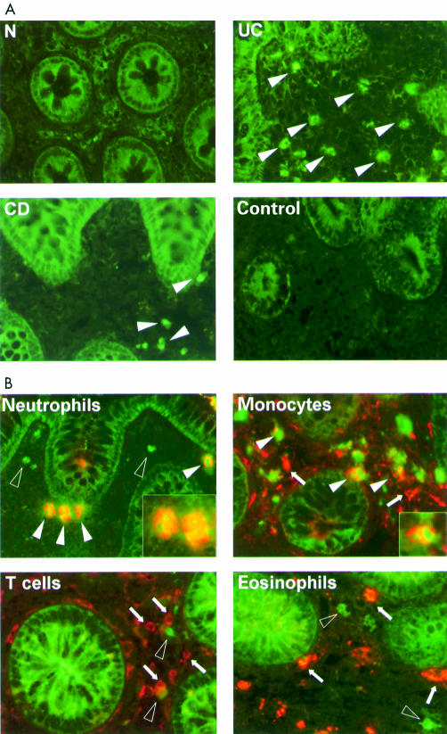
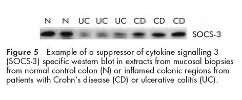
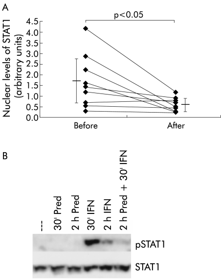
Results: Western blots and immunohistochemical staining revealed an increase in STAT1 expression and activation in mucosal samples from ulcerative colitis and to a lesser extend in Crohn's disease patients. High levels of suppressor of cytokine signalling (SOCS)-3 expression, an inhibitor of STAT activation, were observed in Crohn's disease patients and normal controls in western blot experiments whereas no differences were observed for SOCS-1 expression. Phosphorylated (p) STAT1 was mainly detected in monocytic cells and neutrophils in the inflamed mucosa. Induction of remission by systemic glucocorticoids led to a decrease in levels of pSTAT1. In vitro studies indicated a direct effect of steroid treatment on STAT1 activation.
Conclusions: Expression and activation of STAT1 are predominantly heightened in ulcerative colitis and may therefore play an important role in the pathophysiology of colonic inflammation.
Figures
Similar articles
-
Activation of nuclear factor kappa B inflammatory bowel disease.Gut. 1998 Apr;42(4):477-84. doi: 10.1136/gut.42.4.477. Gut. 1998. PMID: 9616307 Free PMC article.
-
CIS3/SOCS3/SSI3 plays a negative regulatory role in STAT3 activation and intestinal inflammation.J Exp Med. 2001 Feb 19;193(4):471-81. doi: 10.1084/jem.193.4.471. J Exp Med. 2001. PMID: 11181699 Free PMC article.
-
Constitutive activation of the signal transducer and activator of transcription pathway in celiac disease lesions.Am J Pathol. 2003 Jun;162(6):1845-55. doi: 10.1016/S0002-9440(10)64319-2. Am J Pathol. 2003. PMID: 12759242 Free PMC article.
-
Glucocorticoid resistance in inflammatory bowel disease.J Endocrinol. 2003 Sep;178(3):339-46. doi: 10.1677/joe.0.1780339. J Endocrinol. 2003. PMID: 12967327 Review.
-
Differential regulation of JAK/STAT-signaling in patients with ulcerative colitis and Crohn's disease.World J Gastroenterol. 2020 Jul 28;26(28):4055-4075. doi: 10.3748/wjg.v26.i28.4055. World J Gastroenterol. 2020. PMID: 32821070 Free PMC article. Review.
Cited by
-
Isoflavones and inflammatory bowel disease.World J Clin Cases. 2020 Jun 6;8(11):2081-2091. doi: 10.12998/wjcc.v8.i11.2081. World J Clin Cases. 2020. PMID: 32548137 Free PMC article. Review.
-
Differentially Expressed Cell Cycle Genes and STAT1/3-Driven Multiple Cancer Entanglement in Psoriasis, Coupled with Other Comorbidities.Cells. 2022 Nov 30;11(23):3867. doi: 10.3390/cells11233867. Cells. 2022. PMID: 36497125 Free PMC article.
-
STAT1-mediated induction of Ly6c-expressing macrophages are involved in the pathogenesis of an acute colitis model.Inflamm Res. 2022 Sep;71(9):1079-1094. doi: 10.1007/s00011-022-01620-z. Epub 2022 Aug 1. Inflamm Res. 2022. PMID: 35913585
-
Application of Proteomics to Inflammatory Bowel Disease Research: Current Status and Future Perspectives.Gastroenterol Res Pract. 2019 Jan 15;2019:1426954. doi: 10.1155/2019/1426954. eCollection 2019. Gastroenterol Res Pract. 2019. PMID: 30774653 Free PMC article. Review.
-
Tofacitinib for ulcerative colitis: results of the prospective Dutch Initiative on Crohn and Colitis (ICC) registry.Aliment Pharmacol Ther. 2020 May;51(9):880-888. doi: 10.1111/apt.15689. Epub 2020 Apr 1. Aliment Pharmacol Ther. 2020. PMID: 32237087 Free PMC article.
References
-
- Hodgson HJ. Pathogenesis of Crohn's disease. Baillieres Clin Gastroenterol 1998;12:1–17. - PubMed
-
- Abreu-Martin MT, Targan SR. Regulation of immune responses of the intestinal mucosa. Crit Rev Immunol 1996;16:277–309. - PubMed
-
- Kuhn R, Lohler J, Rennick D, et al. Interleukin-10-deficient mice develop chronic enterocolitis. Cell 1993;75:263–74 - PubMed
-
- Powrie F, Leach MW, Mauze S, et al. Inhibition of Th1 responses prevents inflammatory bowel disease in scid mice reconstituted with CD45RBhi CD4+ T cells. Immunity 1994;1:553–62. - PubMed
Publication types
MeSH terms
Substances
LinkOut - more resources
Full Text Sources
Medical
Research Materials
Miscellaneous