Parathyroid hormone-related protein (PTHrP) production sites in elasmobranchs
- PMID: 12171475
- PMCID: PMC1570896
- DOI: 10.1046/j.1469-7580.2002.00070.x
Parathyroid hormone-related protein (PTHrP) production sites in elasmobranchs
Abstract
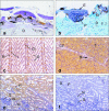
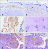
This study describes the distribution of parathyroid hormone-related protein (PTHrP) antigen and its mRNA in seven species of cartilaginous fish from six elasmobranch families. Antigen was detected using antibodies to synthetic human PTHrP and the mRNA with a riboprobe to human PTHrP gene sequence. The distribution pattern of PTHrP in the cartilaginous fish studied, reflected that observed in mammals but PTHrP further occurs in some sites unique to cartilaginous fish. Of particular note was the demonstration of PTHrP in the shark skeleton, which although considered not to contain bone, may form by a process similar to that forming the early stages of mammalian endochondral bone. The distribution of PTHrP in the elasmobranch skeleton resembled the distribution of PTHrP in the developing mammalian skeleton. Differences in the staining pattern between antisera to N-terminal PTHrP and mid-molecule PTHrP in the brain and pituitary suggested that the PTHrP molecule might be post-translationally processed in these tissues. The successful use of antibodies and a probe to human PTHrP in tissues from the early vertebrates examined in this study suggests that the PTHrP molecule is conserved from elasmobranchs to humans.
Figures


Similar articles
-
Parathyroid hormone-related protein (PTHrP) in cartilaginous and bony fish tissues.J Exp Zool. 1999 Oct 1;284(5):541-8. doi: 10.1002/(sici)1097-010x(19991001)284:5<541::aid-jez10>3.3.co;2-v. J Exp Zool. 1999. PMID: 10469992
-
Effects of water temperature and salinity on parathyroid hormone-related protein in the circulation and tissues of elasmobranchs.Comp Biochem Physiol B Biochem Mol Biol. 2001 Jun;129(2-3):327-36. doi: 10.1016/s1096-4959(01)00340-2. Comp Biochem Physiol B Biochem Mol Biol. 2001. PMID: 11399466
-
Parathyroid hormone-related protein in lower vertebrates.Clin Exp Pharmacol Physiol. 1998 Sep;25(9):750-2. doi: 10.1111/j.1440-1681.1998.tb02290.x. Clin Exp Pharmacol Physiol. 1998. PMID: 9750969 Review.
-
In situ hybridization of parathyroid hormone-related protein in normal skin, skin tumors, and gynecological cancers using digoxigenin-labeled probes and antibody enhancement.J Histochem Cytochem. 1995 Jan;43(1):5-10. doi: 10.1177/43.1.7822764. J Histochem Cytochem. 1995. PMID: 7822764
-
Parathyroid hormone-related protein in lower vertebrates.Comp Biochem Physiol B Biochem Mol Biol. 2002 May;132(1):87-95. doi: 10.1016/s1096-4959(01)00536-x. Comp Biochem Physiol B Biochem Mol Biol. 2002. PMID: 11997212 Review.
Cited by
-
Endoskeletal mineralization in chimaera and a comparative guide to tessellated cartilage in chondrichthyan fishes (sharks, rays and chimaera).J R Soc Interface. 2020 Oct;17(171):20200474. doi: 10.1098/rsif.2020.0474. Epub 2020 Oct 14. J R Soc Interface. 2020. PMID: 33050779 Free PMC article.
-
Ultrastructural and developmental features of the tessellated endoskeleton of elasmobranchs (sharks and rays).J Anat. 2016 Nov;229(5):681-702. doi: 10.1111/joa.12508. Epub 2016 Aug 24. J Anat. 2016. PMID: 27557870 Free PMC article.
-
Parathyroid hormone-related protein production in the lamprey Geotria australis: developmental and evolutionary perspectives.Dev Genes Evol. 2005 Nov;215(11):553-63. doi: 10.1007/s00427-005-0015-x. Epub 2005 Nov 4. Dev Genes Evol. 2005. PMID: 16034601
References
-
- Akino K, Ohtsuru A, Nakashima M, Ito M, Ting-Ting Y, Braiden V, et al. Distribution of the parathyroid hormone-related peptide and its receptor in the saccus vasculosus and choroid plexus in the red stingray. Cell. Mol. Neurobiol. 1998;18:362–368. - PubMed
-
- Bancroft JD, Stevens A. Theory and Practice of Histological Techniques. Edinburgh: Churchill Livingstone; 1990.
-
- Bone Q, Marshall NB, Blaxter JHS. Biology of Fishes. Glasgow: Blackie Academic and Professional; 1995.
-
- Caverzasio J, Rizzoli R, Martin TJ, Bonjour JP. Tumoral synthetic parathyroid hormone-related peptide inhibits amiloride sensitive transport in cultured renal epithelia. Pflugers Arch. 1988;413:96–98. - PubMed
-
- Chailleux N, Milet C, Vidal A, Lopez E. Presence of PTH-like and PTH-related peptide-like molecules in submammalian vertebrates. Neth. J. Zool. 1995;45:248–250.
Publication types
MeSH terms
Substances
LinkOut - more resources
Full Text Sources
Research Materials

