. 2002 Aug 15;21(16):4287-96.
doi: 10.1093/emboj/cdf439.

Keren, a new ligand of the Drosophila epidermal growth factor receptor, undergoes two modes of cleavage

Affiliations

Keren, a new ligand of the Drosophila epidermal growth factor receptor, undergoes two modes of cleavage

Aderet Reich et al. EMBO J. .

Abstract

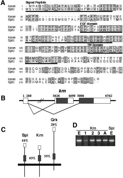
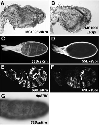
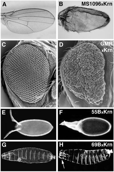
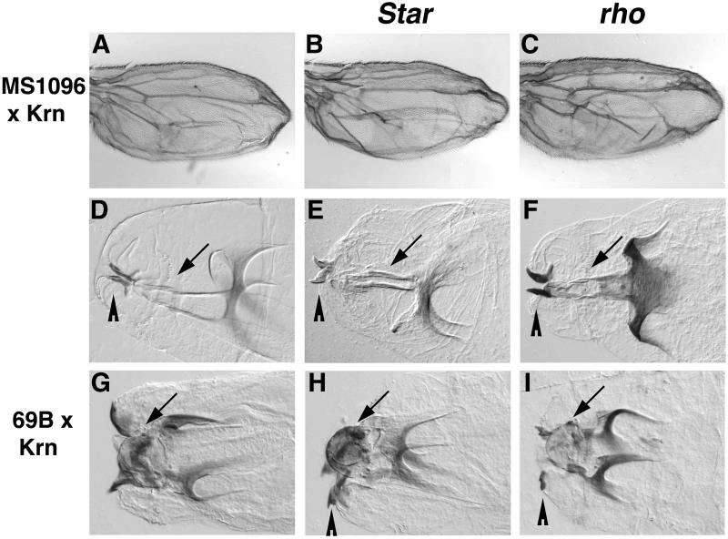
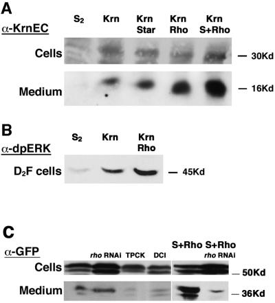
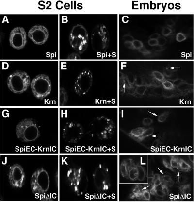
Spitz (Spi) is the most prominent ligand of the Drosophila EGF receptor (DER). It is produced as an inactive membrane precursor which is retained in the endoplasmic reticulum (ER). To allow cleavage, Star transports Spi to the Golgi, where it undergoes cleavage by Rhomboid (Rho). Since some DER phenotypes are not mimicked by any of its known activating ligands, we identified an additional ligand by database searches, and termed it Keren (Krn). Krn is a functional homolog of Spi since it can rescue the spi mutant phenotype in a Rho- and Star-dependent manner. In contrast to Spi, however, Krn also possesses a Rho/Star-independent ability to undergo low-level cleavage and activate DER, as evident both in cell culture and in flies. The difference in basal activity correlates with the cellular localization of the two ligands. While Spi is retained in the ER, the retention of Krn is only partial. Examining Spi/Krn chimeric and deletion constructs implicates the Spi cytoplasmic domain in inhibiting its basal activity. Low-level activity of Krn calls for tightly regulated expression of the Krn precursor.


Figures

None
Fig. 1. Krn is highly homologous to Spi. (A) Alignment of the predicted amino acid sequence of Krn and Spi reveals a similar organization, and significant homology not only in the EGF domain, but also in several stretches of the extracellular region. On the other hand, the cytoplasmic domains of the two proteins are highly divergent. Boxes denote amino acid identity and shadows indicate conservative changes. (B) A schematic of the genomic organization of krn exons. According to the ESTs and RT–PCR, krn’s transcript is comprised of two exons, where the ORF is coded by the second exon. Additional ESTs show that the first exon can be alternatively spliced and joined to a different exon, encoding a different ORF with homology to novel vertebrate proteins. It seems that one promoter is regulating the expression of transcripts encoding two different proteins. The number of ESTs indicates that the transcript that does not include Krn is more prevalent. Thus, a low frequency of splicing may be one way to maintain low levels of the krn transcript. Open reading frames are hatched. (C) A schematic of the homologies of Spi and Grk to Krn. Overall amino acid identities, as well as the identities in the EGF domain (hatched), are shown. Krn is much more similar to Spi in sequence and overall organization. (D) RT–PCR (non-quantitative) of the krn transcript from embryos (E), first, second and third instar larvae (1, 2 and 3), and adults (A) shows the expected band of 0.6 kb. For comparison, see spi RT–PCR from embryos.
None
Fig. 2. Secreted Krn is biologically active. To follow the biological activity of sKrn, the protein was ectopically expressed in several tissues (left panels). The phenotypes obtained displayed striking similarity to those observed following ectopic expression of sSpi (right panels) and are all consistent with hyperactivation of DER. (A and B) Expression in the wing imaginal disc with the MS1096 driver is mostly lethal, and survivors have small, blistered wings with an excess of vein material. See wild-type wing in Figure 4A. (C and D) Expression in the follicle cells with the 55B driver gives rise to excess dorsal appendage material around the circumference of the egg, consistent with dorsalization of the egg. See wild-type egg in Figure 4E. (E and F) Expression in the embryo using the ubiquitous 69B driver generates cuticles where germ band retraction is defective, head structures are missing and the denticle bands are extended dorsally, terminating in a rectangular shape (arrows) instead of the typical wild-type trapezoidal shape. See wild-type cuticle in Figure 4G. (G) Staining of 69B/UAS-sKrn embryos with anti-dpERK antibodies reveals the ubiquitous activation of the DER signaling pathway. For wild-type staining see Figure 3A.
None
Fig. 3. The capacity of Krn and Spi constructs to rescue spi or Star mutant embryos. (A) At stage 10, activation of the DER pathway generates a typical dpERK pattern in the tracheal placodes (TP) and ventral ectoderm (VE). No staining is observed at this stage in spi or Star mutant embryos. (B) Expression of Krn with the 69B driver in spi mutant embryos leads to complete rescue of the dpERK pattern in both tissues. This indicates that like Spi, cleavage of Krn is restricted to the domains of Rho expression. (C) Similar expression of Krn in Star mutant embryos did not lead to rescue, indicating that like Spi, Krn also requires Star for processing. (D) A construct of the Spi precursor in which the residues between the EGF and transmembrane domains have been deleted is unable to rescue spi mutant embryos, indicating that cleavage is necessary for the high level of DER activation. (E) A similar construct of the Krn precursor is unable to rescue spi mutants. (F) A chimeric precursor protein containing the Spi extracellular domain and the Krn intracellular domain is capable of undergoing processing and can rescue the spi mutant. (G) Similarly, a construct of the Spi precursor in which the cytoplasmic domain has been deleted can rescue spi.
None
Fig. 4. Ectopic activity of membrane Krn. In contrast to the precursor of Spi, which does not induce ectopic phenotypes upon overexpression, the precursor of Krn gives rise to phenotypes that are consistent with DER hyperactivation in a variety of tissues, at a level that is significantly lower than that induced by sKrn. (A, C, E and G) The wild-type situation. (B) In the wing, Krn expression by MS1096 generated smaller, blistered wings. The photograph was taken at 1.6× magnification, compared with (A). (D) Rough, disorganized eyes were generated with the GMR driver. (F) Extended dorsal appendages were seen following induction by 55B. (H) Finally, in the embryo induction led to lethality. In contrast to the expression of sKrn, the denticle bands remained normal. However, the structures at the head region appeared wider, with a prevalent lobe on each side (arrow).
None
Fig. 5. Ectopic Krn activity activates the DER pathway. (A) Expression of Krn by GMR-Gal4 gave rise to rough eyes. (B) In a DER heterozygous background, the severity of the phenotype was reduced. A similar reduction was observed in sos heterozygous flies (C), or in ras heterozygous flies (D).
None
Fig. 6. The activation of DER by Krn is Star and Rho independent. (A) Expression of Krn in the wing disc by MS1096-Gal4 gave rise to a bloated wing phenotype. (B and C) This phenotype is not altered by halving the gene dosage of Star or rho. Using head structures as an assay for ectopic Krn activity in the embryo allowed us to test whether Star or Rho is necessary for generating this phenotype. (D) The mouth hooks (arrowhead) and H-piece (arrow) in a wild-type embryo. (E and F) The cephalopharyngeal skeleton is not disrupted in Star or rho mutant embryos. (G) Overexpression of Krn by 69B-Gal4 gave rise to a deformed cephalopharyngeal skeleton, where the mouth hooks are widely separated and the H-piece is displaced exteriorly, forming a lobe between the mouth hooks (arrow). (H and I) A similar phenotype was observed when Krn was expressed in Star or rho mutant embryos identified by the denticle belt defects, demonstrating that the low-level activity of Krn is independent of Star and Rho. Mouth hooks are indicated by an arrowhead.
None
Fig. 7. Krn processing in S2 cells. (A) The upper panel shows comparable levels of Krn expression in stably transfected S2 cell extracts. These cells do not express detectable levels of Star and Rho. The lower panel shows sKrn in the medium collected from each transfection. When Krn was expressed alone, it was cleaved (consistent with the low-level activation observed in flies). When Rho, or Rho and Star were co-expressed with Krn, higher levels of sKrn were detected in the medium. This is likely to reflect the high-level cleavage of Krn, which is Rho and Star dependent. (B) sKrn in the medium was capable of triggering DER in S2 cells expressing the EGF receptor, as monitored by the accumulation of dpERK. The levels of MAPK activation were correlated to the levels of sKrn in the medium. (C) S2 cells were transiently transfected with Krn–GFP, and secreted protein was detected in the medium. Co-transfection of a rho RNAi construct did not reduce low-level cleavage, but dramatically reduced the cleavage in the presence of Star and Rho. Low-level cleavage was sensitive to the serine protease inhibitors TPCK and DCI (100 µM).
None
Fig. 8. Low-level cleavage of Krn in flies and in S2 cells. The chimeric and deletion constructs of Krn and Spi are illustrated. A summary of the high and low cleavage profile of each ligand is presented. All constructs, except those where the cleavage site has been truncated, were capable of rescuing the spi dpERK pattern, e.g. undergoing high-level cleavage (Figure 3). Low-level cleavage of the constructs was assayed in vivo by misexpression in the wing with the MS1096 driver. (A) Spi misexpression did not alter the wing. (B) Krn misexpression gave rise to smaller bloated wings. (C) Deletion construct of 16 amino acids that are essential for cleavage in Spi (comprising the sequence between the EGF and transmembrane domains) had no effect upon misexpression. (D) Deletion of this sequence in Krn abolished the capacity to induce the wing phenotype. This indicates that the low-level activation of Krn is dependent on cleavage. (E) A chimera containing the Spi extracellular domain and the Krn transmembrane and intracellular domains was capable of inducing the wing phenotype. (F) Deletion of the Spi intracellular domain allowed the protein to induce the wing phenotype. The wings in (B), (E) and (F) are shown at a magnification of 1.6× compared with (A), (C) and (D). (G) The upper panel shows sSpi in medium collected from cells transfected with different Spi constructs. Low levels of cleavage could be seen in cells transfected either with SpiΔIC alone or with SpiEC–KrnIC alone, indicating that the intracellular domain of Spi inhibits its low-level cleavage. SpiΔ16aa did not show cleavage under these conditions. The lower panel shows expression levels of each protein in cell extracts.
None
Fig. 9. Low-level cleavage in S2 cells and in embryos correlates with vesicular localization. Each of the proteins was constructed to contain GFP after its signal peptide. (A) Spi was shown to have perinuclear localization identified with the ER, in S2 cells. (B) Star was shown to transport Spi out of the ER. Spi is visualized in vesicles instead of the perinuclear ring. (C) After injection of the construct to embryos, Spi shows exclusive perinuclear localization. (D) Krn localization in S2 cells was predominantly perinuclear, but a punctate vesicular distribution, indicating exit from the ER, was also detected. (E) Star led to complete depletion of Krn from the ER. (F) In embryos, Krn also showed some vesicular localization (arrows), in addition to its perinuclear localization. (G) SpiEC–KrnIC showed similar cellular distribution as Krn, indicating that the intracellular domain is the key to ER retention levels. (H) Star caused complete transport of SpiEC–KrnIC from the ER. (I) In embryos, SpiEC–KrnIC mimicked the Krn pattern, showing some vesicular localization. (J) SpiΔIC showed perinuclear localization together with vesicular distribution, indicating that the intracellular domain of Spi inhibits it from exiting the ER. (K) SpiΔIC was depleted from the ER when co-expressed with Star. (L) Embryonic localization of SpiΔIC was similar to that of Krn.

Similar articles

Cited by

References

    1. Bang A.G. and Kintner,C. (2000) Rhomboid and Star facilitate presentation and processing of the Drosophila TGF-α homolog Spitz. Genes Dev., 14, 177–186. - PMC - PubMed
    1. Baonza A., Casci,T. and Freeman,M. (2001) A primary role for the epidermal growth factor receptor in ommatidial spacing in the Drosophila eye. Curr. Biol., 11, 396–404. - PubMed
    1. Bier E., Jan,L.Y. and Jan,Y.N. (1990) Rhomboid, a gene required for dorsoventral axis establishment and peripheral nervous system development in Drosophila melanogaster. Genes Dev., 4, 190–203. - PubMed
    1. Freeman M. (2000) Feedback control of intercellular signalling in development. Nature, 408, 313–319. - PubMed
    1. Gabay L., Scholz,H., Golembo,M., Klaes,A., Shilo,B.Z. and Klambt,C. (1996) EGF receptor signaling induces pointed P1 transcription and inactivates Yan protein in the Drosophila embryonic ventral ectoderm. Development, 122, 3355–3362. - PubMed

Publication types