. 2002 Jul 15;21(14):3852-62.
doi: 10.1093/emboj/cdf353.

Dissolution of the maskin-eIF4E complex by cytoplasmic polyadenylation and poly(A)-binding protein controls cyclin B1 mRNA translation and oocyte maturation

Affiliations

Dissolution of the maskin-eIF4E complex by cytoplasmic polyadenylation and poly(A)-binding protein controls cyclin B1 mRNA translation and oocyte maturation

Quiping Cao et al. EMBO J. .

Abstract

Cytoplasmic polyadenylation stimulates the translation of several dormant mRNAs during oocyte maturation in Xenopus. Polyadenylation is regulated by the cytoplasmic polyadenylation element (CPE), a cis-acting element in the 3'-untranslated region of responding mRNAs, and its associated factor CPEB. CPEB also binds maskin, a protein that in turn interacts with eIF4E, the cap-binding factor. Here, we report that based on antibody and mRNA reporter injection assays, maskin prevents oocyte maturation and the translation of the CPE-containing cyclin B1 mRNA by blocking the association of eIF4G with eIF4E. Dissociation of the maskin-eIF4E complex is essential for cyclin B1 mRNA translational activation, and requires not only cytoplasmic polyadenylation, but also the poly(A)-binding protein. These results suggest a molecular mechanism by which CPE- containing mRNA is activated in early development.


Figures

None
Fig. 1. Translational control by maskin. (A) Extracts prepared from oocytes exposed to progesterone for 0–9 h were supplemented with GTP, chromatographed on 7mGTP–Sepharose (cap column), eluted with SDS sample buffer and analyzed by western blotting for maskin, eIF4E, CPEB and eIF4G. The maskin, CPEB and eIF4G in the starting material (load) were also examined. The oocytes exposed to progesterone for 7 h reached metaphase I, while those exposed to progesterone for 9 h reached metaphase II; the oocytes not exposed to progesterone remained at prophase I. The lower part of the figure shows a quantification of the western blot results; relative amount refers to the percentage of maximum of each particular protein retained on the column. (B) Affinity-purified maskin antibody or IgG was injected into oocytes, some of which were also injected with 32P-labeled cyclin B1 3′-UTR. One-half of each group of oocytes was then stimulated with progesterone. The oocytes were analyzed for cyclin B1 accumulation by western blotting and for polyadenylation by urea–acrylamide gel electrophoresis and phosphoimaging. (C) Affinity-purified maskin antibody was injected into oocytes as above, or mixed with E.coli-expressed maskin prior to injection. Other oocytes were injected with IgG. An extract was then prepared and analyzed for cyclin B1 protein accumulation by a western blot. (D) Oocytes injected with IgG or maskin antibody were incubated with [35S]methionine and the resulting radioactive proteins were displayed on an SDS gel.
None
Fig. 2. Maskin antibody activates the translation of CPE-containing mRNAs. (A) The salient 3′-UTR sequences of cyclin B1, Mos and Wee1 mRNAs are depicted (top); the CPEs (boxed) and polyadenylation hexanucleotides (ovals) are designated. Maskin antibody or IgG was injected into oocytes, which were then cultured overnight (no progesterone). Extracts were then prepared and three concentrations of protein were analyzed by western blots probed for cyclin B1, Mos, Wee1, MAP kinase and eIF4E. (BIn vitro synthesized CAT reporter RNAs that contained a 5′ cap (CAP CAT) or the EMCV IRES (IRES CAT) were appended with 3′-UTRs composed of either a polylinker sequence or a polylinker sequence harboring three repeated CPE sequences (3CPE) (de Moor and Richter, 1999). The RNAs were injected into oocytes followed by a second injection of IgG or maskin antibody; extracts were then prepared and CAT activity was assessed. Each experiment was performed five times. The bottom portion of the figure shows that injected maskin antibody, but not injected IgG, induced endogenous cyclin B1 synthesis in the same oocytes that were injected with the reporter RNAs.
None
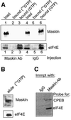
Fig. 3. Maskin antibody induces the dissociation of maskin from eIF4E. (A) Extracts from oocytes injected with maskin antibody or IgG were supplemented with GTP only, or GTP plus 7mGTP, and applied to a cap column. Following extensive washing, the bound material was eluted with SDS and, together with the load material, was analyzed for maskin and eIF4E by western blotting. (B) In a separate experiment, oocyte material bound to a cap column was eluted with free cap and then probed for maskin and eIF4E. (C) Oocyte extract was subjected to immunoprecipitation with either IgG or maskin antibody; the bound material was then probed for CPEB and eIF4E.
None
Fig. 4. Maskin–eIF4E dissociation requires cytoplasmic polyadenylation. (A) Oocytes were incubated in the absence or presence of progesterone or, in some cases, with progesterone and cordycepin. When the oocytes exposed to progesterone only had matured, extracts were prepared from all the oocytes and were applied to a cap column. The relative amounts of maskin and eIF4E on the column and in the initial load solution were analyzed by a western blot. Some oocytes injected with 32P-labeled cyclin B1 3′-UTR and treated with progesterone and/or cordycepin were used to examine cytoplasmic polyadenylation. (B) Oocytes were incubated with progesterone and/or cordycepin, some of which were injected with E.coli-expressed cyclin B1 protein. Extracts were then prepared and applied to a cap column; maskin and eIF4E in the bound fractions, as well as in the load fraction, were analyzed by western blotting. The extracts were also assessed for MPF activity as determined by the phosphorylation of histone H1. Finally, some oocytes treated with the agents noted above were also injected with labeled cyclin B1 3′-UTR and examined for cytoplasmic polyadenylation.
None
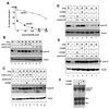
Fig. 5. PABP regulates the dissociation of maskin from eIF4E. (A) Several concentrations of poly(A) or poly(C) were injected into oocytes that were then stimulated with progesterone. Maturation was scored by the appearance of a white spot at the animal pole. (B) Oocytes were injected with poly(A) or poly(C) and some were then incubated with progesterone. Extracts were then analyzed for cyclin B1 accumulation by western blotting. In this and subsequent panels, the blots were also probed for tubulin, which served as a loading control. (C) Oocytes were injected with poly(A) and, in some cases, with E.coli-expressed S-PABP. Some of the oocytes were then incubated with progesterone and scored for the accumulation of cyclin B1 by western blotting. (D) Oocytes were injected with poly(A) and, in some cases, with E.coli-expressed S-PABP or E-PABP and scored for the accumulation of cyclin B1 by western blotting. (E) Oocytes were injected with poly(C) and, in some cases, with E.coli-expressed S-PABP or E-PABP and scored for the accumulation of cyclin B1 by western blotting. (F) RNA was extracted from S-PABP- and E-PABP-injected oocytes and used for a northern blot probed for cyclin B1. The rRNA subunits served as loading controls.
None
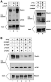
Fig. 6. Poly(A), PABP and the maskin–eIF4E interaction. (A) Oocytes injected with poly(A) and/or S-PABP were incubated in the absence or presence of progesterone. Extracts were then prepared from these oocytes and applied to a cap column as described in Figure 3. Maskin and eIF4E were analyzed by western blotting. (B) Oocytes were injected with combinations of poly(A), poly(C), S-PABP and E-PABP and incubated in the absence or presence of progesterone, followed by extract preparation, cap column chromatography and western blotting for maskin and eIF4E.
None
Fig. 7. An eIF4G–PABP complex displaces maskin from eIF4E. (A) A peptide derived from eIF4G that is necessary for the binding of this protein to PABP was added to an egg extract, as was a control peptide of irrelevant sequence. The extract was then applied to an affinity column containing (lanes 1 and 2) or lacking (lane 4) recombinant His-tagged PABP. Following extensive washing, the columns were eluted with SDS and probed on a western blot for eIF4G and PABP. Lane 3 shows the profile of eIF4G in the extract prior to chromatography. (B) Various concentrations of the eIF4G-derived peptide and the control peptide were injected into oocytes that were subsequently incubated with progesterone. The incidence of oocyte maturation was then scored by the appearance of a white spot at the animal pole. (C) Oocytes injected with various concentrations of the control peptide or the eIF4G-derived peptide were incubated with progesterone, scored for the appearance of the white spot and then analyzed for cyclin B1 accumulation by western blotting. The control peptide-injected oocytes had matured whereas the eIF4G-derived peptide-injected oocytes had not. The asterisk denotes a non-specific band that serves as a loading control. (D) Oocytes injected with the control and eIF4G-derived peptides were incubated with progesterone, homogenized, mixed with free GTP and applied to a cap column. Following extensive washing, the column was eluted with SDS and probed on western blots for maskin and eIF4E. Maskin in the load fraction was also analyzed on a western blot. The control peptide-injected oocytes had matured whereas the eIF4G-derived peptide-injected oocytes had not. (E) Oocytes injected with poly(A) or the eIF4G peptide were also injected with maskin antibody and subsequently used for a cyclin B1 western blot. The asterisk denotes a non-specific band.
None
Fig. 8. Model of polyadenylation-induced translation. Dormant CPE-containing mRNAs (e.g. cyclin B1) in immature oocytes are bound by CPEB, which in turn is bound to maskin, which in turn is bound to eIF4E, the cap-binding factor. The binding of maskin to eIF4E precludes the binding of eIF4G to eIF4E, thus inhibiting the formation of the initiation complex. The cleavage and polyadenylation specificity factor (CPSF) may or may not be loosely associated with the hexanucleotide AAUAAA at this time. Following progesterone stimulation, the kinase aurora is activated and phosphorylates CPEB Ser174, an event that causes CPEB to bind and recruit CPSF into an active cytoplasmic polyadenylation complex, presumably helping it to associate with the AAUAAA. CPSF recruits poly(A) polymerase (PAP) to the end of the mRNA, where it catalyzes poly(A) addition. The newly elongated poly(A) tail is then bound by poly(A)-binding protein (PABP), which in turn associates with eIF4G. eIF4G, when associated with PABP, then displaces maskin from, and binds to, eIF4E, thereby initiating translation. eIF4G, through eIF3, interacts with the 40S ribosomal subunit.

Similar articles

Cited by

References

    1. Andresson T. and Ruderman,J.V. (1998) The kinase Eg2 is a component of the Xenopus oocyte progesterone-activated signaling pathway. EMBO J., 17, 5627–5637. - PMC - PubMed
    1. Bayaa M., Booth,R.A., Sheng,Y. and Liu,X.J. (2000) The classical progesterone receptor mediates Xenopus oocyte maturation through a nongenomic mechanism. Proc. Natl Acad. Sci. USA, 97, 12607–12612. - PMC - PubMed
    1. Bilger A., Fox,C.A., Wahle,E. and Wickens,M. (1994) Nuclear poly adenylation factors recognize cytoplasmic polyadenylation elements. Genes Dev., 8, 1106–1116. - PubMed
    1. Cornelis S., Bruynooghe,Y., Denecker,G., Van Huffel,S., Tinton,S. and Beyaert,R. (2000) Identification and characterization of a novel cell cycle-regulated internal ribosome entry site. Mol. Cell, 5, 597–605. - PubMed
    1. de Moor C.H. and Richter,J.D. (1997) The Mos pathway regulates cytoplasmic polyadenylation in Xenopus oocytes. Mol. Cell. Biol., 17, 6419–6426. - PMC - PubMed

Publication types