

Persistent mitochondrial hyperpolarization, increased reactive oxygen intermediate production, and cytoplasmic alkalinization characterize altered IL-10 signaling in patients with systemic lupus erythematosus
- PMID: 12097418
- PMCID: PMC4020441
- DOI: 10.4049/jimmunol.169.2.1092
Persistent mitochondrial hyperpolarization, increased reactive oxygen intermediate production, and cytoplasmic alkalinization characterize altered IL-10 signaling in patients with systemic lupus erythematosus
Abstract
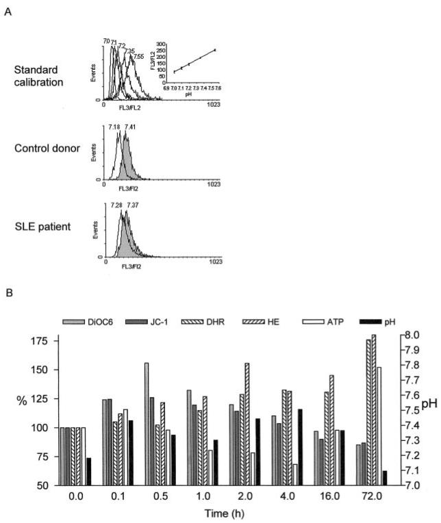
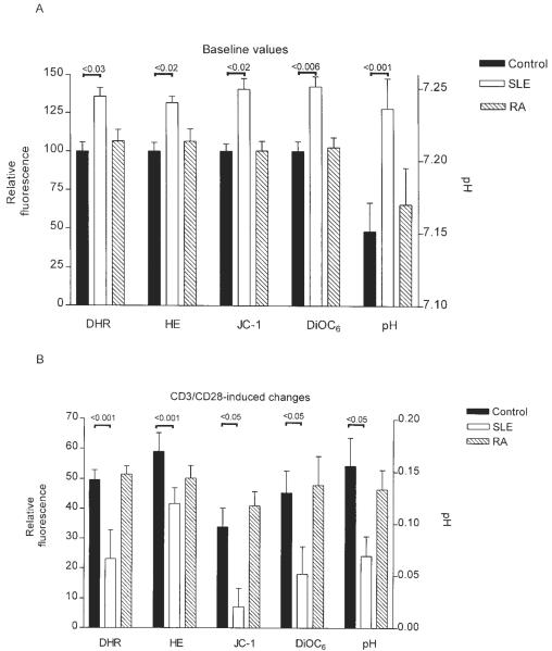
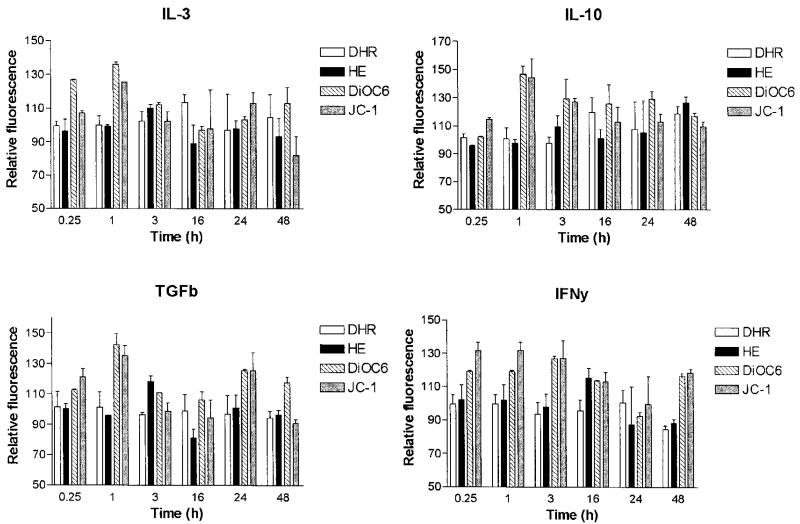
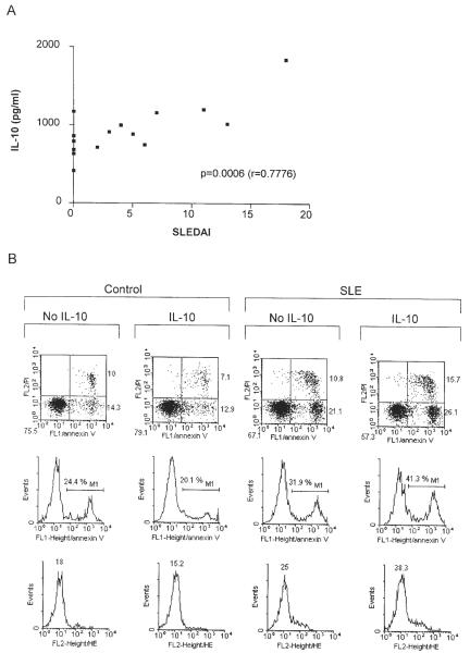
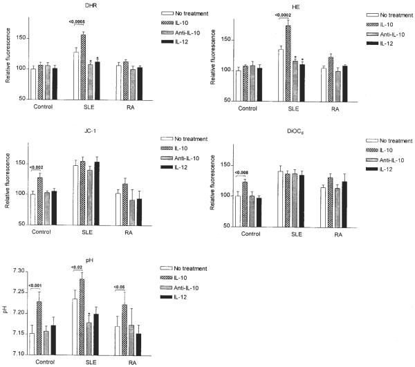
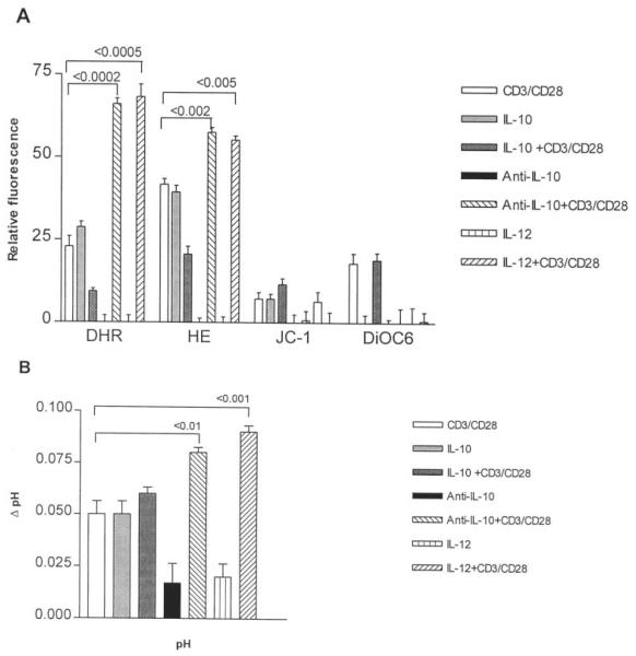
Abnormal death signaling in lymphocytes of systemic lupus erythematosus (SLE) patients has been associated with elevation of the mitochondrial transmembrane potential (Delta psi(m)) and increased production of reactive oxygen intermediates (ROI). The resultant ATP depletion sensitizes T cells for necrosis that may significantly contribute to inflammation in patients with SLE. In the present study, the role of mitochondrial signal processing in T cell activation was investigated. CD3/CD28 costimulation of PBL elicited transient mitochondrial hyperpolarization and intracellular pH (pH(i)) elevation, followed by increased ROI production. Baseline Delta psi(m), ROI production, and pH(i) were elevated, while T cell activation-induced changes were blunted in 15 patients with SLE in comparison with 10 healthy donors and 10 rheumatoid arthritis patients. Similar to CD3/CD28 costimulation, treatment of control PBL with IL-3, IL-10, TGF-beta(1), and IFN-gamma led to transient Delta psi(m) elevation. IL-10 had diametrically opposing effects on mitochondrial signaling in lupus and control donors. Unlike healthy or rheumatoid arthritis PBL, cells of lupus patients were resistant to IL-10-induced mitochondrial hyperpolarization. By contrast, IL-10 enhanced ROI production and cell death in lupus PBL without affecting ROI levels and survival of control PBL. Ab-mediated IL-10 blockade or stimulation with antagonistic lymphokine IL-12 normalized baseline and CD3/CD28-induced changes in ROI production and pH(i) with no impact on Delta psi(m) of lupus PBL. The results suggest that mitochondrial hyperpolarization, increased ROI production, and cytoplasmic alkalinization play crucial roles in altered IL-10 responsiveness in SLE.
Figures
Similar articles
-
Nitric oxide-dependent mitochondrial biogenesis generates Ca2+ signaling profile of lupus T cells.J Immunol. 2004 Sep 15;173(6):3676-83. doi: 10.4049/jimmunol.173.6.3676. J Immunol. 2004. PMID: 15356113 Free PMC article.
-
Mitochondrial hyperpolarization and ATP depletion in patients with systemic lupus erythematosus.Arthritis Rheum. 2002 Jan;46(1):175-90. doi: 10.1002/1529-0131(200201)46:1<175::AID-ART10015>3.0.CO;2-H. Arthritis Rheum. 2002. PMID: 11817589 Free PMC article.
-
Mitochondrial dysfunction in T cells of patients with systemic lupus erythematosus.Int Rev Immunol. 2004 May-Aug;23(3-4):293-313. doi: 10.1080/08830180490452576. Int Rev Immunol. 2004. PMID: 15204090 Review.
-
T cell activation-induced mitochondrial hyperpolarization is mediated by Ca2+- and redox-dependent production of nitric oxide.J Immunol. 2003 Nov 15;171(10):5188-97. doi: 10.4049/jimmunol.171.10.5188. J Immunol. 2003. PMID: 14607919 Free PMC article.
-
Metabolic control of T cell activation and death in SLE.Autoimmun Rev. 2009 Jan;8(3):184-9. doi: 10.1016/j.autrev.2008.07.041. Epub 2008 Aug 21. Autoimmun Rev. 2009. PMID: 18722557 Free PMC article. Review.
Cited by
-
Oxidative Stress and Treg and Th17 Dysfunction in Systemic Lupus Erythematosus.Oxid Med Cell Longev. 2016;2016:2526174. doi: 10.1155/2016/2526174. Epub 2016 Aug 11. Oxid Med Cell Longev. 2016. PMID: 27597882 Free PMC article. Review.
-
Activation of the Mechanistic Target of Rapamycin in SLE: Explosion of Evidence in the Last Five Years.Curr Rheumatol Rep. 2016 Dec;18(12):73. doi: 10.1007/s11926-016-0622-8. Curr Rheumatol Rep. 2016. PMID: 27812954 Free PMC article. Review.
-
Nitric oxide-dependent mitochondrial biogenesis generates Ca2+ signaling profile of lupus T cells.J Immunol. 2004 Sep 15;173(6):3676-83. doi: 10.4049/jimmunol.173.6.3676. J Immunol. 2004. PMID: 15356113 Free PMC article.
-
Mitochondria, Oxidative Stress and Innate Immunity.Front Physiol. 2018 Oct 18;9:1487. doi: 10.3389/fphys.2018.01487. eCollection 2018. Front Physiol. 2018. PMID: 30405440 Free PMC article. Review.
-
T Cell Homeostatic Proliferation Promotes a Redox State That Drives Metabolic and Epigenetic Upregulation of Inflammatory Pathways in Lupus.Antioxid Redox Signal. 2022 Mar;36(7-9):410-422. doi: 10.1089/ars.2021.0078. Epub 2021 Nov 9. Antioxid Redox Signal. 2022. PMID: 34328790 Free PMC article. Review.
References
-
- Elkon KB. Apoptosis in SLE: too little or too much? Clin. Exp. Rheumatol. 1994;12:553. - PubMed
-
- Perl A, Banki K. Molecular mimicry, altered apoptosis, and immuno-modulation as mechanisms of viral pathogenesis in systemic lupus erythematosus. In: Kammer GM, Tsokos GC, editors. Lupus: Molecular and Cellular Pathogenesis. Humana Press; Totowa: 1999. p. 43.
-
- Cohen JJ, Duke RC, Fadok VA, Sellins KS. Apoptosis and programmed cell death in immunity. Annu. Rev. Immunol. 1992;10:267. - PubMed
-
- Thompson CB. Apoptosis in the pathogenesis and treatment of disease. Science. 1995;267:1456. - PubMed
-
- Emlen W, Niebur JA, Kadera R. Accelerated in vitro apoptosis of lymphocytes from patients with systemic lupus erythematosus. J. Immunol. 1994;152:3685. - PubMed
Publication types
MeSH terms
Substances
Grants and funding
LinkOut - more resources
Full Text Sources
Other Literature Sources
Medical
Molecular Biology Databases