. 2002 Jul 1;21(13):3317-26.
doi: 10.1093/emboj/cdf324.

Cellular prion protein transduces neuroprotective signals

Affiliations

Cellular prion protein transduces neuroprotective signals

Luciana B Chiarini et al. EMBO J. .

Abstract

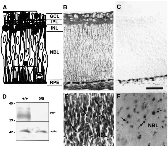
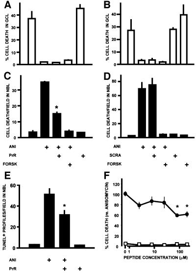
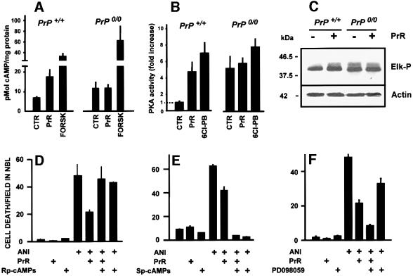
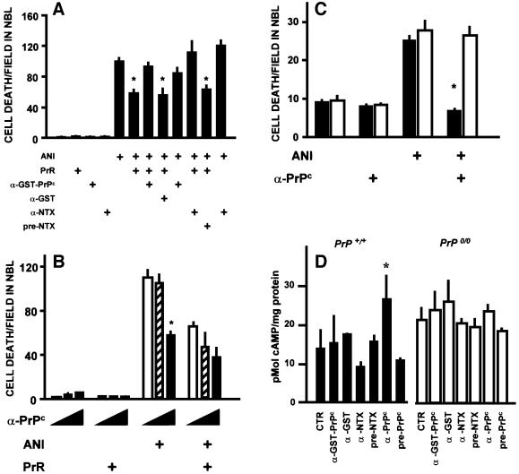
To test for a role for the cellular prion protein (PrP(c)) in cell death, we used a PrP(c)-binding peptide. Retinal explants from neonatal rats or mice were kept in vitro for 24 h, and anisomycin (ANI) was used to induce apoptosis. The peptide activated both cAMP/protein kinase A (PKA) and Erk pathways, and partially prevented cell death induced by ANI in explants from wild-type rodents, but not from PrP(c)-null mice. Neuroprotection was abolished by treatment with phosphatidylinositol-specific phospholipase C, with human peptide 106-126, with certain antibodies to PrP(c) or with a PKA inhibitor, but not with a MEK/Erk inhibitor. In contrast, antibodies to PrP(c) that increased cAMP also induced neuroprotection. Thus, engagement of PrP(c) transduces neuroprotective signals through a cAMP/PKA-dependent pathway. PrP(c) may function as a trophic receptor, the activation of which leads to a neuroprotective state.


Figures

None
Fig. 1. (A) Diagram of the retina of neonatal rodents. The cellular and plexiform strata are indicated, together with a rough schematic representation of the morphology of existing cell types at that stage of development. Dark profiles represent relatively differentiated cells. A mitotic figure is depicted at the lower right corner of the diagram. Elongated profiles with vertically oriented oval nuclei represent both proliferating and early post-mitotic cells, which comprise most of the population within the neuroblastic layer. (B and C) Immunohistochemistry of transverse sections of the retina in a newborn rat, stained either with (B) or without (C) a monoclonal antibody to PrPc. The beaded dark deposits that appear at the bottom of both figures correspond to the melanin-containing retinal pigment epithelium. (D) Example of a western blot of protein extracts from the retinas of wild-type or PrP0/0 mice, probed with an antiserum to PrPc raised in knockout mouse (upper panel), then stripped and re-probed with an antibody to actin as a loading control (lower panel). The monoclonal antibody used for immunohistochemistry in (B) and (C) produced a result similar to (D) in western blots of mouse retinal tissue (data not shown). (E and F) Photomicrographs from the neuroblastic layer of explants from the retina of neonatal rats, treated with anisomycin (1 µg/ml) for 24 h, stained with either neutral red (E) or with the TUNEL technique (F). The arrows indicate degenerating profiles. GCL = ganglion cell layer; IPL = inner plexiform layer; INL = inner nuclear layer; NBL = neuroblastic layer; RPE = retinal pigment epithelium. Bar = 50 µm.
None
Fig. 2. Effect of the PrPc-binding peptide (PrR) upon retinal cell death. The data are shown as means ± SEM of either the percentage of degenerating cells in the single-cell thick ganglion cell layer (GCL), or the densitiy of degenerating profiles in the neuroblastic layer (NBL), in retinal explants from neonatal rats. Treatments in (A–E) are: ANI = anisomycin 1 µg/ml; PrR = PrR peptide 80 µM; SCRA = scrambled peptide 80 µM; FORSK = forskolin 10 µM. The rates of cell death evaluated by counts of pyknotic profiles in the GCL (A and B) and NBL (C and D) are shown for two representative experiments made in triplicate with either the PrR (A and C) or the scrambled control (B and D) peptides. *P < 0.01 versus anisomycin alone. In this and the following illustrations, statistical significance is indicated only for the most relevant among the multiple comparisons tabulated in the Duncan’s test. (E) Blockade of anisomycin-induced apoptosis in the NBL detected with the TUNEL technique (triplicate explants), following treatment with the PrR peptide. *P < 0.01 versus anisomycin alone. (F) Dose–response curve for the PrR peptide, either in the presence (filled circles) or in the absence of anisomycin 1 µg/ml. Each data point is the mean ± SEM of 8–16 explants from a total of five experiments, and normalized with respect to the rate of cell death (100%) induced by anisomycin. *P < 0.01 versus anisomycin alone.
None
Fig. 3. Neuroprotection depends on the cellular prion protein. (A and B) Hydrolysis of GPI anchors prevents neuroprotection by the PrR peptide. Explants from rat retinas were pre-treated (PRE) with either vehicle or PI-PLC, then washed and treated with various combinations of anisomycin (1 µg/ml), the PrR peptide (80 µM) and forskolin (10 µM). Note that PI-PLC treatment prevents the neuroprotection by the PrR peptide in the NBL. *P < 0.01 versus anisomycin alone. (C and D) Neuroprotection by the PrR peptide occurs in the retina of wild-type (C), but not Prnp-knockout mice (D). Data are means ± SEM counts of pyknotic profiles from triplicate explants. *P < 0.01 versus anisomycin alone.
None
Fig. 4. Peptide binding and further evidence that neuroprotection depends on PrPc. (A) PrPc–PrR binding curves. Biotinylated BSA–PrR or biotinylated BSA alone were incubated with either wild-type His6-moPrPc or His6-moPrPc Δ105–128. Non-specific binding to His6-moPrPc (open squares) was subtracted from total binding (diamonds) to yield PrPc-specific binding to PrR (circles, continuous line). Lack of specific binding to moPrPΔ105–128 is also shown (crosses). All error bars were <5% and were omitted for clarity. (B) Competition assay. Results were expressed as a percentage of the absorbance values corresponding to specific PrPc–PrR binding. Note that both cold PrR (filled bars) and neurotoxic (hatched bars) peptides, but not the scrambled peptide (open bars), displace PrPc–PrR binding. (C) Neurotoxic peptide has no effect in either the presence (filled triangles) or absence (open triangles) of anisomycin 1 µg/ml, while the PrR peptide, that has no effect by itself (open circle), had a neuroprotective effect in the presence of anisomycin (filled circle). Data are means ± SEM of triplicate explants in one representative experiment out of three with similar results made with retinal explants from neonatal rats. (D) Co-incubation with equimolar (80 µM) neurotoxic peptide blocks neuroprotection by the PrR peptide. Data are means ± SEM of triplicate experiments with retinal explants from neonatal rats. *P < 0.01 versus anisomycin alone.
None
Fig. 5. PrPc-mediated neuroprotective signaling. (A and B) Responses of the cAMP/PKA signaling pathway to the PrR peptide (PrR) in the retina of either wild-type or PrP0/0 mice. Positive controls were either forskolin (FORSK) or a D1-like dopaminergic receptor agonist (6-Chloro-PB). Note the cAMP (A) and PKA (B) responses restricted to wild-type retinal tissue, and the higher basal values in knockout retinas. (C) Western blots for phospho-Elk (top) and loading control with actin (bottom), following treatment of either wild-type or PrP0/0 mouse retinal tissue with the PrR peptide. Note the activation of the Erk pathway restricted to wild-type tissue, as well as the higher basal activity in knockout tissue. (DF) PrPc-mediated neuroprotective signaling through the cAMP-PKA pathway. Retinal explants from neonatal rats were treated with anisomycin (1 µg/ml), the PrR peptide (PrR 80 µM) in the presence of either 100 µM Rp-cAMP-s (D) or 100 µM Sp-cAMP-s (E), respectively an inhibitor and an activator of cAMP-dependent protein kinase, or in the presence of 30 µM PD98059, an inhibitor of the Erk-activating MEK enzyme (F). Note the reversion of the neuroprotective effect with the PKA inhibitor, and the potentiation of the neuroprotective effect with the Erk pathway inhibitor.
None
Fig. 6. Effects of antibodies raised against PrPc. (A) Both an antiserum to GST–PrPc (1:80) and an antiserum raised against the neurotoxic peptide (α-NTX) (1:50) prevented neuroprotection by the PrR peptide, while their respective controls, anti-GST and pre-immune serum, had no effect. Data are means ± SEM of triplicate experiments with retinal explants from neonatal rats. *P < 0.01 versus anisomycin alone. (B) A polyclonal antiserum raised against PrPc in PrP0/0 mice blocked anisomycin-induced cell death in the neuroblastic layer, and did not revert the neuroprotective effect of the PrR peptide. In each group of three bars, the concentration of the anti-PrPc antiserum was 0 (open bars), 20 (hatched bars) and 100 (filled bars) µg/ml, respectively. Data are means ± SEM of triplicate experiments with retinal explants from neonatal rats. *P < 0.01 versus no antiserum within the same group. (C) The anti-PrPc antibodies raised in knockout mice (same as in B) had a neuroprotective effect upon retinal explants from wild-type (filled bars), but not from PrP0/0 (open bars) mice. Data are means ± SEM of five replicates in each group. *P < 0.01 versus anisomycin alone among the same genotype. (D) Antibodies that induced neuroprotection increased the intracellular concentration of cAMP in wild-type, but not in PrP0/0 mouse retinal tissue. Results are means ± SEM of three experiments done in triplicate. *P < 0.01 versus control among the same genotype.

Similar articles

Cited by

References

    1. Aguzzi A. and Weissmann,C. (1997) Prion research: the next frontiers. Nature, 389, 795–798. - PubMed
    1. Aronoff-Spencer E. et al. (2000) Identification of the Cu2+ binding sites in the N-terminal domain of the prion protein by EPR and CD spectroscopy. Biochemistry, 39, 13760–13771. - PMC - PubMed
    1. Boquet D., Dery,O., Frobert,Y., Grassi,J. and Couraud,J.Y. (1995) Is hydropathic complementarity involved in antigen–antibody binding? Mol. Immunol., 32, 303–308. - PubMed
    1. Bost K.L., Smith,E.M. and Blalock,J.E. (1985) Similarity between the corticotropin (ACTH) receptor and a peptide encoded by an RNA that is complementary to ACTH mRNA. Proc. Natl Acad. Sci. USA, 82, 1372–1375. - PMC - PubMed
    1. Bounhar Y., Zhang,Y., Goodyear,C. and LeBlanc,A. (2001) Prion protein protects human neurons against Bax-mediated apoptosis. J. Biol. Chem., 276, 39145–39149. - PubMed

Publication types

MeSH terms

Substances