. 2002 Jul;22(13):4902-13.
doi: 10.1128/MCB.22.13.4902-4913.2002.

Rad23 promotes the targeting of proteolytic substrates to the proteasome

Affiliations

Rad23 promotes the targeting of proteolytic substrates to the proteasome

Li Chen et al. Mol Cell Biol. 2002 Jul.

Abstract

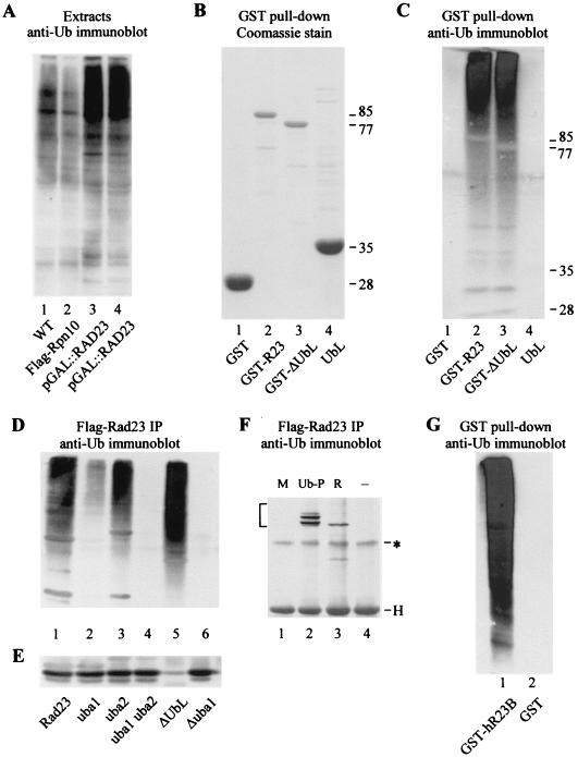
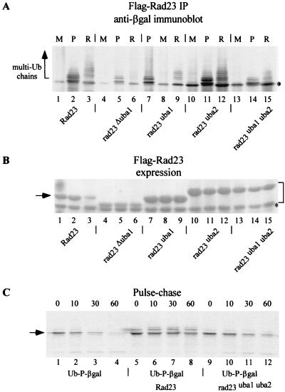
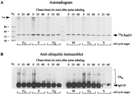
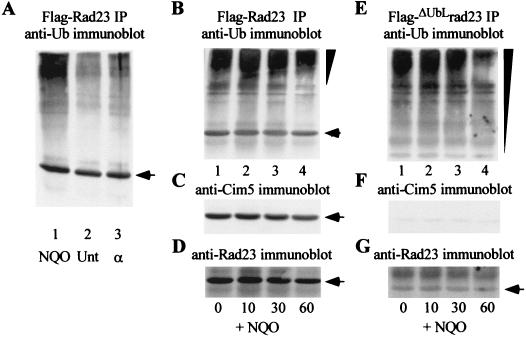
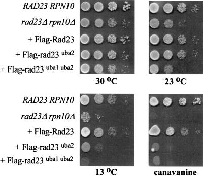
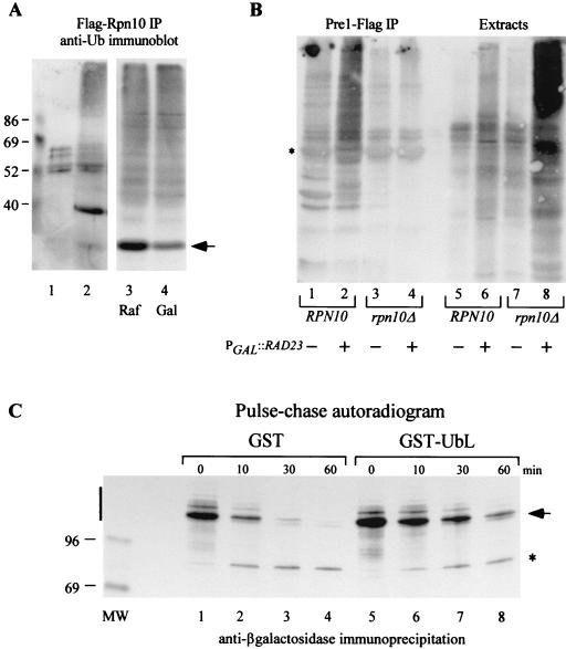
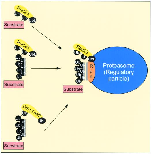
Rad23 contains a ubiquitin-like domain (UbL(R23)) that interacts with catalytically active proteasomes and two ubiquitin (Ub)-associated (UBA) sequences that bind Ub. The UBA domains can bind Ub in vitro, although the significance of this interaction in vivo is poorly understood. Rad23 can interfere with the assembly of multi-Ub chains in vitro, and high-level expression caused stabilization of proteolytic substrates in vivo. We report here that Rad23 interacts with ubiquitinated cellular proteins through the synergistic action of its UBA domains. Rad23 plays an overlapping role with Rpn10, a proteasome-associated multi-Ub chain binding protein. Mutations in the UBA domains prevent efficient interaction with ubiquitinated proteins and result in poor suppression of the growth and proteolytic defects of a rad23 Delta rpn10 Delta mutant. High-level expression of Rad23 revealed, for the first time, an interaction between ubiquitinated proteins and the proteasome. This increase was not observed in rpn10 Delta mutants, suggesting that Rpn10 participates in the recognition of proteolytic substrates that are delivered by Rad23. Overexpression of UbL(R23) caused stabilization of a model substrate, indicating that an unregulated UbL(R23)-proteasome interaction can interfere with the efficient delivery of proteolytic substrates by Rad23. Because the suppression of a rad23 Delta rpn10 Delta mutant phenotype required both UbL(R23) and UBA domains, our findings support the hypothesis that Rad23 encodes a novel regulatory factor that translocates ubiquitinated substrates to the proteasome.


Figures

FIG. 1.
FIG. 1.
Rad23 binds ubiquitinated proteins through UBA domains. (A) Protein extracts were prepared from yeast cells that expressed Flag-Rpn10 from the copper-inducible CUP1 promoter (lane 2) or Rad23 from the galactose-inducible GAL1 promoter (lanes 3 and 4, independent preparations). Extracts were also prepared from a wild-type control strain (WT), which did not overexpress either protein (lane 1). Equal amounts of protein were resolved in an SDS-10% polyacrylamide gel, transferred to nitrocellulose (Bio-Rad), and incubated with anti-Ub antibodies (Sigma). The immunoblot (representing the entire SDS-polyacrylamide gel) was developed with enhanced chemiluminescence (NEN/Dupont). (B) GST (lane 1) and GST fusion proteins that expressed full-length Rad23 (lane 2), ΔUbLrad23 (lane 3), and the UbLR23 domain (lane 4) were purified from yeast extracts on glutathione-Sepharose (Amersham/Pharmacia Biotech) and are shown by Coomassie staining. The sizes of the various proteins are indicated at the right. The faint bands that are visible in lane 4 represent proteasome subunits that were coprecipitated with UbLR23. (C) An immunoblot that contained the purified GST fusion proteins in the order described for panel B was incubated with anti-Ub antibodies. The image represents the entire SDS-10% polyacrylamide gel, and the approximate positions of the fusion proteins are indicated. (D) Flag-Rad23 (lane 1) and derivatives that contained defective UBA domains were expressed in yeast and immunoprecipitated (IP) on Flag-agarose (Sigma). The proteins that were bound to the Rad23 proteins were separated by SDS-PAGE, transferred to nitrocellulose, and incubated with antibodies against Ub. Lanes 2 to 6, Flag-rad23uba1, Flag-rad23uba2, Flag-rad23uba1uba2, Flag-ΔUbLrad23, and Flag-rad23Δuba1, respectively. (E) The blot shown in panel D was stripped and incubated with antibodies against Cim5/Rpt1 to examine the interaction between the Rad23 derivatives and the proteasome. The faint band seen in lane 5 reflects a weak cross-reaction against the heavy chain of the anti-Cim5/Rpt1 antibodies, which comigrated with Cim5. (F) A yeast strain expressing Flag-Rad23 was transformed with plasmids expressing Met-β-Gal (M; lane 1), Ub-Pro-β-Gal (Ub-P; lane 2), or Arg-β-Gal (R; lane 3), and protein extracts were incubated with Flag-agarose. The bound proteins were resolved by SDS-PAGE and examined by immunoblotting with antibodies against β-Gal. Lane 4, yeast extract that contained Flag-Rad23, but none of the β-Gal proteins (control). Bracket, positions of the β-Gal proteins that were copurified with Flag-Rad23; ∗, nonspecific reaction against cellular protein; H, electrophoretic positions of the heavy chain of the anti-β-Gal antibodies. (G) Human hHR23-B was expressed in yeast cells as a fusion to GST and purified on glutathione-Sepharose (lane 1). Lane 2, extracts from a yeast strain that expressed GST. The bound proteins were resolved in a 10% polyacrylamide gel, and an immunoblot was incubated with antibodies against Ub. The image represents the entire SDS-10% polyacrylamide gel.
FIG. 2.
FIG. 2.
The UBA domains in Rad23 are required for binding substrates and inhibiting degradation. (A) Yeast strains expressing Met-β-Gal (M), Ub-Pro-β-Gal (P), and Arg-β-Gal (R) were transformed with plasmids encoding Flag-Rad23, Flag-rad23Δuba1, Flag-rad23uba1, Flag-rad23uba2, and Flag-rad23uba1uba2. All the Rad23 proteins were expressed well in yeast cells, and the levels of Flag-rad23uba1, Flag-rad23uba2, and Flag-rad23uba1uba2 were approximately threefold higher than that of Flag-Rad23 (see panel B). Equal amounts of protein extracts were incubated with Flag-agarose, and the bound material was examined in an immunoblot with antibodies against β-Gal (Promega). Both Ub-Pro-β-Gal and Arg-β-Gal interacted with Flag-Rad23, while the UBA mutants were impaired to various degrees in their interaction with these proteolytic substrates. Weak cross-reaction against a nonspecific protein that comigrated with Met-β-Gal was detected by immunoblotting (asterisk) but not by immunoprecipitation (see panel C). Arrow, positions of multiubiquitinated proteolytic substrates. (B) Expression of Flag-Rad23 and the various UBA mutants in yeast cells. The filter shown in panel A was initially stained with Ponceau-S, and the image revealed that Flag-rad23uba1, Flag-rad23uba2, and Flag-rad23uba1uba2 were expressed well in yeast cells and were present at higher levels than Flag-Rad23 (lanes 1 to 3; arrow). Also, the mutation in UBA2 caused an aberrant decrease in the electrophoretic mobility of Rad23 mutant proteins (bracket), and the reason for this is not known. (C) Pulse-chase measurements were performed to measure the stability of Ub-Pro-β-Gal in strains that expressed either Flag-Rad23 or Flag-rad23uba1uba2. The first set of pulse-chase measurements (lanes 1 to 4) represents the stability of Ub-Pro-β-Gal in a wild-type strain (arrow). The expression of high levels of Rad23 (from the PCUP1 promoter) resulted in stabilization of Ub-Pro-β-Gal (lanes 5 to 8), which was ligated to Ub. In contrast, expression of Flag-rad23uba1uba2 had no effect on Ub-Pro-β-Gal stability (lanes 9 to 12), consistent with its inability to bind this substrate efficiently.
FIG. 3.
FIG. 3.
Rad23 binds transiently to ubiquitinated proteins during the mitotic cell cycle. (A) A yeast bar1-1 mutant was incubated with alpha-factor, and growth was arrested in G1 (27). The cells were washed extensively and resuspended in fresh medium, and aliquots were withdrawn at 0, 30, 60, 90, 120, and 150 min, corresponding approximately to the cell cycle stages indicated at the bottom. Each aliquot was subjected to pulse-chase analysis with 35S label, and native Rad23 was immunoprecipitated from equal amounts of labeled protein by using polyclonal antibodies. The precipitated proteins were separated by SDS-PAGE and transferred to nitrocellulose, and the filter was exposed to X-ray film. The position of 35S-labeled Rad23 is shown. The presence of high-molecular-weight 35S-labeled material, which was bound to Rad23 during the cell cycle, is particularly noticeable in the 0-min sample of each pulse-chase. (B) To confirm that the high-molecular-weight material represented ubiquitinated proteins, the same filter was incubated with antibodies against Ub and detection was by enhanced chemiluminescence. The polyclonal anti-Ub antibodies reacted strongly against the heavy chain of the polyclonal anti-Rad23 antibodies (arrow). IgG-H, immunoglobulin heavy chain. The bracket on the right indicates high-molecular-weight Ub-cross-reacting material. 14C molecular weight protein standards are present in the left lane and are detected by autoradiography (A).
FIG. 4.
FIG. 4.
DNA damage results in increased interaction between Rad23 and ubiquitinated proteins. (A) A bar1Δ mutant was exposed to either 4-NQO (2 μg/ml) or alpha-factor (50 ng/ml) for 2 h, and protein extracts were prepared. Equal amounts of protein were incubated with Flag-agarose to precipitate Flag-Rad23, and the amount of ubiquitinated protein that was recovered was estimated by immunoblotting. Lane 1, extracts prepared from 4-NQO-treated cells; lanes 2 and 3, untreated and alpha-factor-treated cells, respectively. Arrows indicate the position of Flag-Rad23 (A to D) and Flag-ΔUbLrad23 (G). We used the KODAK-1D imaging system to quantitate the levels of Flag-Rad23 to estimate the interaction with ubiquitinated proteins following DNA damage. (B) A wild-type yeast strain expressing Flag-Rad23 was incubated with 4-NQO for 1 h and then washed and resuspended in fresh medium. Aliquots were withdrawn from this culture after 0, 10, 30, and 60 min, extracts were prepared, and Flag-Rad23 was precipitated to examine its interaction with ubiquitinated proteins. Ub-cross-reacting material was detected, and its distribution showed a noticeable shift toward higher-molecular-weight species during the 60-min incubation (indicated by the inverted triangle). The same filter was subsequently incubated with antibodies against Cim5/Rpt1 (C) and Rad23 (D). (E) A strain that expressed Flag-ΔUbLrad23 was examined as indicated for panels B to D. In contrast to the result in panel (B), the ubiquitinated proteins that coprecipitated with Flag-ΔUbLrad23 were more heterogeneous in size distribution (inverted triangle) and the shift toward higher-molecular-weight derivatives was reduced. The same filter was incubated with antibodies against Cim5/Rpt1 (F) and Rad23 (G).
FIG. 5.
FIG. 5.
Suppression of the pleiotropic defects of the rad23Δ rpn10Δ mutant by Rad23 requires an interaction with ubiquitinated proteins. The ability of Flag-Rad23, Flag-rad23uba2, and Flag-rad23uba1uba2 to suppress the growth defect of the rad23Δ rpn10Δ mutant at low temperature and in the presence of amino acid analog canavanine was examined. Yeast cultures were grown to exponential phase, and 10-fold dilutions were spotted on synthetic medium and incubated at the temperatures indicated. A similar plate that contained 1 μg of canavanine/ml was incubated at 30°C. Growth was examined after 3 to 10 days of incubation, depending on the temperature.
FIG. 6.
FIG. 6.
Rad23 and Rpn10 promote the recognition of ubiquitinated proteins by the proteasome. (A) Yeast extracts containing Flag-Rpn10 were incubated with Flag-agarose beads, and the bound proteins were examined by immunoblotting with anti-Ub antibodies (lane 2). A control extract was also prepared from a yeast strain that did not express Flag-Rpn10 (lane 1). To examine the effect of Rad23 on the interaction between Rpn10 and ubiquitinated proteins, we prepared protein extracts from yeast strains that expressed both Flag-Rpn10 and galactose-inducible Rad23 (PGAL1::RAD23). Lanes 3 and 4, extracts prepared from raffinose (uninduced) and galactose (inducing) medium. The interaction between Flag-Rpn10 and cellular ubiquitinated proteins was not affected significantly by high-level expression of Rad23. Flag-Rpn10 (arrow) is visible in lane 2 (∼40 kDa). Lanes 1 to 2 and 3 to 4 are from different immunoblots. (B) Wild-type and rpn10Δ yeast strains expressing epitope-tagged proteasome subunit Pre1-Flag were transformed with a plasmid containing PGAL::RAD23. The cultures were grown in medium containing either glucose or galactose to regulate Rad23 expression. Equal amounts of protein extract were incubated with Flag-agarose to immunoprecipitate the proteasome, and the precipitated proteins were examined by immunoblotting using antibodies against Ub. Lanes 1 to 4, Pre1-Flag immunoprecipitation reactions from wild-type (lanes 1 and 2) and rpn10Δ (lanes 3 and 4) cells that were grown in either glucose- (lanes 1 and 3) or galactose-containing medium (lanes 2 and 4). Following incubation with antibodies against Ub, we found that high-level expression of Rad23 resulted in increased amounts of ubiquitinated proteins in the Pre1-Flag immunoprecipitate (compare lanes 1 and 2). However, this effect was not observed in a strain that lacked Rpn10 (compare lanes 2 and 4). Lanes 5 to 8, samples representing equal amounts of extract from the various cultures and corresponding to the samples examined in lanes 1 to 4. Note the significantly elevated levels of cellular ubiquitinated proteins in rpn10Δ cells that overexpressed Rad23 (lane 8). (The image represents the entire 10% polyacrylamide gel.) ∗, position of the heavy chain of the anti-Flag antibodies. (C) A strain that expressed Ub-Pro-β-Gal was transformed with plasmids expressing either GST or GST-UbLR23. Actively growing cultures were metabolically labeled with [35S]Met-[35S]Cys, and translation was terminated by the addition of cycloheximide and unlabeled methionine and cysteine. To examine the stability of Ub-Pro-β-Gal, equal amounts of TCA-insoluble material were incubated with antibodies against β-Gal, and bound proteins were separated in an SDS-8% polyacrylamide gel and visualized by autoradiography. Vertical bar, region of the autoradiogram that was quantitated to determine the stability of Ub-Pro-β-Gal; asterisk, appearance of a stable ∼90-kDa degradation product, described previously.
FIG. 7.
FIG. 7.
Model. Rad23 translocates ubiquitinated proteins to the proteasome. We provide evidence that Rad23 can interact with substrates that are ligated to multi-Ub chains (black circles). The interaction with ubiquitinated substrates could transiently inhibit further multi-Ub chain assembly and thereby stabilize the protein. However, subsequent delivery of the ubiquitinated substrate to specific proteasome-associated multi-Ub chain binding factors, such as Rpn10 (Rpn), could initiate degradation. Rad23 and Rpn10 may recognize different determinants in a multi-Ub chain, and consequently both proteins could interact simultaneously with substrate-linked multi-Ub chains. The interaction between Rpn10 and the multi-Ub chain is mediated by hydrophobic interactions, indicated by the stripe next to the multi-Ub chain and Rpn10. In contrast, Rad23 might interact with the distal Ub moieties in a multi-Ub chain. We speculate that proteasome-associated E2 and E3 factors could further ubiquitinate the substrate to promote efficient degradation. This scheme also anticipates that other UBA- and UbL-containing proteins, including Ddi1 and Dsk2, perform similar roles in the delivery of proteolytic substrates and regulators to the proteasome.

Similar articles

Cited by

References

    1. Araki, M., C. Masutani, M. Takemura, A. Uchida, K. Sugawawa, J. Kondoh, Y. Okhuma, and F. Hanaoka. 2001. Centrosome protein centrin 2/caltractin 1 is part of the xeroderma pigmentosum group C complex that initiates global genome nucleotide excision repair. J. Biol. Chem. 276:18665-18672. - PubMed
    1. Bachmair, A., D. Finley, and A. Varshavsky. 1986. In vivo half-life of a protein is a function of its amino-terminal residue. Science 234:179-186. - PubMed
    1. Beal, R. E., D. Toscano-Cantaffa, P. Young, M. Rechsteiner, and C. M. Pickart. 1998. The hydrophobic effect contributes to polyubiquitin chain recognition. Biochemistry 37:2925-2934. - PubMed
    1. Bertolaet, B. L., D. J. Clarke, M. Wolff, M. H. Watson, M. Henze, G. Divita, and S. I. Reed. 2001. UBA domains mediate protein-protein interactions between two DNA damage-inducible proteins. J. Mol. Biol. 313:955-963. - PubMed
    1. Bertolaet, B. L., D. J. Clarke, M. Wolff, M. H. Watson, M. Henze, G. Divita, and S. I. Reed. 2001. UBA domains of DNA damage-inducible proteins interact with ubiquitin. Nat. Struct. Biol. 8:417-422. - PubMed

Publication types

MeSH terms