. 2002 May 27;157(5):831-8.
doi: 10.1083/jcb.200203126. Epub 2002 May 28.

LIM kinase and Diaphanous cooperate to regulate serum response factor and actin dynamics

Affiliations

LIM kinase and Diaphanous cooperate to regulate serum response factor and actin dynamics

Olivier Geneste et al. J Cell Biol. .

Abstract

The small GTPase RhoA controls activity of serum response factor (SRF) by inducing changes in actin dynamics. We show that in PC12 cells, activation of SRF after serum stimulation is RhoA dependent, requiring both actin polymerization and the Rho kinase (ROCK)-LIM kinase (LIMK)-cofilin signaling pathway, previously shown to control F-actin turnover. Activation of SRF by overexpression of wild-type LIMK or ROCK-insensitive LIMK mutants also requires functional RhoA, indicating that a second RhoA-dependent signal is involved. This is provided by the RhoA effector mDia: dominant interfering mDia1 derivatives inhibit both serum- and LIMK-induced SRF activation and reduce the ability of LIMK to induce F-actin accumulation. These results demonstrate a role for LIMK in SRF activation, and functional cooperation between RhoA-controlled LIMK and mDia effector pathways.


Figures

Figure 1.
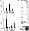
Figure 1.
Serum-induced activation of SRF reporter requires the ROCK–LIMK–cofilin pathway in PC12, SKNMC, and Neuro2A cells. (A) The different cell types were transfected with the SRF reporter together with expression plasmids encoding C3 transferase (50 ng), nonphosphorylatable cofilin S3A (0.5 or 2.0 μg), kinase-inactive LIMK1 kinase domain (K1C.D460A) (0.5 or 1.0 μg), or kinase-inactive ROCKΔ3 K105A (0.5 or 2.0 μg) as indicated, or treated with latrunculin B (LB; 0.5 μM) or Y27632 (10 μM). Serum induction of reporter activity was measured and quantitated as described in the Materials and methods; error bars show SEM (n = 3) for PC12 and Neuro2A, and half range (n = 2) for SKNMC. (B) SRF activation by serum stimulation does not require the ROCK–LIMK–cofilin pathway in NIH3T3 cells. NIH3T3 cells were transfected with the SRF reporter and expression plasmids as in A. Reporter activity was measured and quantitated taking serum-induced level as 100%. Error bars show half range (n = 2). (C) Activation of SRF by Rho GTPases and actin-binding drugs in PC12 cells. Cells were transfected with the SRF reporter and either cotransfected with expression plasmids encoding RhoA.V14 (1.0 μg), Rac1.V12 (0.3 μg), Cdc42.V12 (0.3 μg), or N-WASP minimal F-actin–nucleating VCA domain (0.3 μg), or treated with 2 μM cytochalasin D or 0.5 μM jasplakinolide before reporter activity was measured. (D) Serum-induced activation of the SRF target gene vinculin requires functional ROCK in PC12 cells. Serum-starved PC12 cells were serum stimulated with or without Y27632 pretreatment as indicated. Total RNA was analyzed for vinculin and GAPDH transcripts by RNA'ase protection.
Figure 2.
Figure 2.
Cofilin phosphorylation via the Rho pathway in PC12 cells. The 2D gels are shown with acidic isoelectric point to the right, SDS-PAGE top to bottom. (A) Serum-induced phosphorylation of endogenous cofilin requires ROCK activity. PC12 cells were maintained in 0.5% serum before stimulation with 15% serum with or without Y27632 (Y) pretreatment. Cell extracts were prepared and fractionated by 2D gel electrophoresis; cofilin was detected by immunoblotting. Where indicated, extracts were treated with alkaline phosphatase (P'tase) before analysis. (B) FLAG–cofilin reporter assay. PC12 cells transfected with the indicated FLAG–cofilin expression plasmids (0.2 μg) were analyzed by 2D gel electrophoresis and immunoblotting with anti-FLAG antibody; phosphatase treatment was performed on anti-FLAG immunoprecipitates. (C) Phosphorylation status of FLAG–cofilin under different growth conditions. Cells transfected with wild-type FLAG–cofilin expression plasmids were maintained as indicated and analyzed as in part B. Only the cofilin species are shown; the positions of unphosphorylated and phosphorylated forms are indicated above the figure. (D) Serum-induced phosphorylation of FLAG–cofilin requires RhoA and ROCK activity. Cells transfected with expression plasmids encoding wild-type FLAG–cofilin and C3 transferase (50 ng), as indicated, were serum starved and restimulated with 15% serum for 5 min; Y27632 treatment (Y) was as indicated.
Figure 3.
Figure 3.
LIMK1 requires an additional RhoA-dependent event to activate SRF. (A) Sensitivity of SRF activation to C3 transferase. PC12 cells were transfected with the SRF reporter and expression plasmids encoding RhoA.V14 (1.0 μg), LIMK1 (1.0 μg), or ROCKΔ3 (60 ng), and C3 transferase (50 ng) as indicated, and reporter activity was measured. (B) FLAG–cofilin phosphorylation does not require RhoA. Cells were transfected with expression plasmids encoding wild-type FLAG–cofilin (0.2 μg) alone or with LIMK1 (1.0 μg), ROCKΔ3 (30 ng), LIMK1 kinase domain K1.C (0.1 μg), or ROCK-insensitive LIMK1 kinase domain mutant K1.C/T508V (1.0 μg), and C3 transferase (50 ng) as indicated, before processing by 2D gel electrophoresis followed by anti-FLAG immunoblot. Only the FLAG–cofilin species are shown; the positions of unphosphorylated and phosphorylated forms are indicated above the figure. (C) SRF activation by LIMK1 T508V requires RhoA. Cells were transfected with the SRF reporter and expression plasmids encoding LIMK1 kinase domain K1.C (0.1 μg) or ROCK-insensitive LIMK1 kinase domain mutant K1.C/T508V (0.1, 0.3, or 1.0 μg), with C3 transferase (50 ng) as indicated, and reporter activity was measured. (D) LIMK1 derivatives. The LIMK1 coding region is indicated at the top with LIM, PDZ, and kinase domains shown as boxes; the K1.C kinase domain (residues 293–646) is shown below and its ROCK-insensitive mutant T508V is also indicated.
Figure 4.
Figure 4.
mDia activity is required for SRF activation in PC12 cells. (A) mDia1 derivatives. The mDia1 coding region is indicated at the top with RhoA-binding domain as a circle and formin homology regions 1–3 as boxes; below are shown the activated mDia1 derivative Dia1* (residues 256–1255), the inactive interfering derivative dnDia1 (residues 571–1181, lacking 750–771), and the control inactive noninterfering derivative ΔDia1 (residues 256–567). (B) SRF activation by mDia1 in PC12 cells. Cells were transfected with the SRF reporter and expression plasmids encoding Dia1* (0.3 μg) with C3 transferase (50 ng), as indicated, and reporter activity was measured. The effect of C3 transferase on reporter activation by the synthetic activator SRF-VP16 (50 ng) is shown for comparison. Error bars show half range (n = 2). (C) mDia1 does not promote cofilin phosphorylation. Cells were transfected with expression plasmids encoding wild-type FLAG–cofilin (0.2 μg) alone or with Dia1* (0.3 μg), dnDia1 (2.0 μg), or LIMK1 (1.0 μg). After maintenance in 0.5% serum with serum stimulation as indicated, cells were analyzed for FLAG–cofilin phosphorylation by 2D gel electrophoresis followed by anti-FLAG immunoblot. Only the FLAG–cofilin species are shown; the positions of unphosphorylated and phosphorylated forms are indicated above the figure. (D) Serum-induced SRF activation requires functional mDia. Left, cells were transfected with the SRF reporter and an expression plasmid encoding dnDia1 (2.0 μg), as indicated, and either maintained in 0.5% serum or serum stimulated before measurement of reporter activity. Serum-stimulated reporter activity is taken as 100%. Error bars show SEM (n = 4). (E) Cofilin S3A potentiates the effect of interfering mDia. Cells were transfected with the SRF reporter together with expression plasmids encoding dnDia1 (2.0 μg) and/or cofilin S3A (0.5 or 2.0 μg), as indicated, and either was maintained in 0.5% serum or was serum stimulated before quantitation of reporter activity. Serum-induced reporter activity is taken as 100%. Error bars show half range (n = 2).
Figure 5.
Figure 5.
mDia is required for LIMK1-induced SRF activation and F-actin assembly. (A) LIMK1-induced SRF activation requires mDia. PC12 cells were transfected with expression plasmids encoding LIMK1 (1.0 μg), inactive mDia derivative ΔDia1 (Dia1 256–567; 3.0 μg), or interfering mDia1 derivative dnDia1 (Figure 4 A; 3.0 μg), as indicated, together with the SRF reporter plasmid and maintained in 0.5% serum before quantitation of reporter activity, taking LIMK1+ΔDia1 as 100%. Error bars show SEM (n = 3). (B) dnDia1 does not affect LIMK1- induced cofilin phosphorylation. Cells were transfected with expression plasmids encoding wild-type FLAG–cofilin (0.2 μg) alone or with LIMK1 (1.0 μg) and dnDia1 (2.0 μg) as indicated. After maintenance in 0.5% serum, cells were analyzed for FLAG–cofilin phosphorylation by 2D gel electrophoresis and anti-FLAG immunoblot. Only the FLAG–cofilin species are shown; the positions of unphosphorylated and phosphorylated forms are indicated above the figure. (C) LIMK-induced F-actin accumulation requires mDia activity. PC12 cells were transfected with expression plasmids encoding LIMK1 (1.0 μg), inactive mDia derivative ΔDia1 (Dia1 256–567; 3.0 μg), or interfering mDia1 derivative dnDia1 (Figure 4 A; 3.0 μg). The mean F-actin content in transfected cells was determined using FACS® analysis and expressed relative to the mean F-actin content of untransfected cells from the same population, indicated by the dotted line (Materials and methods). The chart summarizes three independent experiments of this type, presenting the average of the mean F-actin content in each population ± SEM; statistical significance was estimated using the unpaired t test.
Figure 6.
Figure 6.
Regulatory pathways to SRF via actin dynamics. The actin treadmilling cycle is shown schematically with actin monomer as a wedge, profilin as a rectangle, and cofilin as a circle. The treadmilling cycle is regulated by two Rho effector pathways. The Dia proteins promote F-actin accumulation via an uncharacterized mechanism, perhaps involving recruitment of profilin–actin to the Dia FH1 domain. The ROCK–LIMK–cofilin cascade negatively regulates cofilin activity, thereby increasing the F-actin level by inhibiting monomer dissociation and F-actin severing. Serum-induced SRF activation in PC12 cells is dependent on Rho signaling and actin polymerization, consistent with previous findings that SRF activity responds to depletion of the cellular G-actin pool or subpopulation of it (Sotiropoulos et al., 1999; unpublished data). Maximal serum-induced activity in PC12 cells requires operation of both ROCK–LIMK–cofilin and Dia pathways. The ability of LIMK both to activate SRF and to increase the F-actin level in PC12 cells is dependent on Dia proteins, suggesting that the activity of LIMK is targeted to a pool of Dia-dependent F-actin.

Similar articles

Cited by

References

    1. Alberts, A.S. 2001. Identification of a carboxy-terminal diaphanous-related formin homology protein autoregulatory domain. J. Biol. Chem. 276:2824–2830. - PubMed
    1. Amano, M., M. Ito, K. Kimura, Y. Fukata, K. Chihara, T. Nakano, Y. Matsuura, and K. Kaibuchi. 1996. Phosphorylation and activation of myosin by Rho-associated kinase (Rho-kinase). J. Biol. Chem. 271:20246–20249. - PubMed
    1. Amano, M., Y. Fukata, and K. Kaibuchi. 2000. Regulation and functions of Rho-associated kinase. Exp. Cell Res. 261:44–51. - PubMed
    1. Arber, S., F.A. Barbayannis, H. Hanser, C. Schneider, C.A. Stanyon, O. Bernard, and P. Caroni. 1998. Regulation of actin dynamics through phosphorylation of cofilin by LIM-kinase. Nature. 393:805–809. - PubMed
    1. Arsenian, S., B. Weinhold, M. Oelgeschlager, U. Ruther, and A. Nordheim. 1998. Serum response factor is essential for mesoderm formation during mouse embryogenesis. EMBO J. 17:6289–6299. - PMC - PubMed

Publication types